

A high-affinity, high-stability photoacoustic agent for imaging gastrin-releasing peptide receptor in prostate cancer
- PMID: 24850845
- PMCID: PMC4121111
- DOI: 10.1158/1078-0432.CCR-13-3405
A high-affinity, high-stability photoacoustic agent for imaging gastrin-releasing peptide receptor in prostate cancer
Abstract
Purpose: To evaluate the utility of targeted photoacoustic imaging (PAI) in providing molecular information to complement intrinsic functional and anatomical details of the vasculature within prostate lesion.
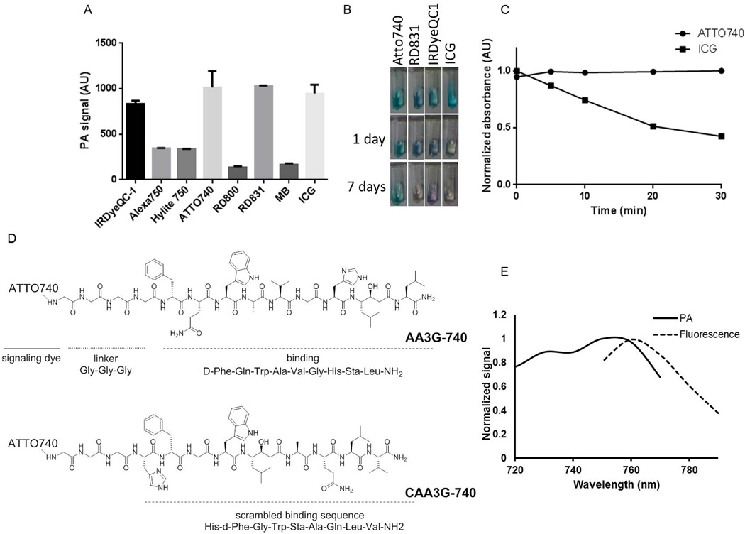
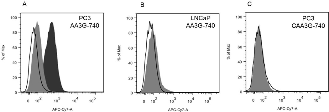
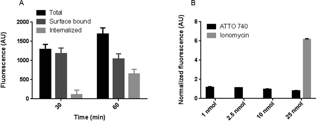
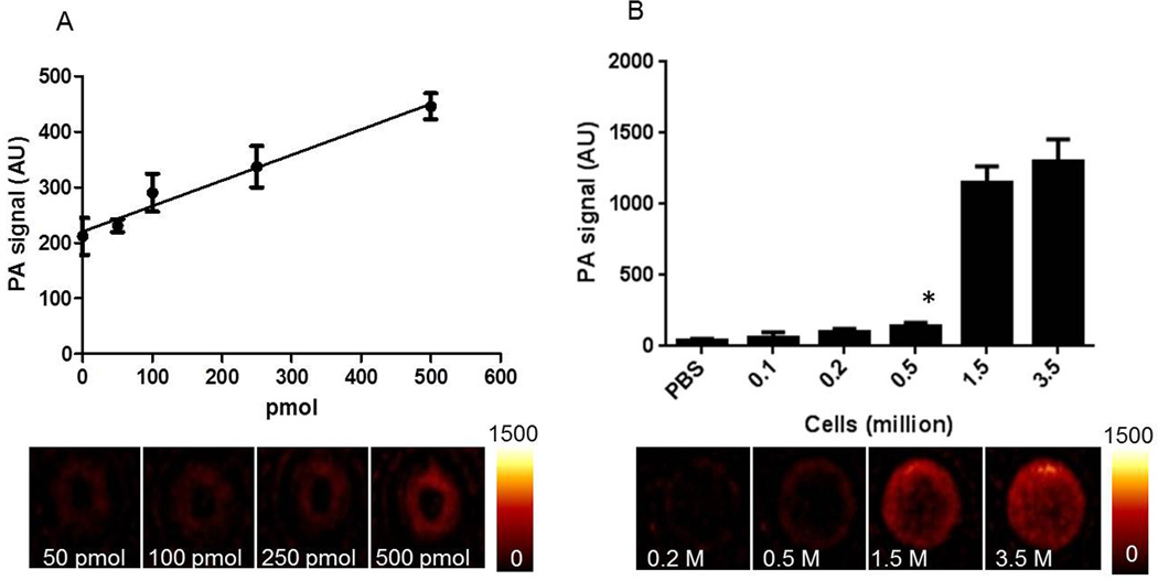
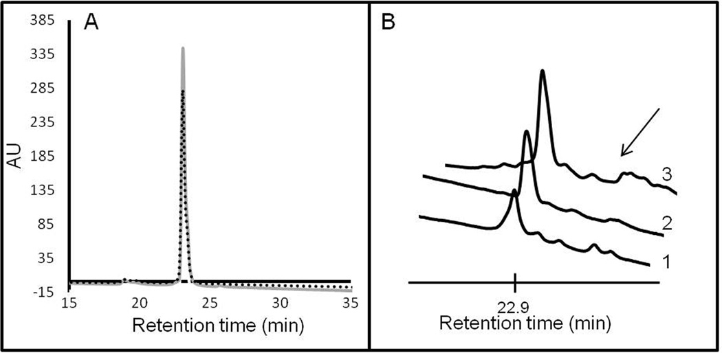
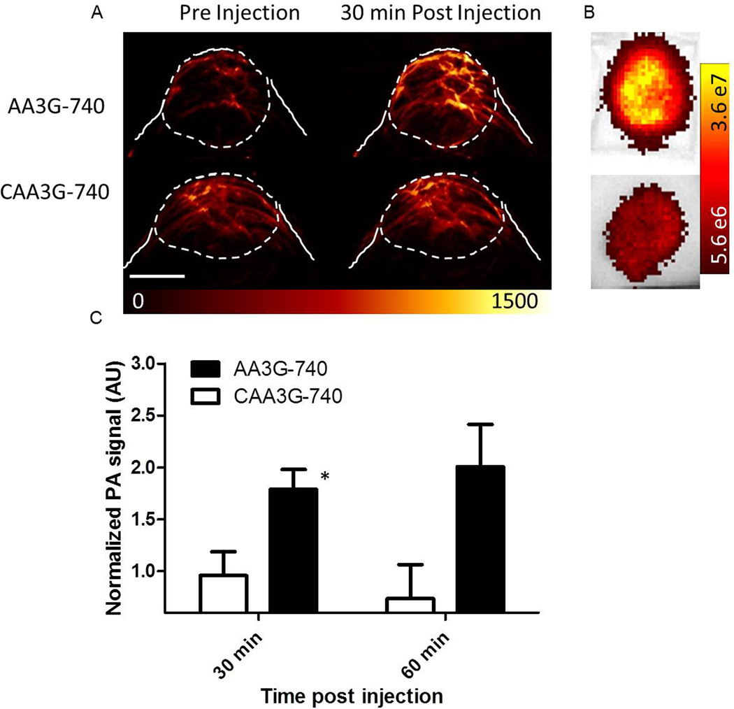
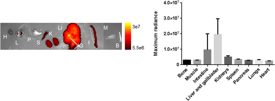
Experimental design: We developed a PAI agent, AA3G-740, that targets gastrin-releasing peptide receptor (GRPR), found to be highly overexpressed in prostate cancer. The binding specificity of the agent was evaluated in human prostate cancer cell lines, PC3 and LNCaP, and antagonist properties determined by cell internalization and intracellular calcium mobilization studies. The imaging sensitivity was assessed for the agent itself and for the PC3 cells labeled with agent. The in vivo stability of the agent was determined in human plasma and in the blood of living mice. The in vivo binding of the agent was evaluated in PC3 prostate tumor models in mice, and was validated ex vivo by optical imaging.
Results: AA3G-740 demonstrated strong and specific binding to GRPR. The sensitivity of detection in vitro indicated suitability of the agent to image very small lesions. In mice, the agent was able to bind to GRPR even in poorly vascularized tumors leading to nearly 2-fold difference in photoacoustic signal relative to the control agent.
Conclusions: The ability to image both vasculature and molecular profile outside the blood vessels gives molecular PAI a unique advantage over currently used imaging techniques. The imaging method presented here can find application both in diagnosis and in image-guided biopsy.
©2014 American Association for Cancer Research.
Figures
Similar articles
-
Synthesis and radiopharmacological evaluation of a high-affinity and metabolically stabilized 18F-labeled bombesin analogue for molecular imaging of gastrin-releasing peptide receptor-expressing prostate cancer.Nucl Med Biol. 2013 Nov;40(8):1025-34. doi: 10.1016/j.nucmedbio.2013.07.005. Epub 2013 Aug 19. Nucl Med Biol. 2013. PMID: 23969085
-
18F-labeled bombesin analog for specific and effective targeting of prostate tumors expressing gastrin-releasing peptide receptors.J Nucl Med. 2011 Feb;52(2):270-8. doi: 10.2967/jnumed.110.081620. Epub 2011 Jan 13. J Nucl Med. 2011. PMID: 21233180
-
Bombesin antagonist-based radioligands for translational nuclear imaging of gastrin-releasing peptide receptor-positive tumors.J Nucl Med. 2011 Dec;52(12):1970-8. doi: 10.2967/jnumed.111.094375. Epub 2011 Nov 11. J Nucl Med. 2011. PMID: 22080443
-
Nuclear imaging of prostate cancer with gastrin-releasing-peptide-receptor targeted radiopharmaceuticals.Curr Pharm Des. 2008;14(28):3033-47. doi: 10.2174/138161208786404335. Curr Pharm Des. 2008. PMID: 18991717 Review.
-
Peptide receptor imaging of prostate cancer with radiolabelled bombesin analogues.Methods. 2009 Jun;48(2):200-4. doi: 10.1016/j.ymeth.2009.04.002. Epub 2009 May 3. Methods. 2009. PMID: 19398012 Review.
Cited by
-
Water-Soluble Chitosan Conjugated DOTA-Bombesin Peptide Capped Gold Nanoparticles as a Targeted Therapeutic Agent for Prostate Cancer.Nanotechnol Sci Appl. 2021 Mar 18;14:69-89. doi: 10.2147/NSA.S301942. eCollection 2021. Nanotechnol Sci Appl. 2021. PMID: 33776426 Free PMC article.
-
Review of cost reduction methods in photoacoustic computed tomography.Photoacoustics. 2019 Jul 26;15:100137. doi: 10.1016/j.pacs.2019.100137. eCollection 2019 Sep. Photoacoustics. 2019. PMID: 31428558 Free PMC article. Review.
-
Advances in Diagnostic and Intraoperative Molecular Imaging of Pancreatic Cancer.Pancreas. 2018 Jul;47(6):675-689. doi: 10.1097/MPA.0000000000001075. Pancreas. 2018. PMID: 29894417 Free PMC article. Review.
-
Emerging Intraoperative Imaging Modalities to Improve Surgical Precision.Mol Imaging Biol. 2018 Oct;20(5):705-715. doi: 10.1007/s11307-018-1227-6. Mol Imaging Biol. 2018. PMID: 29916118 Review.
-
Photoacoustic and Fluorescence Imaging of Cutaneous Squamous Cell Carcinoma in Living Subjects Using a Probe Targeting Integrin αvβ6.Sci Rep. 2017 Feb 9;7:42442. doi: 10.1038/srep42442. Sci Rep. 2017. PMID: 28181579 Free PMC article.
References
-
- [Accessed on May31st 2013]; http://www.uptodate.com/contents/clinical-presentation-and-diagnosis-of-....
-
- Hricak H, Choyke PL, Eberhardt SC, Leibel SA, Scardino PT. Imaging prostate cancer: a multidisciplinary perspective. Radiology. 2007;243:28–53. - PubMed
Publication types
MeSH terms
Substances
Grants and funding
LinkOut - more resources
Full Text Sources
Other Literature Sources
Medical
