

MIF antagonist (CPSI-1306) protects against UVB-induced squamous cell carcinoma
- PMID: 24850900
- PMCID: PMC4284200
- DOI: 10.1158/1541-7786.MCR-14-0255-T
MIF antagonist (CPSI-1306) protects against UVB-induced squamous cell carcinoma
Abstract
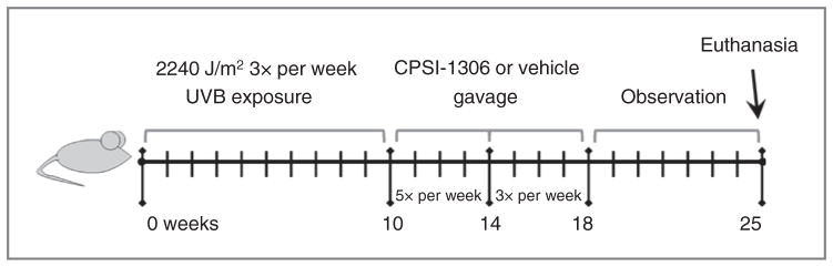
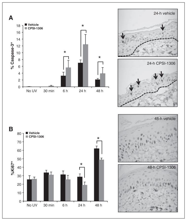
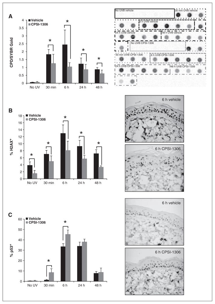
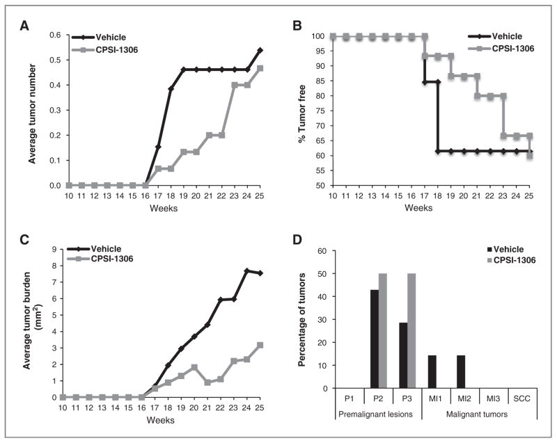
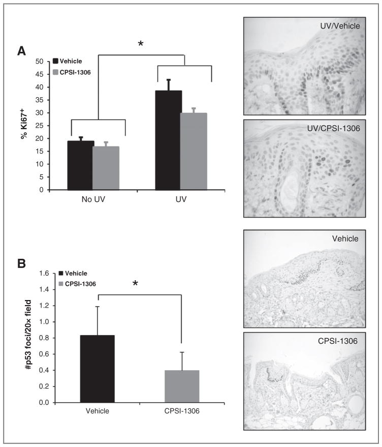
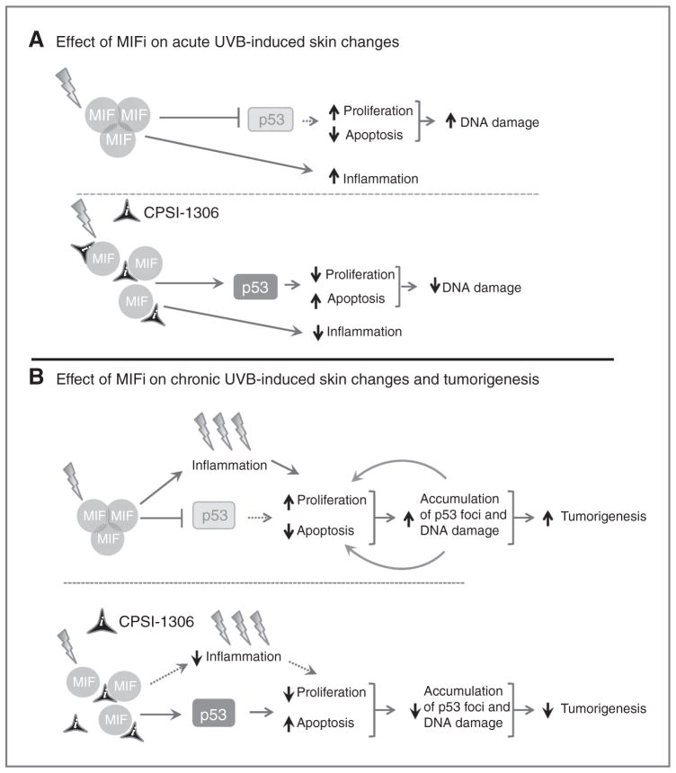
Macrophage migration inhibitory factor (MIF) is a homotrimeric proinflammatory cytokine implicated in chronic inflammatory diseases and malignancies, including cutaneous squamous cell carcinomas (SCC). To determine whether MIF inhibition could reduce UVB light-induced inflammation and squamous carcinogenesis, a small-molecule MIF inhibitor (CPSI-1306) was utilized that disrupts homotrimerization. To examine the effect of CPSI-1306 on acute UVB-induced skin changes, Skh-1 hairless mice were systemically treated with CPSI-1306 for 5 days before UVB exposure. In addition to decreasing skin thickness and myeloperoxidase (MPO) activity, CPSI-1306 pretreatment increased keratinocyte apoptosis and p53 expression, decreased proliferation and phosphohistone variant H2AX (γ-H2AX), and enhanced repair of cyclobutane pyrimidine dimers. To examine the effect of CPSI-1306 on squamous carcinogenesis, mice were exposed to UVB for 10 weeks, followed by CPSI-1306 treatment for 8 weeks. CPSI-1306 dramatically decreased the density of UVB-associated p53 foci in non-tumor-bearing skin while simultaneously decreasing the epidermal Ki67 proliferation index. In addition to slowing the rate of tumor development, CPSI-1306 decreased the average tumor burden per mouse. Although CPSI-1306-treated mice developed only papillomas, nearly a third of papillomas in vehicle-treated mice progressed to microinvasive SCC. Thus, MIF inhibition is a promising strategy for prevention of the deleterious cutaneous effects of acute and chronic UVB exposure.
Implications: Macrophage migration inhibitory factor is a viable target for the prevention of UVB-induced cutaneous SSCs.
©2014 American Association for Cancer Research.
Conflict of interest statement
T. Sielecki has ownership interest (including patents) in Cytokine PharmaSciences. No potential conflicts of interest were disclosed by the other authors.
Figures
Similar articles
-
Macrophage migration inhibitory factor inhibition as a novel therapeutic approach against triple-negative breast cancer.Cell Death Dis. 2020 Sep 17;11(9):774. doi: 10.1038/s41419-020-02992-y. Cell Death Dis. 2020. PMID: 32943608 Free PMC article.
-
Macrophage migration inhibitory factor (MIF) plays a critical role in pathogenesis of ultraviolet-B (UVB) -induced nonmelanoma skin cancer (NMSC).FASEB J. 2009 Mar;23(3):720-30. doi: 10.1096/fj.08-119628. Epub 2008 Oct 24. FASEB J. 2009. PMID: 18952710
-
Deficient deletion of apoptotic cells by macrophage migration inhibitory factor (MIF) overexpression accelerates photocarcinogenesis.Carcinogenesis. 2009 Sep;30(9):1597-605. doi: 10.1093/carcin/bgp160. Epub 2009 Jul 7. Carcinogenesis. 2009. PMID: 19584138
-
Effect of caffeine on UVB-induced carcinogenesis, apoptosis, and the elimination of UVB-induced patches of p53 mutant epidermal cells in SKH-1 mice.Photochem Photobiol. 2008 Mar-Apr;84(2):330-8. doi: 10.1111/j.1751-1097.2007.00263.x. Epub 2008 Jan 7. Photochem Photobiol. 2008. PMID: 18179623 Review.
-
[Apoptosis, UV-radiation, precancerosis and skin tumors].Acta Med Croatica. 2009 Oct;63 Suppl 2:53-8. Acta Med Croatica. 2009. PMID: 19999548 Review. Croatian.
Cited by
-
Bioinspired polydopamine nanoparticles as efficient antioxidative and anti-inflammatory enhancers against UV-induced skin damage.J Nanobiotechnology. 2023 Sep 30;21(1):354. doi: 10.1186/s12951-023-02107-7. J Nanobiotechnology. 2023. PMID: 37775761 Free PMC article.
-
Macrophage migration inhibitory factor inhibition as a novel therapeutic approach against triple-negative breast cancer.Cell Death Dis. 2020 Sep 17;11(9):774. doi: 10.1038/s41419-020-02992-y. Cell Death Dis. 2020. PMID: 32943608 Free PMC article.
-
Macrophage migration inhibitory factor: a potential driver and biomarker for head and neck squamous cell carcinoma.Oncotarget. 2017 Feb 7;8(6):10650-10661. doi: 10.18632/oncotarget.12890. Oncotarget. 2017. PMID: 27788497 Free PMC article. Review.
-
Sex differences in skin carotenoid deposition and acute UVB-induced skin damage in SKH-1 hairless mice after consumption of tangerine tomatoes.Mol Nutr Food Res. 2015 Dec;59(12):2491-501. doi: 10.1002/mnfr.201500317. Epub 2015 Oct 20. Mol Nutr Food Res. 2015. PMID: 26394800 Free PMC article.
-
Targeted Knockdown of Macrophage Migration Inhibitory Factor Enhances UVB Irradiation-Induced Apoptosis Via Increasing ROS Generation in Oral Squamous Cell Carcinoma.Technol Cancer Res Treat. 2023 Jan-Dec;22:15330338231163436. doi: 10.1177/15330338231163436. Technol Cancer Res Treat. 2023. PMID: 37272017 Free PMC article.
References
-
- Candido J, Hagemann T. Cancer-related inflammation. J Clin Immunol. 2013;33 (Suppl 1):S79–84. - PubMed
-
- Mantovani A, Allavena P, Sica A, Balkwill F. Cancer-related inflammation. Nature. 2008;454:436–44. - PubMed
-
- Bucala R, Donnelly SC. Macrophage migration inhibitory factor: a probable link between inflammation and cancer. Immunity. 2007;26:281–5. - PubMed
-
- Sanchez-Zamora Y, Terrazas LI, Vilches-Flores A, Leal E, Juarez I, Whitacre C, et al. Macrophage migration inhibitory factor is a therapeutic target in treatment of non-insulin-dependent diabetes mellitus. FASEB J. 2010;24:2583–90. - PubMed
Publication types
MeSH terms
Substances
Grants and funding
LinkOut - more resources
Full Text Sources
Other Literature Sources
Medical
Molecular Biology Databases
Research Materials
Miscellaneous
