. 2014 May;4(5):140041.
doi: 10.1098/rsob.140041.

Titin kinase is an inactive pseudokinase scaffold that supports MuRF1 recruitment to the sarcomeric M-line

Affiliations

Titin kinase is an inactive pseudokinase scaffold that supports MuRF1 recruitment to the sarcomeric M-line

Julijus Bogomolovas et al. Open Biol. 2014 May.

Abstract

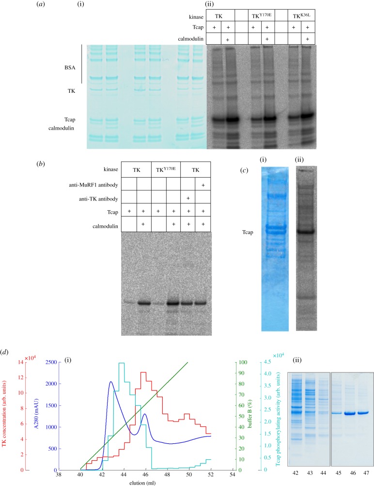
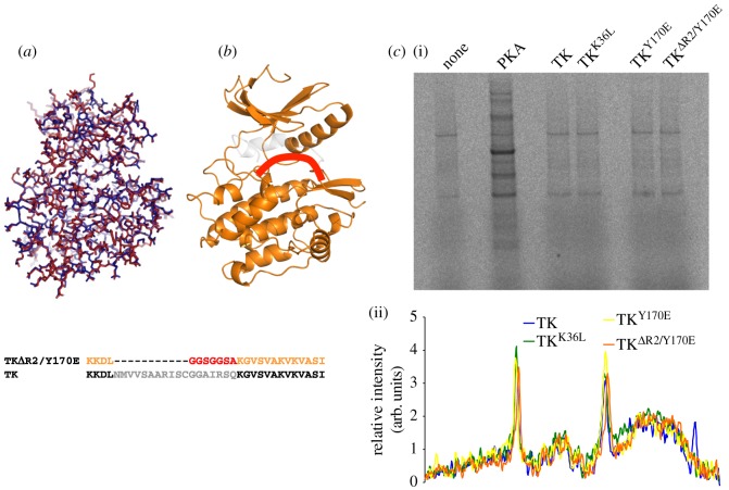
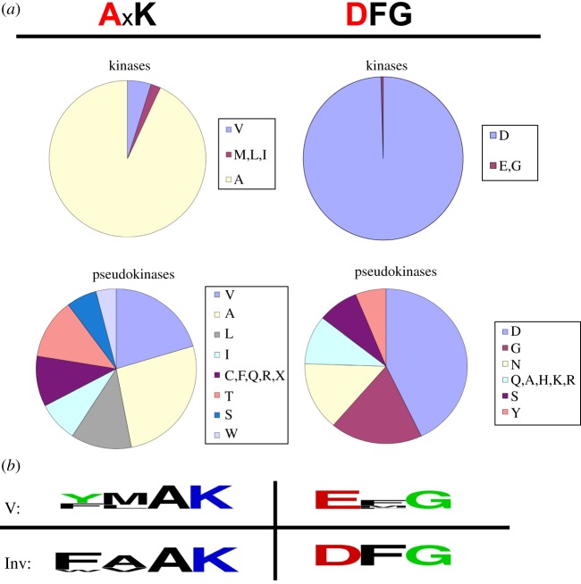
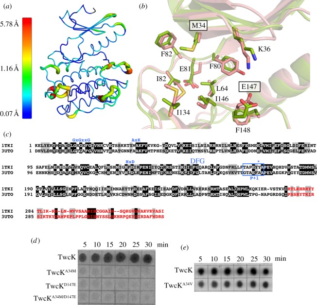
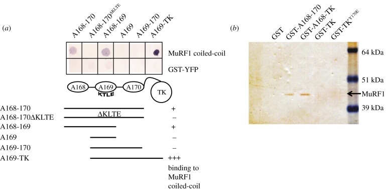
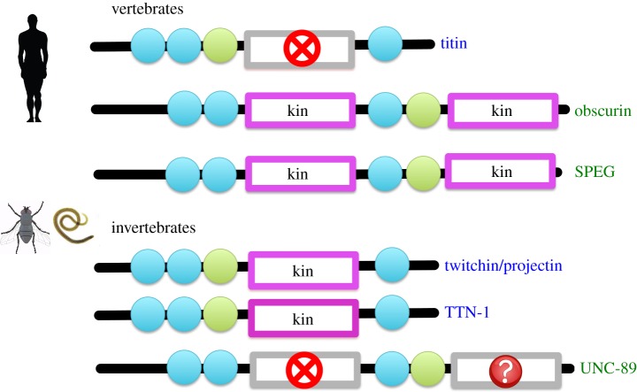
Striated muscle tissues undergo adaptive remodelling in response to mechanical load. This process involves the myofilament titin and, specifically, its kinase domain (TK; titin kinase) that translates mechanical signals into regulatory pathways of gene expression in the myofibril. TK mechanosensing appears mediated by a C-terminal regulatory tail (CRD) that sterically inhibits its active site. Allegedly, stretch-induced unfolding of this tail during muscle function releases TK inhibition and leads to its catalytic activation. However, the cellular pathway of TK is poorly understood and substrates proposed to date remain controversial. TK's best-established substrate is Tcap, a small structural protein of the Z-disc believed to link TK to myofibrillogenesis. Here, we show that TK is a pseudokinase with undetectable levels of catalysis and, therefore, that Tcap is not its substrate. Inactivity is the result of two atypical residues in TK's active site, M34 and E147, that do not appear compatible with canonical kinase patterns. While not mediating stretch-dependent phospho-transfers, TK binds the E3 ubiquitin ligase MuRF1 that promotes sarcomeric ubiquitination in a stress-induced manner. Given previous evidence of MuRF2 interaction, we propose that the cellular role of TK is to act as a conformationally regulated scaffold that functionally couples the ubiquitin ligases MuRF1 and MuRF2, thereby coordinating muscle-specific ubiquitination pathways and myofibril trophicity. Finally, we suggest that an evolutionary dichotomy of kinases/pseudokinases has occurred in TK-like kinases, where invertebrate members are active enzymes but vertebrate counterparts perform their signalling function as pseudokinase scaffolds.

Keywords: mutagenesis; phosphorylation assay; pseudokinase; titin.


Figures

Figure 1.
Figure 1.
Tcap phosphorylation assays using TK preparations from insect cells. (a) Preparations of wild-type TK, the activated TKY170E phosphomimic and the constitutively inactive TKK36L phosphorylate Tcap comparably and stimulated by Ca2+/calmodulin. (i) SDS-PAGE and (ii) autoradiogram of catalysis by samples after Ni2+-NTA are shown. (b) Phosphorylation assay of TK sterically inhibited by immuno-complexation with an antibody raised against the P+1 loop vicinal to the active site. An antibody (anti-MuRF1) that does not complex TK is included for comparison. (c) Untransfected Sf21 cell extracts supplemented with Tcap (but not Ca2+/calmodulin) display phosphorylating activity (the stimulation of catalysis upon addition of calmodulin was approx. 25%, this modest activation is likely due to the presence of endogenous calmodulin in the extract). (i) SDS-PAGE and (ii) autoradiogram revealing Tcap phosphorylation. (d) (i) Chromatogram and (ii) corresponding SDS-PAGE of Sf21 cell crude extract containing recombinant TKK36L eluted from a Ni2+-NTA column. Segregation of phosphorylating activity (cyan) and TK (red) during purification is observed. Bound proteins were eluted with a linear gradient of imidazole (100% buffer B = 0.3 M imidazole; green line) and monitored by A280; the resultant chromatogram is in blue. The content of TKK36L in eluted fractions was determined by spot-blot immunoassay using anti-TK P+1 loop antibody. The amount of coloured product quantified densitometrically was proportional to the amount of TKK36L in each fraction (red). Phosphorylation of a Tcap-derived peptide substrate in the presence of calmodulin was quantified in each fraction densitometrically by our standard phosphorylation assay that used [γ-33P]ATP and spotting on P81 paper (cyan). The data show that Tcap phosphorylation segregated from TKK36L.
Figure 2.
Figure 2.
Structural and functional characterization of TK produced in E. coli. (a) Superposition of the crystal structures of TK expressed in bacteria (red) and insect cells (PDB entry 1TKI) (blue). The overall RMSD for Cα atoms is 0.29 Å. (b) Representation of the activated variant TKΔR2/Y170E, where the deleted fraction is in grey and the added loop is shown schematically in red. The sequence exchanges in this variant are shown below. (c) Identification of potential TK substrates in differentiating C2C12 cell extracts depleted of endogenous kinases by treatment with FSBA. Protein kinase A (PKA) was used as positive control. (i) Autoradiogram and (ii) densitogram of phosphor-image are provided. The data show no significant differences in labelling pattern when comparing cell extract alone or supplemented with activated forms of TK.
Figure 3.
Figure 3.
TK contains atypical residues in catalytic motifs. (a) Distribution of residues in position 2 of the VAIK motif and position 1 of the DFG motif in protein kinases of the human kinome. The classification of kinases and pseudokinases was taken from [32]. TK, the only human kinase containing an EFG motif, is misclassified as active kinase due to previous reports of catalysis [10,13,16]. Other kinases with deviant catalytic motifs are: CASK (GFG motif); ATR (xMxK motif); LMR2, NEK8 and RIPK1 (IxK); and DNAPK, FRAP and SMG1 (LxK). (b) Commonly occurring residues in the VAIK and DFG motifs of titin kinases from vertebrate (V) and invertebrate titin-like kinases (Inv). (Sequences for representative kinases of each group are given in the electronic supplementary material, figure S5.)
Figure 4.
Figure 4.
Comparison of TK and TwcK active sites. (a) Structural superposition of TK and ceTwcK (PDB entry 3UTO). Ribbon thickness and colouring indicate the RMSD values of the superposition as given in the accompanying scale (minimum, maximum and average values are shown). The structural agreement is excellent overall, including active site regions; divergences only occur in peripheral loop areas. (b) Detailed comparison of the ATP-binding pockets of TK (green) and ceTwcK (pink) (numbering corresponds to TK). Boxed labels indicate TK residues that were trans-engineered into ceTwcK. (c) Structure-based sequence alignment of the catalytic domains of human TK and ceTwcK corresponding to the superposition displayed in (a,b). Identical residues are highlighted in black and closest conservation is in grey. The canonical composition of functional motifs is shown in blue, the P+1 loop is boxed and the tyrosine residue undergoing phosphorylation in TK is indicated with an asterisk. The CRD is in red. (d) Comparative autoradiogram of catalysis by ecTwck and its variants TwcKA34M, TwcKD147E and TwcKA34M/D147E carrying TK residues in their ATP-binding pockets. The time course shows phosphorylation of a MLC-derived peptide. (e) Comparative autoradiogram of the catalysis from ecTwck and TwcKA34V. The latter carries the non-inactivating valine residue commonly found in the ATP-binding pocket of TwcK from molluscs.
Figure 5.
Figure 5.
Dissection of TK/MuRF1 molecular interactions. (a) Identification of the MuRF1–titin interaction region by filter-binding assay. The helical domain of MuRF1 interacts with its established docking site A168–A170 and the C-terminally truncated version A168–A169, but not with the loop mutant A168–A170ΔKTLE, the N-terminally truncated A169–A170 or the single domain A169. Binding to A169–A170 is restored and enhanced when TK is included in the construct (A169-TK). The latter is used here in 10-fold lower quantity than the other samples. (b) Pull-down assay using skeletal muscle extract demonstrates that A168–A170 and A168-TK bind endogenous MuRF1 efficiently, but that the binding is stronger in the presence of TK. Neither GST nor TK alone are capable of pulling down endogenous MuRF1.
Figure 6.
Figure 6.
Distribution of active kinases and inactive pseudokinases in titin-like filaments from vertebrate and invertebrate muscle. For each filament, the following domains are shown: Ig (blue circles), Fn3 (green circles), active kinases (magenta boxes) and inactive pseudokinases (grey boxes). Pseudokinases where inactivity is tentatively suggested but cannot be as reliably predicted are indicated with a question mark.

References

    1. Granzier HL, Labeit S. 2005. Titin and its associated proteins: the third myofilament system of the sarcomere. Adv. Protein Chem. 71, 89–119. (doi:10.1016/S0065-3233(04)71003-7) - DOI - PubMed
    1. Knoll R, et al. 2011. Telethonin deficiency is associated with maladaptation to biomechanical stress in the mammalian heart. Circ. Res. 109, 758–769. (doi:10.1161/CIRCRESAHA.111.245787) - DOI - PMC - PubMed
    1. Lange S, Auerbach D, McLoughlin P, Perriard E, Schafer BW, Perriard JC, Ehler E. 2002. Subcellular targeting of metabolic enzymes to titin in heart muscle may be mediated by DRAL/FHL-2. J. Cell Sci. 115, 4925–4936. (doi:10.1242/jcs.00181) - DOI - PubMed
    1. Miller MK, et al. 2003. The muscle ankyrin repeat proteins: CARP, ankrd2/Arpp and DARP as a family of titin filament-based stress response molecules. J. Mol. Biol. 333, 951–964. (doi:10.1016/j.jmb.2003.09.012) - DOI - PubMed
    1. Ojima K, et al. 2010. Dynamic distribution of muscle-specific calpain in mice has a key role in physical-stress adaptation and is impaired in muscular dystrophy. J. Clin. Invest. 120, 2672–2683. (doi:10.1172/JCI40658) - DOI - PMC - PubMed

Publication types

MeSH terms

Associated data

LinkOut - more resources