Review
. 2013 Jan 15;257(2):541-563.
doi: 10.1016/j.ccr.2012.04.028.

Structure and function of atypically coordinated enzymatic mononuclear non-heme-Fe(II) centers

Affiliations
Review

Structure and function of atypically coordinated enzymatic mononuclear non-heme-Fe(II) centers

Daniela Buongiorno et al. Coord Chem Rev. .

Abstract

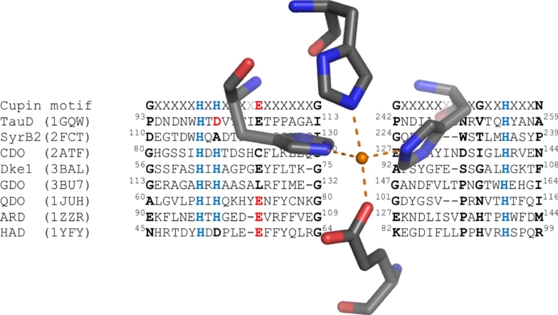
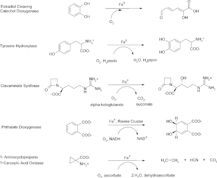
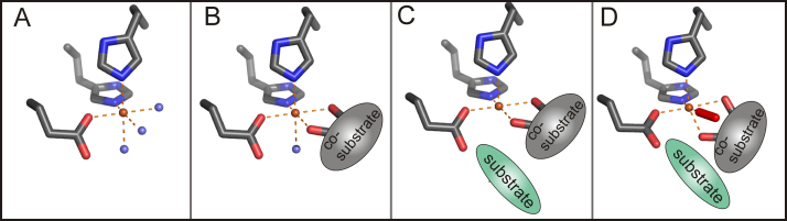
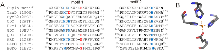
Mononuclear, non-heme-Fe(II) centers are key structures in O2 metabolism and catalyze an impressive variety of enzymatic reactions. While most are bound via two histidines and a carboxylate, some show a different organization. A short overview of atypically coordinated O2 dependent mononuclear-non-heme-Fe(II) centers is presented here Enzymes with 2-His, 3-His, 3-His-carboxylate and 4-His bound Fe(II) centers are discussed with a focus on their reactivity, metal ion promiscuity and recent progress in the elucidation of their enzymatic mechanisms. Observations concerning these and classically coordinated Fe(II) centers are used to understand the impact of the metal binding motif on catalysis.

Keywords: 1,3-bis(2-pyridylimino)isoindoline, ind; 2OH-1,3-Ph2PD, 2-hydroxy-1,3-diphenylpropanedione; 6-Ph2TPA, N,N-bis[(6-phenyl-2-pyridyl)methyl]-N-[(2-pyridyl)-methyl]amine; ADO, cysteamine dioxygenase; AO, apocarotenoid 15,15′-oxygenase; ARD, aci-reductone dioxygenase; BsQDO, quercetin 2,3-dioxygenase from Bacillus subtilis; CD, circular dichroism; CDO, cysteine dioxygenase; CGDO, 5-chloro-gentisate 1,2-dioxygenase; CS2, clavaminate synthase; CarOs, carotenoid oxygenases; DFT, density functional theory; Dioxygen activation; Dioxygenase; Dke1, diketone dioxygenase; EPR, electron paramagnetic resonance; EXAFS, extended X-ray absorption fine structure spectroscopy; Enzyme catalysis; Facial triad; GDO, gentisate 1,2-dioxygenase; HADO, 3-hydroxyanthranilate 3,4-dioxygenase; HGDO, homogentisate 1,2-dioxygenase; HNDO, hydroxy-2-naphthoate dioxygenase; MCD, magnetic circular dichroism; MNHEs, mononuclear non-heme-Fe(II) dependent enzymes; Metal binding motif; NRP, nonribosomal peptide; OTf-, trifluormethanesulfonate; PDB, protein data bank; QDO, quercetin 2,3-dioxygenase; SDO, salicylate 1,2-dioxygenase; Structure–function relationships; TauD, taurine hydroxylase; XAS, X-ray absorption spectroscopy; acac, acetylacetone (2,4-pentanedione); fla, flavonolate; α-KG, α-ketoglutarate.


Figures

None
Graphical abstract
Fig. 1
Fig. 1
Prototypical reactions of MNHEs that show the 2-His-1-carboxylate ‘facial triad’ Fe(II)-binding motif.
Fig. 2
Fig. 2
Mechanistic paradigm for dioxygen activation at the 2-His-1-carboxylate facial triad MNHEs as proposed by Solomon , as exemplified by the active site of deacetoxycephalosporin C synthase (PDB: 1RXF).
Fig. 3
Fig. 3
Conserved metal binding motif in cupins. (a.) An alignment of O2 dependent cupins that use Fe(II) as a cofactor is displayed. Metal binding residues are in blue (His) and red (carboxylate), moieties in ‘conserved’ positions are in bold. Variable residues in the cupin motif are in light grey. Abbreviations indicate the MNHEs taurine hydroxylase (TauD), SyrB2, cysteine dioxygenase (CDO), diketone dioxygenase (Dke1), gentisate dioxygenase (GDO), quercetin 2,3-dioxygenase (QDO), aci-reductone dioxygenase (ARD), 3-hydroxyanthranilate3,4-dioxygenase (HADO) and homogentisate dioxygenase (HGDO). The respective protein data bank (PDB) numbers are given in brackets. (b.) The prototypical metal center organization of a cupin-metal center is shown. (For interpretation of the references to color in this figure legend, the reader is referred to the web version of the article.)
Fig. 4
Fig. 4
Schematic representation of biosynthesis of a halogenated threonyl-moiety in the biosynthetic pathway of the NRP syringomycin E. The threonyl moiety is tethered to the peptidyl-carrier protein domain (T) in the two-domain protein SyrB1 via a phosphopantetheinly arm. Holo-SyrB1 is then used as a substrate for the halogenase SyrB2.
Fig. 5
Fig. 5
Metal binding center of SyrB2 in the presence of chloride and α-KG (PDB: 2FCT) showing six-coordinate geometry. Fe(II) is shown in orange, the chloride anion is depicted in green, water molecules are in slate blue. Oxygen and nitrogen atoms are shown in red and blue. (For interpretation of the references to color in this figure legend, the reader is referred to the web version of the article.)
Fig. 6
Fig. 6
The principle reaction mechanism of non-heme-Fe(II) dependent halogenations as proposed in analogy to the mechanisms of α-KG dependent dioxygenases .
Fig. 7
Fig. 7
Bioinorganic complexes of Fe(II) with chloride, the bulky α-keto-acid 2,6-dimesitylbenzoyl formate and (A) N,N,N′,N′-tetramethylpropylendiamine or (B) 6,6′-dimethyl-2,2′-bipyridine mimick the active site geometry of cofactor bound Fe(II) dependent halogenases but are inert towards O2. (C) A complex, where unsubstituted benzoyl formate is used and the chloride ion cannot be stabilized at the Fe(II) center shows α-ketoacid decarboxylation in the presence of O2.
Fig. 8
Fig. 8
Active site of (A) resting CDO (PDB:2B5H), (B) L-cysteine bound CDO (PDB:2IC1) and (C) persulfenate coordinated CDO (PDB:3ELN) . Metal binding amino acids and the covalently linked outer sphere residues Cys93 and Tyr157 are shown. Note that the resting CDO structure contains an Fe(III) ion, while substrate and intermediate bound structures shown have an Fe(II) as active site metal ion. The iron ion is in orange, water molecules are slate blue; oxygen, nitrogen and sulfur-atoms are depicted in red, blue and yellow, respectively. H-bonds are indicated as slate blue dashed lines. (For interpretation of the references to color in this figure legend, the reader is referred to the web version of the article.)
Fig. 9
Fig. 9
Reaction mechanism of cysteine oxidation as proposed based on the mouse crystal structure, adapted from ref. .
Fig. 10
Fig. 10
Reaction mechanism of cysteine oxidation as proposed based on the human, substrate bound crystal structure, adapted from ref. .
Fig. 11
Fig. 11
Reaction mechanism of CDO involving attack of the proximal oxygen atom at the metal coordinated sulfur atom of the substrate, as proposed based on a crystal-structure which has a persulfenate bound to the Fe(II) center.
Fig. 12
Fig. 12
Biomimetic complexes with the potential to oxygenate thiols. (A) The Fe(II)(LN3S)(OTf) complex (Fig. 12A, left) has the thiol group covalently linked to the chelating ligand and incubation with O2 leads to the sulfonate product. By analogy, (B) the non-covalently linked thiolate ligand in cis-position of complex Fe(II)(iPrBIP)(SPh)(OTf) is oxygenated, while (C) for the analogous complex Fe(II)(iPrBIP)(SPh)(Cl), which has the thiol ligand in trans position, iron oxygenation to Fe(IV)formula imageO is observed instead.
Fig. 13
Fig. 13
3-His metal center of Dke1 and hydrophilic active site residues, Tyr70, Arg80, Glu98 and Thr107 (PDB: 3BAL) . The metal ion in the structure, which is Zn(II), is shown in orange, water molecules are in slate blue, oxygen atoms and nitrogen atoms are in red and blue, respectively. H-bonds are indicated as slate blue dashed lines. (For interpretation of the references to color in this figure legend, the reader is referred to the web version of the article.)
Fig. 14
Fig. 14
Reaction mechanism of diketone cleavage by Dke1. As the initially proposed reaction mechanism via a dioxetane intermediate (path 1, dashed arrows) is high in energy, an alternative, energetically more feasible C—C bond cleavage mechanism has been proposed based on DFT calculations (path 2) .
Fig. 15
Fig. 15
Structural representation of an Fe(II) complex mimicking β-dicarbonyl cleavage. Hydridotris(3,5-dimethylpyrazol-1-yl)borate is the chelating ligand and diethyl 3-phenylmalonate is the β-dicarbonyl ligand, which undergoes cleavage in the presence of O2.
Fig. 16
Fig. 16
Ring cleavage reactions of the facial triad enzymes (A) homogentisate dioxygenase (HGDO) and (B) 3-hydroxyanthranilate 3,4-dioxygenase (HADO) and their 3-His coordinated counterparts (C) gentisate dioxygenase (GDO), (D) 1-hydroxy 2-naphthoate dioxygenase (HNDO), (E) salicylate 1,2-dioxygenase (SDO) and (F) 5-chloro-gentisate 1,2-dioxygenase (CGDO).
Fig. 17
Fig. 17
Proposed gentisate cleavage mechanism, adapted from references , . The principle mechanism of Harpel and coworkers suggest initial activation of O2 by electron transfer from Fe(II), followed by attack of the activated oxygen species at C2 of the substrate, resulting in the peroxidate intermediate. The 3-His metal binding motif and metal coordinated water molecules as well as outer shell residues that assist in catalysis are inferred based on the crystal structure , .
Fig. 18
Fig. 18
Iron-center of GDO from S. pomeroy (PDB: 3BU7) in the N-terminal domain of the bicupin structure. The metal ion is coordinated by the metal binding residues His119, His121 and His160. Outer-shell residues Gln108, His162 and Asp175, which have been suggested to partake in acid–base catalysis are also shown. Note that the metal ion is presumably present in its ferric form in the crystal structure. Oxygen atoms and nitrogen atoms are shown in red and blue, respectively. Water molecules are shown in slate blue. H-bonds are indicated as slate blue dashed lines. (For interpretation of the references to color in this figure legend, the reader is referred to the web version of the article.)
Fig. 19
Fig. 19
Iron center and substrate binding pocket of SDO from P. salicylatoxidans in its (A) free (PDB: 2PHD) and (B) gentisate chelated (PDB: 3NL1) forms. Some residues are omitted for clarity. Oxygen atoms and nitrogen atoms are shown in red and blue, respectively. The substrate ligand is in teal. Water molecules are shown in slate blue. H-bonds are indicated as slate blue dashed lines. Note that substrate binding leads to a movement of the Met46 residue away from the substrate binding site, while Arg83 moves closer above the plane of the substrate ligand. Residues, Gln108, Asp174, His162, Arg127 Trp72 are repositioned due to substrate binding. Further note that residues analogous to Gln108, Asp174, His162 have been suggested to promote acid–base catalysis in GDO. (For interpretation of the references to color in this figure legend, the reader is referred to the web version of the article.)
Fig. 20
Fig. 20
Homogentisate mechanism as deduced from DFT calculations , which were calculated in the presence of putative catalytic outer shell residues His365 and His292 .
Fig. 21
Fig. 21
Substrate bound metal center of Cu(II)-QDO from A. japonicus (PDB: 1H1I) The first coordination sphere is shown. Oxygen atoms and nitrogen atoms are shown in red and blue, respectively. The substrate ligand is in teal. (For interpretation of the references to color in this figure legend, the reader is referred to the web version of the article.)
Fig. 22
Fig. 22
Proposed catalytic pathway of QDOs, consolidated from the mechanistic scenarios suggested for the Mn(II)/Fe(II) enzyme and the Cu(II) enzyme . Two alternative cleavage mechanisms, a nucleophilic ring formation (red, pathway 1) and a Criegee rearrangement (green, pathway 2) are depicted. (The alternative redox-independent dioxygen reduction mechanism, which proceeds via peroxidate formation in one step, is not shown.). (For interpretation of the references to color in this figure legend, the reader is referred to the web version of the article.)
Fig. 23
Fig. 23
Biomimetic complexes of QDO(A) Cu(II)(fla)(ind) and (B) Fe(III)(fla)(salen) adapted from refs. and .
Fig. 24
Fig. 24
Proposed catalytic pathways for Fe(II)-ARD (upper cycle) and Ni(II)-ARD (lower cycle), adapted from ref. .
Fig. 25
Fig. 25
Active site of (A) Fe(II)-ARD (PDB:1HJI) and (B) Ni(II)-ARD (PDB: 1ZZR). The 6 Å sphere surrounding the metal center of a representative NMR structure from the deposited data-set of NMR structures is shown for each variant. Additionally, for Ni(II)-ARD C-terminal residues in the vicinity of the metal center (Asp157-Ile163) are shown in teal. Note that for the Fe(II)-ARD structure the N-terminus is disordered and hence not present in the deposited structure. Generally, the Ni(II) variant appears more tightly packed around the metal center. Fe(II) is shown in orange, Ni(II) is depicted in green. Oxygen atoms and nitrogen atoms are shown in red and blue, respectively. Metal bound waters have been omitted for clarity. (For interpretation of the references to color in this figure legend, the reader is referred to the web version of the article.)
Fig. 26
Fig. 26
(A) Structure of the Ni(II) (2OH-1,3-Ph2PD)(6-Ph2TPA) complex as proposed based on 1H NMR, electronic absorption and infrared spectroscopy . (B) One layer of the hexanickel enediolate cluster, which is the actual reactive species in the previously described Ni(II)-ARD model .
Fig. 27
Fig. 27
(A) β-Carotene Structure and (B) (3R)-3-hydroxy-8′-apocarotenol cleavage by the structurally characterized AO from Synechocystis sp. PCC 6803.
Fig. 28
Fig. 28
4-His metal binding site of AO (PDB: 2BIW) with bound substrate. Note that the 15-15′ double bond of the apocarotenoid substrate displays cis-configuration. Oxygen atoms and nitrogen atoms are shown in red and blue, respectively. An iron bound water molecule is shown in slate blue. The protein contains Fe(III) in the active site, which is shown in orange. (For interpretation of the references to color in this figure legend, the reader is referred to the web version of the article.)
Fig. 29
Fig. 29
Reaction mechanisms of CarO as proposed based on DFT calculations, adapted from . Only the catalytically relevant substructure of the polyene substrate is shown. The lowest energy pathways found proceed via a dioxetane- (path 1) and an epoxide-intermediate (path 2), respectively. The pathway in the presence of water is depicted. Both reaction pathways have been calculated in the presence and absence of metal-bound water, with similar results, however, in the absence of water the side on addition of O2 porceeds via one concerted step (path 1a*, dashed arrow).

References

    1. Solomon E.I., Brunold T.C., Davis M.I., Kemsley J.N., Lee S.K., Lehnert N., Neese F., Skulan A.J., Yang Y.S., Zhou J. Chem. Rev. 2000;100:235. - PubMed
    1. Costas M., Mehn M.P., Jensen M.P., Que L., Jr. Chem. Rev. 2004;104:939. - PubMed
    1. Schenk G., Pau M.Y., Solomon E.I. J. Am. Chem. Soc. 2004;126:505. - PubMed
    1. Neidig M.L., Solomon E.I. Chem. Commun. 2005:5843. - PubMed
    1. Mirica L.M., Klinman J.P. Proc. Natl. Acad. Sci. U. S. A. 2008;105:1814. - PMC - PubMed