. 2014 Jan;24(1):6-12.
doi: 10.4103/0971-3026.130684.

Functional MRI: Genesis, State of the art and the Sequel

Affiliations

Functional MRI: Genesis, State of the art and the Sequel

Rose Dawn Bharath. Indian J Radiol Imaging. 2014 Jan.
No abstract available


Conflict of interest statement

Conflict of Interest: None declared.

Figures

Figure 1
Figure 1
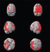
Activation analysis. Surface rendered group analysis using SPM [P < 0.05 with FWE (Familywise error rate) corrected] during encoding of working memory in 53 subjects reveal increased activations in the bilateral prefrontal cortex left more than right, bilateral parieto occipital and orbitofrontal lobes
Figure 2(A-E)
Figure 2(A-E)
Resting state analysis.(A) denotes motor network with bilateral symmetric signal in the precentral gyrus along the motor cortex, (B) is the auditory network in the temporal and inferior parietal lobes, (C) is the visual network in the medial occipital lobes bilaterally, (D) reveals left Fronto parietal executive network, (E) is the default mode network with bilaterally symmetric signal in the medial superiofrontal, anterior cingulate, posterior cingulate, and bilateral parietal and temporal lobes
Figure 3Connectivity analysis
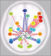
PCC seed to voxel based connectivity using the same data during Encoding of working memory. Pink color arrows indicate significant positive correlation (P < 0.05 FDR) brain regions connection with bilateral frontal lobes left more than right, bilateral supramarginal cortex and visual cortex with source seed (white circle). Blue color arrows indicate significant negative correlation of bilateral-inferior parietal cortex, medial frontal lobes (P < 0.05 FDR) with source seed

References

    1. Supekar K, Uddin LQ, Khouzam A, Phillips J, Gaillard WD, Kenworthy LE, et al. Brain Hyperconnectivity in Children with Autism and its Links to Social Deficits. Cell Rep. 2013;5:738–47. - PMC - PubMed
    1. Balthazar ML, de Campos BM, Franco AR, Damasceno BP, Cendes F. Whole Whole cortical and default mode network mean functional connectivity as potential biomarkers for mild Alzheimer's disease. Psychiatry Res. 2014;221:37–42. - PubMed
    1. Sharp DJ, Beckmann CF, Greenwood R, Kinnunen KM, Bonnelle V, De Boissezon X, et al. Default mode network functional and structural connectivity after traumatic brain injury. Brain. 2011;134:2233–47. - PubMed
    1. Grefkes C, Nowak DA, Wang LE, Dafotakis M, Eickhoff SB, Fink GR. Modulating cortical connectivity in stroke patients by rTMS assessed with fMRI and dynamic causal modeling. NeuroImage. 2010;50:233–42. - PMC - PubMed
    1. Golestani AM, Tymchuk S, Demchuk A, Goodyear BG, VISION-2 Study Group Longitudinal Evaluation of Resting-State fMRI After Acute Stroke With Hemiparesis. Neurorehabil Neural Repair. 2013;27:153–63. - PubMed