

Brainstem Congestion due to Dural Ateriovenous Fistula at the Craniocervical Junction
- PMID: 24851151
- PMCID: PMC4024815
- DOI: 10.3340/jkns.2014.55.3.152
Brainstem Congestion due to Dural Ateriovenous Fistula at the Craniocervical Junction
Abstract
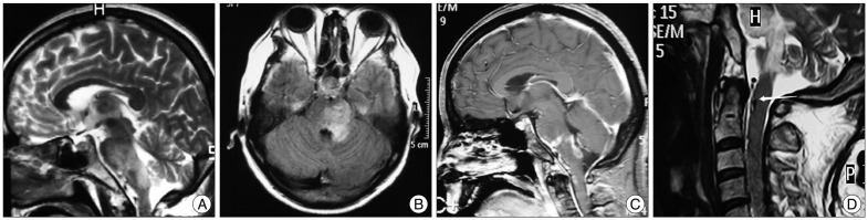
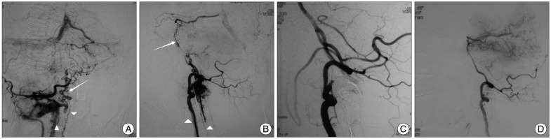
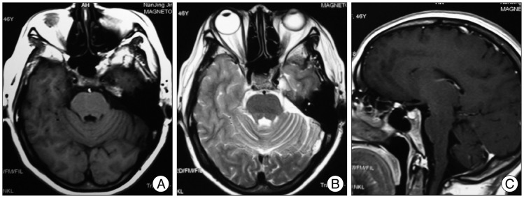
Dural ateriovenous fistula (DAVF) at the craniocervical junction is rare. We report a patient presenting with brainstem dysfunction as an uncommon onset. Brainstem lesion was suggested by magnetic resonance image study. Angiogram revealed a DAVF at a high cervical segment supplied by the meningeal branch of the right vertebral artery, with ascending and descending venous drainage. Complete obliteration of the fistula was achieved via transarterial Onyx embolization. Clinical cure was achieved in the follow-up period; meanwhile, imaging abnormalities of this case disappeared. Accordingly, we hypothesize that a brainstem lesion of this case was caused by craniocervical DAVF, which induced venous hypertension. Thus, venous drainage patterns should be paid attention to because they are important for diagnosis and theraputic strategy.
Keywords: Brainstem dysfunction; Diagnosis; Dural arteriovenous fistula; Venous congestion.
Figures
Similar articles
-
An Isolated Unilateral Pontomedullary Lesion Due to An Intracranial Dural Arteriovenous Fistula Mimicking A Brain Tumor - Case and Review.J Nippon Med Sch. 2019;86(1):48-54. doi: 10.1272/jnms.JNMS.2019_86-9. J Nippon Med Sch. 2019. PMID: 30918157
-
Brainstem Congestion Due to Dural Arteriovenous Fistula at the Craniocervical Junction: Case Report and Review of the Literature.World Neurosurg. 2018 Oct;118:181-187. doi: 10.1016/j.wneu.2018.06.243. Epub 2018 Aug 7. World Neurosurg. 2018. PMID: 30010077 Review.
-
Medullary Hemorrhage Caused by Foramen Magnum Dural Arteriovenous Fistula Successfully Obliterated using Combination of Endovascular and Surgical Treatments: A Case Report and Literature Review.Asian J Neurosurg. 2019 Nov 25;14(4):1256-1267. doi: 10.4103/ajns.AJNS_259_19. eCollection 2019 Oct-Dec. Asian J Neurosurg. 2019. PMID: 31903375 Free PMC article.
-
Far Lateral Approach for Disconnection of Craniocervical Junction Dural Arteriovenous Fistula Presented with Myelopathy and Hydrocephalus.J Neurol Surg B Skull Base. 2021 Feb;82(Suppl 1):S45-S47. doi: 10.1055/s-0040-1714402. Epub 2020 Nov 26. J Neurol Surg B Skull Base. 2021. PMID: 33717817 Free PMC article.
-
Complete Obliteration of a Foramen Magnum Dural Arteriovenous Fistula by Microsurgery After Failed Endovascular Treatment Using Onyx: Case Report and Literature Review.World Neurosurg. 2020 Dec;144:43-49. doi: 10.1016/j.wneu.2020.08.077. Epub 2020 Aug 15. World Neurosurg. 2020. PMID: 32805464 Review.
Cited by
-
A Rare Case of Subarachnoid Hemorrhage caused by Ruptured Venous Varix Due to Dural Arteriovenous Fistula at the Foramen Magnum Fed Solely by the Ascending Pharyngeal Artery.J Cerebrovasc Endovasc Neurosurg. 2018 Jun;20(2):120-126. doi: 10.7461/jcen.2018.20.2.120. Epub 2018 Jun 30. J Cerebrovasc Endovasc Neurosurg. 2018. PMID: 30370246 Free PMC article.
-
Acute Tetraparesis with Respiratory Failure after Steroid Administration in a Patient with a Dural Arteriovenous Fistula at the Craniocervical Junction.Intern Med. 2018 Feb 15;57(4):591-594. doi: 10.2169/internalmedicine.9115-17. Epub 2017 Dec 8. Intern Med. 2018. PMID: 29225249 Free PMC article.
-
Intracranial Dural Arteriovenous Fistulas With Brainstem Engorgement: An Under-Recognized Entity in Diagnosis and Treatment.Front Neurol. 2020 Sep 25;11:526550. doi: 10.3389/fneur.2020.526550. eCollection 2020. Front Neurol. 2020. PMID: 33101168 Free PMC article.
-
A rare case of spinal dural arteriovenous fistula mimicking malignant glioma of the medulla oblongata: Significance of cerebral angiography for accurate diagnosis of brain stem region.Surg Neurol Int. 2020 Sep 12;11:287. doi: 10.25259/SNI_437_2020. eCollection 2020. Surg Neurol Int. 2020. PMID: 33033649 Free PMC article.
-
The impact of different imaging modalities in diagnosis and management of patient with dural arteriovenous fistula: A rare case report.Trauma Case Rep. 2024 Jun 7;52:101044. doi: 10.1016/j.tcr.2024.101044. eCollection 2024 Aug. Trauma Case Rep. 2024. PMID: 38952476 Free PMC article.
References
-
- Bussière M, Lownie SP, Pelz DM, Nicolle D. Direct carotid-cavernous fistula causing brainstem venous congestion. J Neuroophthalmol. 2009;29:21–25. - PubMed
-
- Chen G, Wang Q, Tian Y, Gu Y, Xu B, Leng B, et al. Dural arteriovenous fistulae at the craniocervical junction : the relation between clinical symptom and pattern of venous drainage. Acta Neurochir Suppl. 2011;110(Pt 2):99–104. - PubMed
-
- Fassett DR, Rammos SK, Patel P, Parikh H, Couldwell WT. Intracranial subarachnoid hemorrhage resulting from cervical spine dural arteriovenous fistulas : literature review and case presentation. Neurosurg Focus. 2009;26:E4. - PubMed
-
- Guo LM, Zhou HY, Xu JW, Wang GS, Tian X, Wang Y, et al. Dural arteriovenous fistula at the foramen magnum presenting with subarachnoid hemorrhage : case reports and literature review. Eur J Neurol. 2010;17:684–691. - PubMed
-
- Hamada J, Yano S, Kai Y, Koga K, Morioka M, Ishimaru Y, et al. Histopathological study of venous aneurysms in patients with dural arteriovenous fistulas. J Neurosurg. 2000;92:1023–1027. - PubMed
Publication types
LinkOut - more resources
Full Text Sources
Other Literature Sources
