Review
. 2014 May;19(3):317-31.
doi: 10.1111/adb.12101.

The cerebellum and addiction: insights gained from neuroimaging research

Review

The cerebellum and addiction: insights gained from neuroimaging research

Eric A Moulton et al. Addict Biol. 2014 May.

Abstract

Although cerebellar alterations have been consistently noted in the addiction literature, the pathophysiology of this link remains unclear. The cerebellum is commonly classified as a motor structure, but human functional neuroimaging along with clinical observations in cerebellar stroke patients and anatomical tract tracing in non-human primates suggests its involvement in cognitive and affective processing. A comprehensive literature search on the role of the cerebellum in addiction was performed. This review article (1) considers the potential role of the cerebellum in addiction; (2) summarizes the cerebellar structural alterations linked to addiction; (3) presents the functional neuroimaging evidence linking the cerebellum with addiction; and (4) proposes a model for addiction that underscores the role of the cerebellum. The data implicate the cerebellum as an intermediary between motor and reward, motivation and cognitive control systems, as all are relevant etiologic factors in addiction. Furthermore, consideration of these findings could contribute to deeper and more sophisticated insights into normal reward and motivational function. The goal of this review is to spread awareness of cerebellar involvement in addictive processes, and to suggest a preliminary model for its potential role.


Figures

Figure 1
Figure 1
Cerebellar anatomy. A) Top-down axial view. B) Coronal view of the deep cerebellar nuclei. C) Sagittal view highlighting the major afferent pathways into the cerebellum. Mossy fibers project to granule cells in the cerebellar cortex, and send collaterals to the deep cerebellar nuclei. Climbing fibers extend to Purkinje cells, and also have collateral projections to deep cerebellar nuclei. The axons of the deep cerebellar nuclei form the primary output channels away from the cerebellum, and to the brainstem and cerebral cortex.
Figure 2
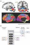
Figure 2. Cerebellar functional connectivity to cerebral resting state networks
A) Colors in the cerebellum correspond to areas functionally correlated to cerebral resting state networks. Coordinates correspond to MNI atlas space. Figure adapted from Buckner et al., 2011. B) Schematic of cerebral connectivity in cerebellar areas with gray matter deficits in addiction, and fMRI activation in response to acute drug exposure and drug craving. Colored lines correspond to the colored resting state networks in panel A. Each cerebral area is classified based on its contribution to different functional processes (i.e. cognitive control, salience, etc.). ACC, anterior cingulate cortex; aINS, anterior insula; DLPFC, dorsolateral prefrontal cortex; DMPFC, dorsomedial prefrontal cortex; M1, primary motor cortex; pINS, posterior insula; S1, primary somatosensory cortex; S2, secondary somatosensory cortex.
Figure 3
Figure 3. Model of cerebellar modulation of circuitry related to aversion and addiction
Aversive stimuli in the form of pain (red) and unpleasant pictures (yellow) produce overlapping fMRI activation (blue) in posterior cerebellar hemispheres Lobule VI, VIIb, and Crus I. Activation in these overlapping cerebellar areas is inversely related to activation in limbic areas in the brain, including the hypothalamus (Hypo), parahippocampal gyrus (PHG), and subgenual anterior cingulate cortex (sACC). These limbic structures have been associated with networks for salience, memory, and motivational drive. Figure adapted from Moulton et al., 2011.
Figure 4
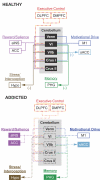
Figure 4. A cerebellar model for addiction
The top panel highlights the cerebellum's role as a modulator of affective and cognitive function in the healthy brain. This schematic highlights the four major brain networks proposed to be affected by addiction (adapted from Volkow et al., 2010). The components within each network are color-coded to match the cerebral resting state networks in Figure 2 (adapted from Buckner et al., 2011). Arrows show the proposed direction of cerebellar down-modulation of specific brain regions within each circuit (Moulton et al., 2011). The bottom panel proposes how cerebellar dysfunction may impact the addicted brain. The black dashed boxes highlight structural degradation of specifically localized cerebellar structures with addiction. The dashed lines show disrupted functional connectivity and cerebellar modulation of specific brain regions related to executive control, reward/salience, motivational drive, and memory. Filled colored boxes and bold type represent brain circuits released from cerebellar modulation, leading to an uninhibited and sensitized neurological state. Note that all cerebellar outputs project through the deep cerebellar nuclei (refer to text for details). ACC, anterior cingulate cortex; aINS, anterior insula; DLPFC, dorsolateral prefrontal cortex; DMPFC, dorsomedial prefrontal cortex; Hypo, hypothalamus; M1, primary motor cortex; PHG, parahippocampal gyrus; sACC, subgenual anterior cingulate cortex; Verm, vermis; VI, cerebellar hemispheric lobule VI; VIIb, cerebellar hemispheric lobule VIIb.

Similar articles

Cited by

References

    1. Anderson CM, Maas LC, Frederick B. deB, Bendor JT, Spencer TJ, Livni E, Lukas ES, Fischman AJ, Madras BK, Renshaw PF, Kaufman MJ. Cerebellar vermis involvement in cocaine-related behaviors. Neuropsychopharmacology. 2006;31(6):1318–1326. - PubMed
    1. Ashton JC, Appleton I, Darlington CL, Smith PF. Immunohistochemical localization of cannabinoid CB1 receptor in inhibitory interneurons in the cerebellum. Cerebellum. 2004;3(4):222–226. - PubMed
    1. Barrós-Loscertales A, Garavan H, Bustamante JC, Ventura-Campos N, Llopis JJ, Belloch V, Parcet MA, Avila C. Reduced striatal volume in cocaine-dependent patients. NeuroImage. 2011;56(3):1021–1026. - PubMed
    1. Bauer DJ, Kerr AL, Swain RA. Cerebellar dentate nuclei lesions reduce motivation in appetitive operant conditioning and open field exploration. Neurobiology of learning and memory. 2011;95(2):166–175. - PubMed
    1. Benegal V, Antony G, Venkatasubramanian G, Jayakumar PN. Gray matter volume abnormalities and externalizing symptoms in subjects at high risk for alcohol dependence. Addiction biology. 2007;12(1):122–132. - PubMed

Publication types