

The complex ultrastructure of the endolysosomal system
- PMID: 24851870
- PMCID: PMC4176003
- DOI: 10.1101/cshperspect.a016857
The complex ultrastructure of the endolysosomal system
Abstract
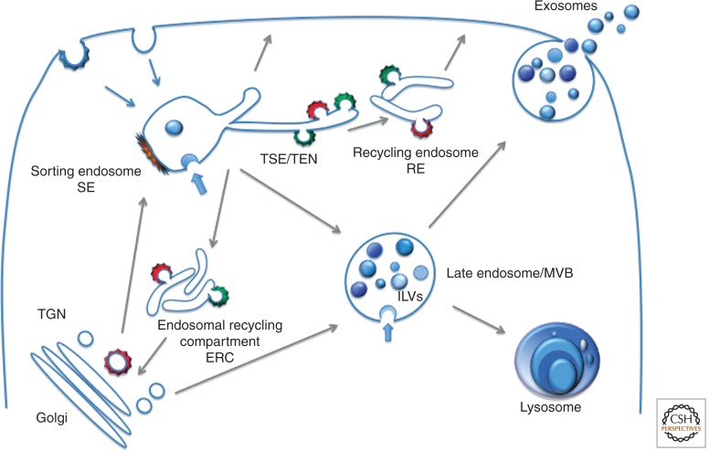
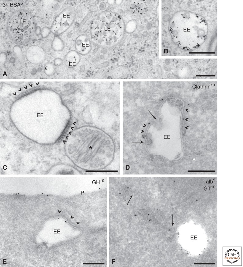
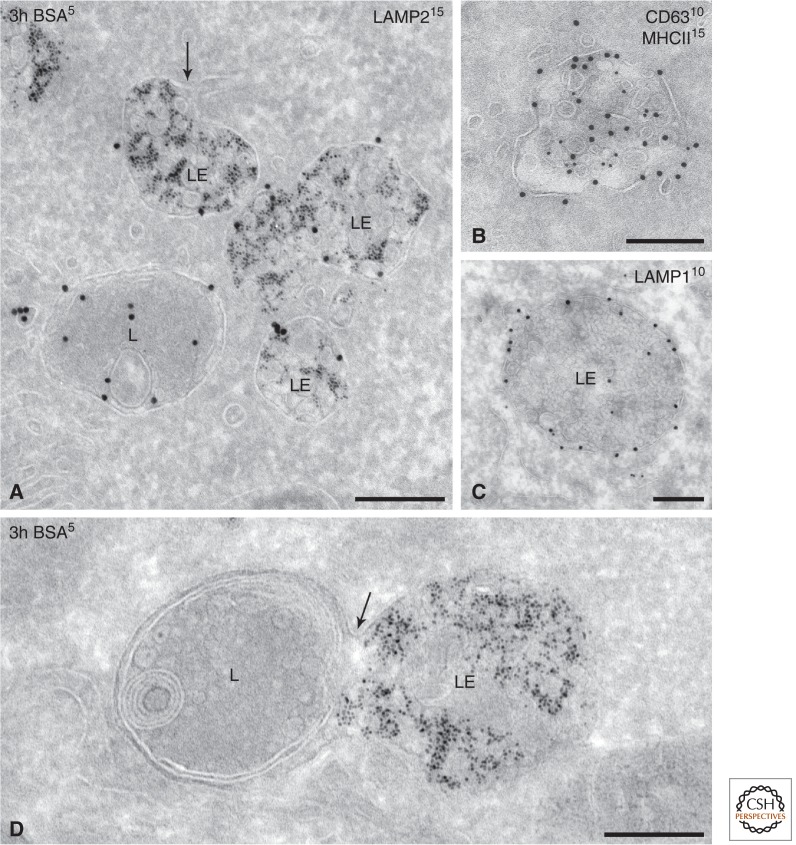
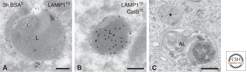
Live-cell imaging reveals the endolysosomal system as a complex and highly dynamic network of interacting compartments. Distinct types of endosomes are discerned by kinetic, molecular, and morphological criteria. Although none of these criteria, or combinations thereof, can capture the full complexity of the endolysosomal system, they are extremely useful for experimental purposes. Some membrane domain specializations and specific morphological characteristics can only be seen by ultrastructural analysis after preparation for electron microscopy (EM). Immuno-EM allows a further discrimination of seemingly identical compartments by their molecular makeup. In this review we provide an overview of the ultrastructural characteristics and membrane organization of endosomal compartments, along with their organizing machineries.
Copyright © 2014 Cold Spring Harbor Laboratory Press; all rights reserved.
Figures
References
-
- Allen RD, Fok AK 1980. Membrane recycling and endocytosis in Paramecium confirmed by horseradish peroxidase pulse-chase studies. J Cell Sci 45: 131–145 - PubMed
-
- Almeida CG, Yamada A, Tenza D, Louvard D, Raposo G, Coudrier E 2011. Myosin 1b promotes the formation of post-Golgi carriers by regulating actin assembly and membrane remodelling at the trans-Golgi network. Nat Cell Biol 13: 779–789 - PubMed
Publication types
MeSH terms
Grants and funding
LinkOut - more resources
Full Text Sources
Other Literature Sources