. 2014 May 22;8(5):e2885.
doi: 10.1371/journal.pntd.0002885. eCollection 2014 May.

Intra-subtype variation in enteroadhesion accounts for differences in epithelial barrier disruption and is associated with metronidazole resistance in Blastocystis subtype-7

Affiliations

Intra-subtype variation in enteroadhesion accounts for differences in epithelial barrier disruption and is associated with metronidazole resistance in Blastocystis subtype-7

Zhaona Wu et al. PLoS Negl Trop Dis. .

Abstract

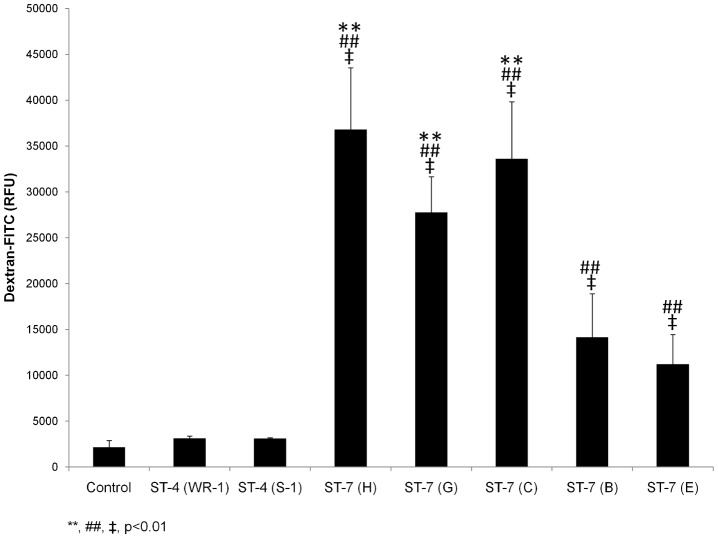
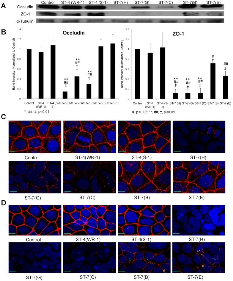
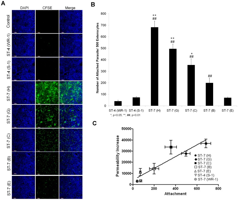
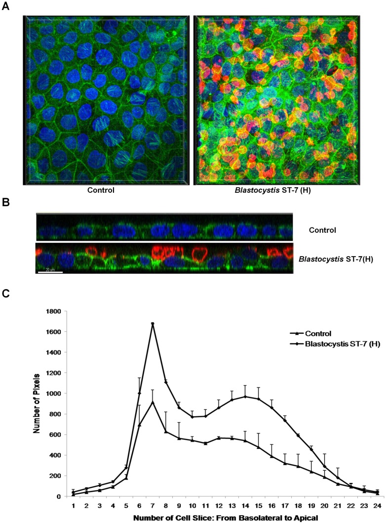
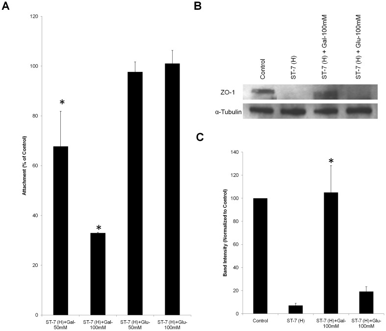
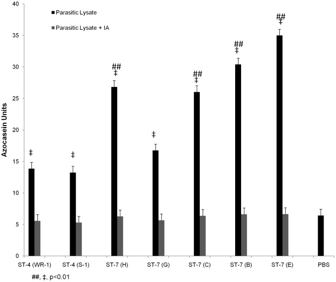
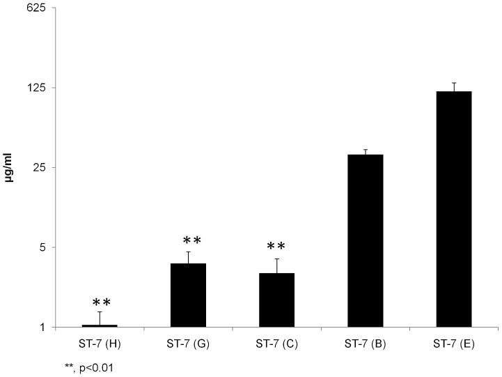
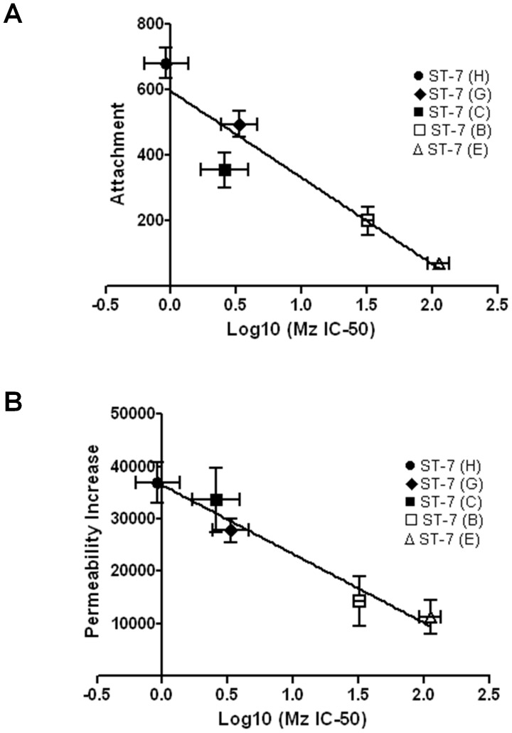
Blastocystis is an extracellular, enteric pathogen that induces intestinal disorders in a range of hosts including humans. Recent studies have identified potential parasite virulence factors in and host responses to this parasite; however, little is known about Blastocystis-host attachment, which is crucial for colonization and virulence of luminal stages. By utilizing 7 different strains of the parasite belonging to two clinically relevant subtypes ST-4 and ST-7, we investigated Blastocystis-enterocyte adhesion and its association with parasite-induced epithelial barrier disruption. We also suggest that drug resistance in ST-7 strains might result in fitness cost that manifested as impairment of parasite adhesion and, consequently, virulence. ST-7 parasites were generally highly adhesive to Caco-2 cells and preferred binding to intercellular junctions. These strains also induced disruption of ZO-1 and occludin tight junction proteins as well as increased dextran-FITC flux across epithelial monolayers. Interestingly, their adhesion was correlated with metronidazole (Mz) susceptibility. Mz resistant (Mzr) strains were found to be less pathogenic, owing to compromised adhesion. Moreover, tolerance of nitrosative stress was also reduced in the Mzr strains. In conclusion, the findings indicate that Blastocystis attaches to intestinal epithelium and leads to epithelial barrier dysfunction and that drug resistance might entail a fitness cost in parasite virulence by limiting entero-adhesiveness. This is the first study of the cellular basis for strain-to-strain variation in parasite pathogenicity. Intra- and inter-subtype variability in cytopathogenicity provides a possible explanation for the diverse clinical outcomes of Blastocystis infections.


Conflict of interest statement

The authors have declared that no competing interests exist.

Figures

Figure 1
Figure 1. Intra-and inter-subtype variation among Blastocystis ST-4 and ST-7 strains in inducing permeability increase.
Graph representing epithelial permeability of Caco-2 monolayers after co-incubation with live cells of Blastocystis ST-7 and ST-4 for 24 h. All strains in ST-7 induced significant increase in flux of dextran–FITC across epithelial monolayer of Caco-2 cells compared with control monolayers (p<0.01). No significant change in permeability was observed in Blastocystis ST-4-infected Caco-2.Within ST-7, an intra-subtype variation in the capability of inducing permeability change was observed. Isolates C, G and H induced much higher permeability increase compared to isolates B and E within the same subtype (p<0.01). **, p<0.01 vs. ST-7 (B, E)-infected cells; ##, p<0.01 vs. ST-4-infected cells; ‡, p<0.01 vs. non-infected cells. Each value represents mean of twelve samples, taken from three independent experiments, four samples from each. Error bars represent the standard errors.
Figure 2
Figure 2. Differential effects on tight junction protein degradation in Caco-2 cell monolayers by different strains of Blastocystis.
(A) Representative Western blot analysis of occludin and ZO-1 level in Caco-2 epithelium. Monolayers were harvested after infection with Blastocystis ST-4 and ST-7 isolates; normal culture media was used as the negative control. No obvious change in occludin and ZO-1 was observed in ST-4-treated samples. Within ST-7, a more prominent decrease or loss of occludin band could be seen in cells treated by isolates C, G and H, while the changes in B-, E-treated cells were not obvious; for ZO-1 tight junction protein, loss of band was observed in Caco-2 cells after coincubation with ST-7 (C, G, H), whereas a decrease in band intensity was also noticed in B- and E-treated cells. (B) Quantification of tight junction levels through densitometry analysis of Western blot radiographs. Densitometric values of occludin and ZO-1 signals were quantified and expressed as the ratio to α-Tubulin. ST-4-treated samples did not differ significantly from control in occludin and ZO-1 level; ST-7 (C, G, H) induced significant occludin and ZO-1 degradation compared with ST-4 and ST-7 (B, E) isolates (p<0.01). ST-7 (B, E), however, were able to induce significant ZO-1 degradation compared with control and ST-4, but not for occludin. **, p<0.01 vs. ST-7 (B, E)-infected cells; #, p<0.05 vs. ST-4-infected cells; ##, p<0.01 vs. ST-4-infected cells; ‡, p<0.01 vs. non-infected cells. Results were from three independent experiments. Error bars represent the standard errors. (C) Representative confocal micrographs illustrating occludin integrity in Caco-2 monolayers. Caco-2 cells are labelled with DAPI (Blue) and an antibody targeting tight junction protein occludin and Cy3® goat anti-mouse IgG (Red). Corresponding with Western blot results, in ST-4 and ST-7 (B, E)-treated samples, no obvious change in occludin was observed. Infection with ST-7 (C, G, H), however, induced focal disruptions and degradation in occludin, as shown by the decreased occludin staining intensity in Caco-2 cell line. Scale bar = 10 µm. (D) Representative confocal micrographs illustrating ZO-1 integrity in Caco-2 monolayers. Caco-2 cells are labelled with DAPI (Blue) and an antibody targeting tight junction protein ZO-1 and Cy3® goat anti-mouse IgG (Red). Compared with the negative control and ST-4-treated samples, where ZO-1 appeared continuous with sharp pericellular staining patterns, all ST-7 isolates resulted in focal disruption as well as degradation of ZO-1 in Caco-2 cells. In ST-7 (C, G, H)-treated samples, ZO-1 staining almost became invisible, indicating more degradation of the protein, which corresponded with the western blot results. Notably, treatments with ST-7 (B, E) resulted in both focal disruptions (green arrows) as well as reorganizations of ZO-1 (yellow arrows). Scale bar = 10 µm.
Figure 3
Figure 3. Blastocystis exhibits intra- and inter-subtype variation in attachment to Caco-2 cells.
(A) Representative confocal micrographs illustrating intra- and inter-subtype variation in Blastocystis attachment to Caco-2 cells. Caco-2 monolayers were grown to confluency on glass converslips and were then co-incubated with the same number of parasites of different strains of Blastocystis pre-stained with CFSE (green). Normal culture media was used as a negative control. After co-incubation, the non-attached parasites were washed away. The Caco-2 monolayers were then stained with DAPI and then were viewed using confocal microscope (Olympus Fluoview FV1000; Olympus, Japan). More green in the field represents more parasites attached to the monolayer. Both ST-4 strains adhere with a negligible number. An intra-subtype variation in the number of attachment within ST-7 is obvious. Isolates C, G, H appeared to attach at a much higher level than B and E to Caco-2 cells. Scale bar = 100 µm. (B) Graph representing number of Blastocystis parasites attached to host cells. ST-7 strains C, G and H exhibited a significantly higher number of attached parasites than ST-4 strains and ST-7 isolates B and E. *, p<0.05 vs. ST-7 (B, E); **, p<0.01 vs. ST-7 (B, E); ##, p<0.01 vs. ST-4. Each value represents a mean of six readings derived from 3 independent experiments. Error bar represents standard error. (C) Relationship between attachment and permeability increase by Blastocystis ST-4 and ST-7 parasites. The data points indicate individual strains. x and y error bars indicate the standard error for the respective measurements (n = 3). The R2 for the trend line shown is 0.8506, and the p value is 0.0031. There is a positive correlation between the level of attachment and permeability increase (R = 0.9223).
Figure 4
Figure 4. Blastocystis ST-7 isolate H attaches to an intestinal monolayer.
Caco-2 monolayers were grown to confluency on glass coverslips and were then coincubated with Blastocystis ST-7 (H). Normal culture media was used as the negative control. (A) Representative confocal micrographs illustrating attachment of Blastocystis ST-7 (H) to Caco-2 monolayers. Blastocystis was labelled with legumain antibody-mAb1D5and secondary AlexaFluro 594 goat anti-mouse IgM (red). Phalloidin-FITC (green) was used to label F-actin of Caco-2 cells and DAPI (blue) for nuclei. Compared with the negative control, attachment of Blastocystis ST-7 to Caco-2 apical side could be seen clearly. (B) Expanded section views of epithelium of control and ST-7 (H)-treated-monolayers. Parasites could be seen intimately adhering to the epithelium and it could also be noted that the parasites adhered preferentially to the cell-cell junction site (yellow arrows) and induced an increase in actin polymerization. Compared with the negative control which displayed a properly organized epithelium, there was loss of cellular symmetry in the cells treated by ST-7 (H) (black arrowheads). Scale bar  = 20 µm. (C) Quantification of F-actin staining in Blastocystis-infected Caco-2 monolayers. Each cell slice (1–25) corresponds to series of images from Z-stack sections taken at 1 µm thickness through the cell monolayer. X-axis illustrates cell layers from basolateral to apical. Y-axis illustrates the number of pixels present over the entire area of image. Monolayers treated with ST-7 (H) resulted in marked increase in actin intensity compared to the negative control (p<0.01).
Figure 5
Figure 5. Inhibition of Blastocystis ST-7 (H) adhesion by galactose rescues Blastocystis ST-7 (H)–induced ZO-1 tight junction degradation.
(A) Dose-dependent inhibition of galactose on Blastocystis ST-7 (H) adhesion to Caco-2 monolayers. Blastocystis ST-7 (H) were incubated with epithelial cells in the presence of different concentrations of galactose and glucose (50 and 100 mM, respectively). A value of 100% was assigned to number of binding parasites without addition of sugars as control. The numbers of attached parasites with galactose addition were normalized to control.*, p<0.01 vs. control. (B) Representative western blot analysis of ZO-1 level in Caco-2 epithelium. Caco-2 monolayers were infected with Blastocystis ST-7 (H) in the presence of saccharides galactose and glucose at 100 mM and incubated for 1 h. Monolayers were washed and prepared for western blotting. Normal culture media with no sugar addition was used as the negative control. Galactose rescued Blastocystis–induced ZO-1 tight junction degradation. (C) Quantification of ZO-1 levels through densitometry analysis of Western blot radiographs. Densitometric values of ZO-1 signals were quantified and expressed as the ratio to α-Tubulin. ZO-1 degradation was significantly rescued by addition of galactose. *, p<0.01 vs. ST-7 (H)-treated sample. Values are the means ± standard errors from data of three experiments. Error bars represent the standard errors. Glu, glucose; Gal, Galactose.
Figure 6
Figure 6. Intra- and inter-subtype variations in protease activity in Blastocystis.
Protease activity of Blastocystis ST-4 and ST-7 isolates was determined by azocasein assays. Except isolate G, most of ST-7 strains exhibited significantly higher protease activities than the two ST-4 isolates (p<0.01). PBS as a background control showed activity that was significantly lower when compared to protease activities of all the isolates (p<0.01). Note that cysteine protease inhibitor iodoacetamide (IA) abolished protease activity of all the isolates, which is comparable to PBS. ##, p<0.01 vs. ST-4; ‡, p<0.01 vs. PBS control. Each value represents mean of six samples, taken from three independent experiments. Error bars represent the standard errors.
Figure 7
Figure 7. Blastocystis exhibits intra-subtype variation in susceptibility and resistance to Mz.
Graph representing IC50s of Mz against Blastocystis ST-4 and ST-7 isolates tested in the study using the resazurin assay. Y axis was presented on a logarithmic scale in base 5. The IC50s of Mz against ST-7 isolates C, G and H were found to be significantly lower than those of isolates B, E within the same subtype (p<0.01). **, p<0.01 vs. ST-7 (B, E). Each point represents a mean of nine readings derived from three independent experiments, triplicate each. Error bars represent the standard errors.
Figure 8
Figure 8. Correlation analyses between Mz resistance and (A) attachment, as well as (B) permeability increase in Blastocystis ST-7 parasites.
(A) Relationship between Mz resistance and attachment for Blastocystis ST-7 parasites. The data points indicate individual strains. Error bars indicate the standard error for the respective measurements (n = 3). There was a negative correlation between the level of resistance and attachment (p<0.05, R2 = 0.8908). (B) Relationship between Mz resistance of Blastocystis ST-7 parasites and their ability in inducing permeability increase. The data points indicate individual strains. Error bars indicate standard errors for the respective measurements (n = 3). There was a negative correlation between the level of resistance and permeability increase (p<0.01, R2 = 0.9644).

Similar articles

Cited by

References

    1. Scanlan PD (2012) Blastocystis: past pitfalls and future perspectives. Trends Parasitol 28: 327–334. - PubMed
    1. Tan KS (2008) New insights on classification, identification, and clinical relevance of Blastocystis spp. Clin Microbiol Rev 21: 639–665. - PMC - PubMed
    1. Tan KS, Mirza H, Teo JD, Wu B, Macary PA (2010) Current Views on the Clinical Relevance of Blastocystis spp. Curr Infect Dis Rep 12: 28–35. - PubMed
    1. Poirier P, Wawrzyniak I, Vivares CP, Delbac F, El Alaoui H (2012) New insights into Blastocystis spp.: a potential link with irritable bowel syndrome. PLoS Pathog 8: e1002545. - PMC - PubMed
    1. Alfellani MA, Taner-Mulla D, Jacob AS, Imeede CA, Yoshikawa H, et al. (2013) Genetic diversity of blastocystis in livestock and zoo animals. Protist 164: 497–509. - PubMed

Publication types

MeSH terms

LinkOut - more resources