. 2014 May 22;10(5):e1004356.
doi: 10.1371/journal.pgen.1004356. eCollection 2014 May.

Octopamine neuromodulation regulates Gr32a-linked aggression and courtship pathways in Drosophila males

Affiliations

Octopamine neuromodulation regulates Gr32a-linked aggression and courtship pathways in Drosophila males

Jonathan C Andrews et al. PLoS Genet. .

Abstract

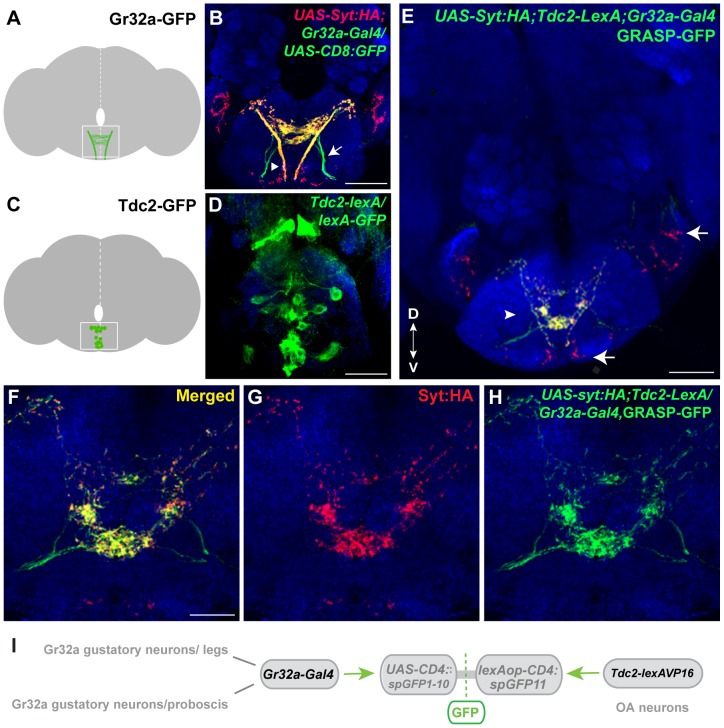
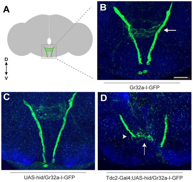
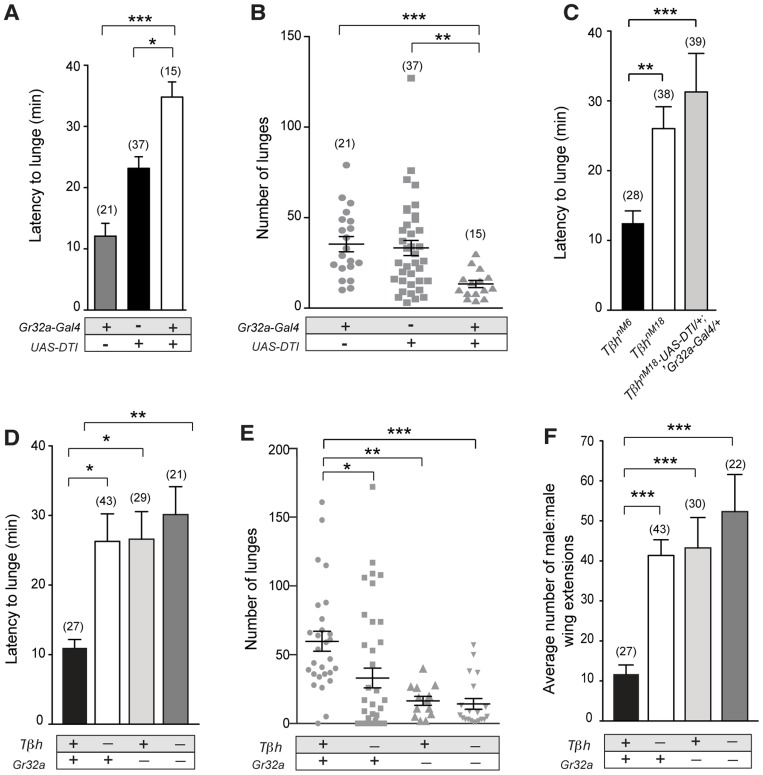
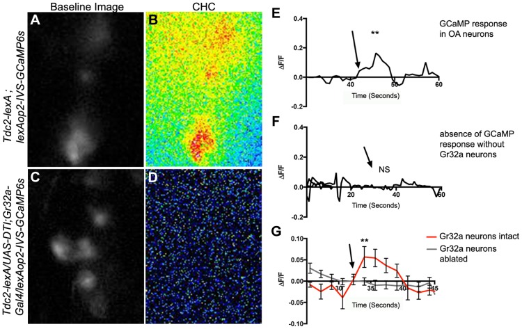
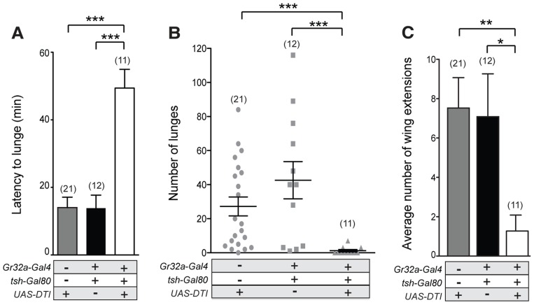
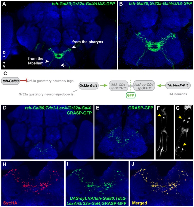
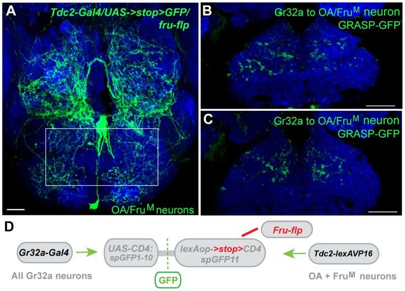
Chemosensory pheromonal information regulates aggression and reproduction in many species, but how pheromonal signals are transduced to reliably produce behavior is not well understood. Here we demonstrate that the pheromonal signals detected by Gr32a-expressing chemosensory neurons to enhance male aggression are filtered through octopamine (OA, invertebrate equivalent of norepinephrine) neurons. Using behavioral assays, we find males lacking both octopamine and Gr32a gustatory receptors exhibit parallel delays in the onset of aggression and reductions in aggression. Physiological and anatomical experiments identify Gr32a to octopamine neuron synaptic and functional connections in the suboesophageal ganglion. Refining the Gr32a-expressing population indicates that mouth Gr32a neurons promote male aggression and form synaptic contacts with OA neurons. By restricting the monoamine neuron target population, we show that three previously identified OA-Fru(M) neurons involved in behavioral choice are among the Gr32a-OA connections. Our findings demonstrate that octopaminergic neuromodulatory neurons function as early as a second-order step in this chemosensory-driven male social behavior pathway.


Conflict of interest statement

The authors have declared that no competing interests exist.

Figures

Figure 1
Figure 1. Gr32a neurons contact OA neurons in the suboesophageal ganglion.
(A) Schematic depicting the SOG region targeted by Gr32a axons visualized in panel B. (B) Axons and presynaptic terminals of Gr32a-expressing neurons identified by immunofluorescence to CD8:GFP and the synaptotagmin:HA fusion protein in UAS-sytHA;;UAS-CD8:GFP/Gr32a-Gal4 progeny (green, anti-CD8, Invitrogen; red, anti-HA, Roche). Sensory neurons from the labellum project through the labial nerve (arrow), mouthpart neurons project through the pharyngeal/accessory nerve, and neurons from thoracic ganglia project via the cervical connective (arrowhead). Scale bar represents 30 µM (C–D) A subset of Tdc2-positive neurons located in the SOG in a schematic (C) and with GFP expression driven by the Tdc2-LexA line (Tdc2-LexA;lexAop-rCD4:GFP. Cell bodies are visible (D) with extensive arborizations apparent in a series of optical sections ventral to the cell bodies (Figure S1). (E) GRASP-mediated GFP reconstitution is observed between Gr32a neurons expressing CD4::spGFP1-10 and UAS-syt:HA (red, anti-HA, Roche) and OA neurons expressing CD4::spGFP11. GRASP reconstitution is detected by immunofluorescence using a rabbit monoclonal GFP antibody (Life Technologies). Regions in the SOG with only syt:HA expression are indicated (arrows) in addition to GFP-reconstitution contacts that show co-localization with syt-HA expression (arrowhead). Scale bar is 50 µM. (F–H) Optical sections of the same brain at higher magnification showing GRASP-mediated GFP reconstituted expression (H), syt:HA localization (G) and clear overlap or close association at synaptic-like puncta in the merged channel (F). Scale bar represents 20 µM. (See also Figure S2.) (I) Schematic representation of the GRASP reporter lines combined with the Gr32a-Gal4 and Tdc2-lexA driver lines.
Figure 2
Figure 2. Removing OA neurons significantly alters Gr32a axonal projections.
(A) Schematic representation of the adult brain with Gr32a-expressing axonal arborizations in the SOG. (B) Gr32a-I-GFP expression in a typical wildtype adult brain. The Gr32a-expressing neurons located in the tarsi, labellum, and mouthparts all terminate in the SOG (arrow). (C) Confocal sections of a UAS-hid UAS-Red Stinger control brain verifying wildtype organization of Gr32a-I-GFP projections (D) Confocal sections of transgenic Tdc2-Gal4/UAS-hid UAS-Red Stinger;Gr32a-I-GFP adult brains. When all OA neurons are eliminated, a range of axonal projection defects was observed including a severe reduction and disorganization of Gr32a leg and labellum termini (arrow, arrowhead). Scale bar represents 30 µM.
Figure 3
Figure 3. Gr32a-expressing neurons promote aggression via OA signaling.
(A–B) Fights between males with Gr32a-expressing neurons removed by expressing Diptheria Toxin (UAS-DTI;Gr32a-Gal4) and individual transgenic controls, UAS-DTI or Gr32a-Gal4. (A) The latency to first lunge was significantly higher in UAS-DTI/+; Gr32a-Gal4/+ males as compared to controls (all statistical tests are Kruskal-Wallis with Dunn's multiple comparison test except where noted, ***p<0.001, *p<0.05). (B) Number of lunges (represented by each dot) performed in a 30 min period after the first lunge by any control or experimental male in a fighting pair. Males without Gr32a neurons exhibited a significant reduction in lunges as compared to controls (***p<0.001, **p<0.01). (C) Fights between control male pairs (revertant tβhM6 allele), experimental males without OA (revertant null mutation, tβhnM18), or experimental males without OA and without Gr32a-expressing neurons (tβhnM18;UAS-DTI/+; Gr32a-Gal4/+). The latency to first lunge was significantly higher in males without OA and in experimental males compared to control males (**p<0.01) and not statistically different between males without OA and experimental tβhnM18;UAS-DTI/+; Gr32a-Gal4/+ males. (D–F) Fights between control male pairs (revertant tβhM6 allele) and three groups of experimental males; without OA = tβhnM18, without Gr32a receptors = tβhM6;;Gr32a−/−, and without OA and Gr32a receptors = tβhnM18;;Gr32a−/−). (D) The latency to first lunge was significantly higher in males without OA (tβhnM18) and in experimental males without OA and the Gr32a receptor (tβhnM18; Gr32a−/−) or without only the Gr32 receptor (tβhM6; Gr32a−/−) males as compared to control tβhM6 males (One way ANOVA, post hoc Tukey's comparison, *p<0.05, **p<0.01). (E) The number of lunges by pairs of experimental males were significantly less than exhibited by control males but not when compared to each other (***p = 0.0002, **p = 0.002, *p = 0.01). (F) The average number of wing extensions directed toward the second male in each aggression assay. The number of wing extensions exhibited by males without the Gr32a receptor and without OA, and males without Gr32a receptors were significantly greater than control tβhM6 males (***p<0.001) but not males without OA (tβhnM18). Error bars denote s.e.m.
Figure 4
Figure 4. Male CHCs evoke intracellular Ca2+ responses in OA neurons that are dependent on Gr32a neurons.
(A) Greyscale image (background subtracted) of GCaMP3 fluorescence in OA neurons located within the SOG in a Tdc2-lexA;lexAop2-IVS-GCaMP6s male. (B) Pseudocolored subtraction image demonstrating an increase in fluorescence in response to male CHC application. (C) Greyscale image (background subtracted) of baseline fluorescence in the SOG of a male with Gr32a neurons eliminated (Tdc2-lexA; UAS-DTI;Gr32a-Gal4/LexAop2-IVS-GCaMP6s). (D) No changes in fluorescence are observed in the pseudocolored subtraction image of OA SOG neurons when male CHC extract is administered to the legs of males lacking Gr32a neurons. (E) A representative calcium signal trace of OA neurons expressing GCaMP6s in panels A–B in response to male CHC extract application (arrow), unpaired t-test **p<0.006. (F) A representative trace demonstrating the lack of calcium response in OA neurons after male CHC extract application (arrow) to the legs of males without Gr32a neurons (Tdc2-LexA;UAS-DTI;Gr32a-LexA/20XLexAop2-IVS-GCaMP6s). (G) The average calcium response of eight regions of interest from five Tdc2-lexA;lexAop2-IVS-GCaMP6s males (red line) before and after male CHC administration (arrow, unpaired t-test, **p<0.0001). The gray line is the average calcium response of ten regions of interest from five Tdc2-LexA;UAS-DTI;Gr32a-LexA/20XLexAop2-IVS-GCaMP6s males in response to male CHC administration. No significant change in response was observed (unpaired t-test, p<0.0788). Error bars denote s.e.m.
Figure 5
Figure 5. Gr32a chemosensory neurons located in the mouth promote aggression without an elevation in male-male courtship.
(A–B) Fights between males with the Gr32a-expressing mouth neuronal population removed by expressing Diptheria Toxin (UAS-DTI) through the Gr32a-Gal4 line with Gal4 activity in the legs blocked by tsh-Gal80. Separate transgenic controls, UAS-DTI/+ and tsh-Gal80/+; Gr32a-Gal4/+ were scored. (A) The latency to first lunge was significantly higher in UAS-DTI/tsh-Gal80; Gr32a-Gal4/+ males as compared to controls (Kruskal-Wallis with Dunn's multiple comparison test, ***p<0.001). (B) Number of lunges performed per 30 min period after the first lunge by controls or experimental UAS-DTI/tsh-Gal80; Gr32a-Gal4/+ males. Each dot represents the numbers of lunges performed by either male in a fighting pair. Males without Gr32a-expressing mouth neurons exhibited a significant reduction in lunges as compared to controls (Kruskal-Wallis test with Dunn's multiple comparison test, ***p<0.001). (C) The average number of wing extensions directed toward the second male in each aggression assay. The number of wing extensions exhibited by males without mouth Gr32a neurons were less than control males (Kruskal-Wallis with Dunn's multiple comparison test, *p<0.05, **p<0.01). Error bars denote s.e.m.
Figure 6
Figure 6. Mouth-specific Gr32a neurons contact OA neurons in the suboesophageal ganglion.
(A) Axons of Gr32a-expressing neurons located in the mouth identified by immunofluorescence to CD8:GFP in tsh-Gal80;UAS-CD8:GFP/Gr32a-Gal4 progeny (green, anti-CD8, Invitrogen). Note the absence of axonal projections from the legs via the thoracic ganglion (arrow, compare to Figure 1A). (B) Higher magnification of Gr32a mouth neurons expressing CD8:GFP. (C) Schematic representation of the GRASP reporter lines combined with the tsh-Gal80;Gr32a-Gal4 and Tdc2-lexA driver lines. Gal80 driven by the tsh-Gal80 line prevents Gal4 activity and subsequent expression of the UAS-CD4::spGFP1-10 GRASP reporter. (D–E) Two different confocal image magnifications of a male brain with the same number of optical sections as in panel A. A reduced amount of GRASP-mediated GFP reconstitution is observed reflecting Gr32a neurons located only in the mouth expressing CD4::spGFP1-10 and OA neurons expressing CD4::spGFP11. GRASP reconstitution is detected by immunofluorescence using rabbit monoclonal GFP antibody (green; Life Technologies). (F–G) tsh-Gal80 blocks GFP expression in Gr32a-expressing leg neurons. Less than one neuron per leg of UAS-nlsGFP; teashirt-Gal80/Gr32a-Gal4 progeny is observed (arrowhead, 0.38 neurons per front leg, n = 8), versus males without Gal80 expression (arrowhead, 5 neurons per front leg, n = 8). (H–J) Optical sections of a female brain (UAS-syt:HA; tsh-Gal80;UAS-CD8:GFP/Gr32a-Gal4) at higher magnification showing GRASP-mediated GFP reconstituted expression (I), syt:HA localization (H) and clear overlap or close association at synaptic-like puncta in the merged channel (J).
Figure 7
Figure 7. Gr32a neurons anatomically contact three FruM-OA neurons.
(A) The morphology of three FruM-OA neurons located in the suboesophageal ganglion identified by immunofluorescence to CD8:GFP in Tdc2-Gal4/UAS->stop>CD8:GFP;fruFLP progeny (green, anti-GFP, Life Technologies). The box outlines the area of putative synaptic connections observed in B and C. Scale bar represents 20 µM. (B, C) Two different optical sections of a male brain exhibiting GRASP-mediated GFP reconstitution as a result of FruM-OA neurons expressing CD4::spGFP11 and the entire Gr32a neuron population expressing CD4::spGFP1-10. GRASP reconstitution is detected by immunofluorescence using rabbit monoclonal GFP antibody (green; Life Technologies). Scale bar represents 30 µM. (D) Schematic representation outlining the GRASP reporter lines combined with fruFLP, Gr32aVP16-Gal4, and Tdc2-lexA. The FLP recombinase enzyme driven by fruFLP excises the stop codon and permits expression of the lexAop2>stop>::spGFP11 GRASP reporter. Scale bar represents 30 µM.

Similar articles

Cited by

References

    1. Dahanukar A, Ray A (2010) Courtship, aggression and avoidance: Pheromones, receptors and neurons for social behaviors in Drosophila. Fly (Austin) 5: 58–63. - PubMed
    1. Ferrero DM, Liberles SD (2010) The secret codes of mammalian scents. Wiley Interdiscip Rev Syst Biol Med 2: 23–33. - PubMed
    1. Matsunami H, Amrein H (2003) Taste and pheromone perception in mammals and flies. Genome Biol 4: 220. - PMC - PubMed
    1. Birmingham JT, Tauck DL (2003) Neuromodulation in invertebrate sensory systems: from biophysics to behavior. J Exp Biol 206: 3541–3546. - PubMed
    1. Farooqui T (2007) Octopamine-mediated neuromodulation of insect senses. Neurochem Res 32: 1511–1529. - PubMed

Publication types

LinkOut - more resources