. 2014 Oct 15;23(20):5402-17.
doi: 10.1093/hmg/ddu258. Epub 2014 May 22.

STRA6 is critical for cellular vitamin A uptake and homeostasis

Affiliations

STRA6 is critical for cellular vitamin A uptake and homeostasis

Jaume Amengual et al. Hum Mol Genet. .

Abstract

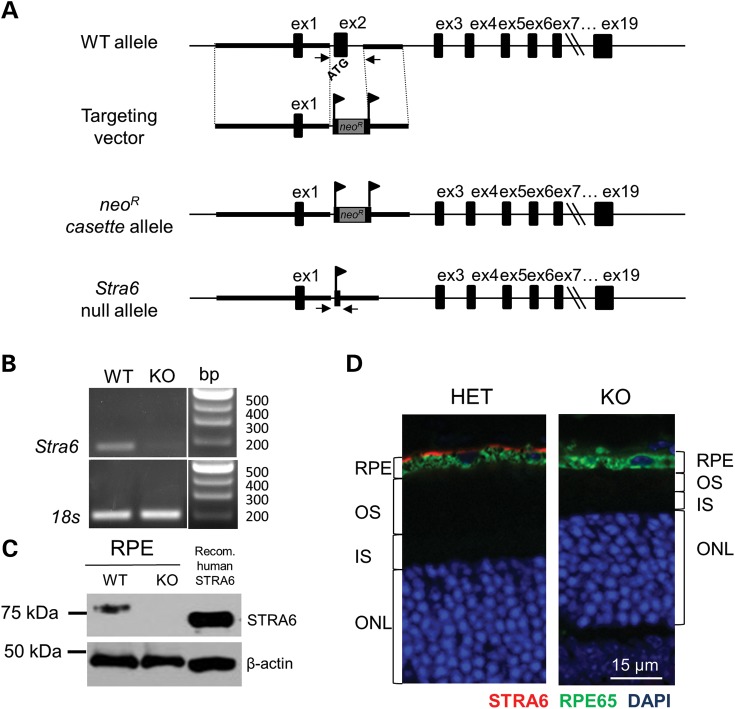
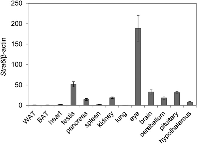
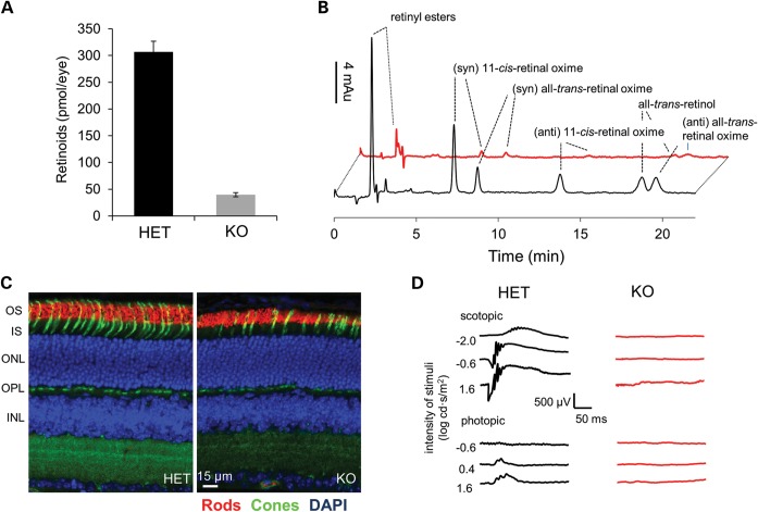
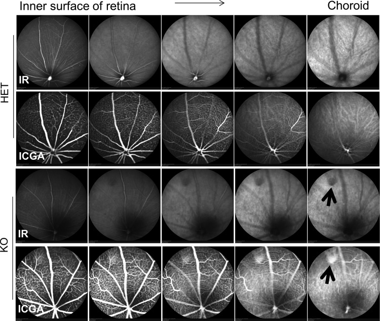
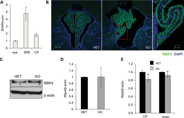
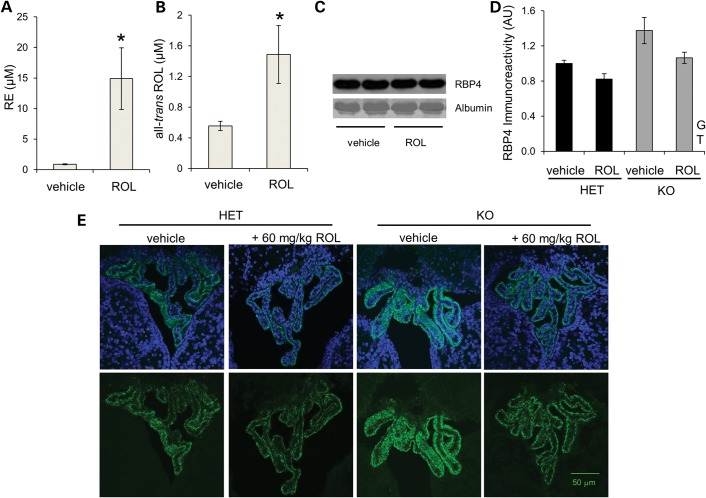
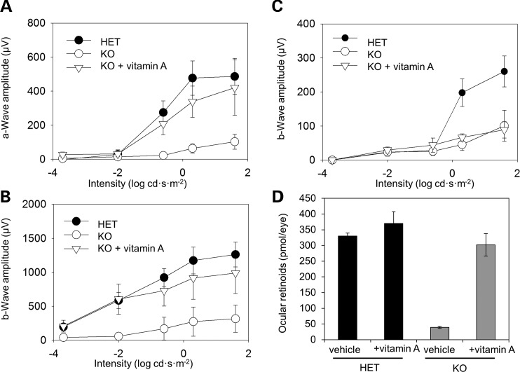
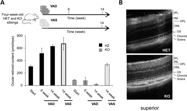
Vitamin A must be adequately distributed within the body to maintain the functions of retinoids in the periphery and chromophore production in the eyes. Blood transport of the lipophilic vitamin is mediated by the retinol-binding protein, RBP4. Biochemical evidence suggests that cellular uptake of vitamin A from RBP4 is facilitated by a membrane receptor. This receptor, identified as the Stimulated by retinoic acid gene 6 (Stra6) gene product, is highly expressed in epithelia that constitute blood-tissue barriers. Here we established a Stra6 knockout mouse model to analyze the metabolic basis of vitamin A homeostasis in peripheral tissues. These mice were viable when bred on diets replete in vitamin A, but evidenced markedly reduced levels of ocular retinoids. Ophthalmic imaging and histology revealed malformations in the choroid and retinal pigmented epithelium, early cone photoreceptor cell death, and reduced lengths of rod outer segments. Similar to the blood-retina barrier in the RPE, vitamin A transport through the blood-cerebrospinal fluid barrier in the brain's choroid plexus was impaired. Notably, treatment with pharmacological doses of vitamin A restored vitamin A transport across these barriers and rescued the vision of Stra6(-/-) mice. Furthermore, under conditions mimicking vitamin A excess and deficiency, our analyses revealed that STRA6-mediated vitamin A uptake is a regulated process mandatory for ocular vitamin A uptake when RBP4 constitutes the only transport mode in vitamin A deficiency. These findings identifying STRA6 as a bona fide vitamin A transporter have important implications for disease states associated with impaired blood vitamin A homeostasis.


Figures

Figure 1.
Figure 1.
Generation of Stra6 knockout mice. (A) Scheme of the WT and mutant Stra6 alleles. The targeting vector was designed to replace exon 2 (ex2) with a neomycin cassette (neoR) flanked by two loxP sites (indicated by banners). This resulted in the Stra6 neomycin resistance (neoR) cassette allele. The neoR cassette was floxed out by Cre recombinase resulting in the Stra6 null allele. Genotyping of mice was performed by PCR with the primer pair Stra6up/down for the wild-type allele (WT) and Stra6up/KOdown for the null allele (KO) as described in the Materials and Methods. Positions of the primers are indicated by the arrows. (B) Total mRNA was isolated from the retinal pigmented epithelium (RPE) of WT and KO mice. RT-PCR for Stra6 mRNA employing a primer pair that spans exon 3 to exon 5 demonstrates the absence of Stra6 mRNA in Stra6−/− mice. RT-PCR for 18 s ribosomal RNA was used as loading control. (C) Immunoblot of protein extracts isolated from the retinal pigmented epithelium (RPE) of WT and KO mice (20 μg total protein was loaded per lane). Hek293 cells overexpressing human STRA6 were used as a positive control (5 μg total protein) and β-actin was used as a loading control. (D) Immunostaining for STRA6 (red) in eye sections of mice heterozygous (HET) and homozygous (KO) for the Stra6 null allele. Staining for RPE65 (green) was used to visualize the RPE and DAPI (blue) was used to stain the nuclei. ex, exon; OS, outer segments; IS, inner segments; ONL, outer nuclear layer.
Figure 2.
Figure 2.
Stra6 mRNA expression in tissues of 28-day-old mice. Tissues were collected from 21-day-old wild-type mice (n = 3). Total RNA was isolated and RT-qPCR analysis for Stra6 mRNA expression levels was carried out using beta-actin as control. Numbers on the ordinate represent fold changes compared with the expression level in white adipose tissue (WAT) expressed as means ± SEM. BAT, brown adipose tissue.
Figure 3.
Figure 3.
Stra6−/− mice evidence blindness and ocular vitamin A deficiency. (A) Ocular retinoid content of 21-day-old mice heterozygous (HET) and homozygous (KO) for the Stra6 null allele. (B) HPLC traces (325 nm) of 21-day-old mice heterozygous (black trace) and homozygous (red trace) for the Stra6 null allele. Identities of individual peaks are indicated in the figure. (C) Immunostaining of cone (green) and rod (red) photoreceptors in superior retinas of 21-day-old mice heterozygous (HET) and homozygous (KO) for the Stra6 null allele. DAPI was used to stain nuclei (blue). Note the reduced thickness of the rod outer segments and inner nuclear layer. (D) Scotopic (upper panel) and photopic (lower panel) electroretinograms from 21-day-old mice heterozygous and homozygous for the Stra6 null allele. OS, outer segment(s); IS, inner segments; ONL, outer nuclear layer; OPL, outer plexiform layer; INL, inner nuclear layer.
Figure 4.
Figure 4.
Stra6−/− mice display impaired choroidal vascularization. Fundus imaging of 28-day-old mice heterozygous (HET) and homozygous (KO) for the Stra6 null allele was performed with a cSLO (SpectralisHRA2, Heidelberg Engineering, Heidelberg, Germany) and a 55° lens. The near infrared reflectance image (IR mode, 820 nm laser) was used to align the fundus camera relative to the pupil and thus acquire evenly illuminated fundus images. The ICGA mode (790 nm laser) was used for angiography. After 10 min, mice were injected intraperitoneally with ICG (15 mg/kg, Acros Organics). Arrows indicate the malformed area in the choroid of KO mice.
Figure 5.
Figure 5.
Stra6−/− mice develop RPE and choroidal malformations in the superior eye. (A) Isolated eye cups of 21-day-old mice heterozygous (HET) and homozygous (KO) for the Stra6 null allele. White arrow indicates an area of discoloration in KO mice. (B) Histological analyses of retinas from 21-day-old heterozygous (HET) and homozygous (KO) for the Stra6 null allele. KO mice display altered morphology of the RPE, choroid and sclera in superior parts of the retina (arrow heads). In the inferior retina of KO mice, the outer nuclear layer reveals an altered stratification (arrow heads). Note that aside from these malformations, the outer segment layer is significantly reduced in the retina of KO mice. (C) (Top panels) Immunostaining for RPE65 (green) of cross-sections of the superior retina of 21-day-old mice HET and KO for the Stra6 null allele. (Bottom panels) Phase contrast pictures of the same area. Both pictures show a reduction of the RPE and a lack of the choroid in the diseased area of the superior retina. OS, outer segment; IS, inner segment; ONL, outer nuclear layer; OPL, outer plexiform layer; INL, inner nuclear layer; IPL, inner plexiform layer.
Figure 6.
Figure 6.
Stra6−/− mice accumulate RBP4 in the CP. (A) Whole eyes, retina pigmented epithelium (RPE) and CP from the fourth ventricle were collected from 21-day-old wild-type mice (n = 3). Total RNA was isolated and RT-qPCR analyses for Stra6 mRNA expression levels were carried out using β-actin as internal control. Numbers on the ordinate represent fold changes compared with the expression level in whole eye. (B) Immunostaining for RBP4 (green) in the CP of 21-day-old Stra6+/− (HET) and Stra6−/− (KO) mice. In the left most panel, a projected image with a higher magnification for RBP4 staining of RBP4 reveals its localization in ependymal cells of the CP. For all fluorography, DAPI was used to visualize the nuclei (blue). (C) Immunoblotting for RBP4 in protein extracts of CP isolated from 21-day-old mice heterozygous (HET) and homozygous (KO) for the Stra6 null allele. Ten micrograms of total protein per lane were loaded and β-actin was used as a loading control. (D) Rbp4 mRNA levels in isolated CP, and Rarβ mRNA levels in CP and whole brain homogenate. β-Actin was used as loading control (n = 3 mice/group). Numbers on the ordinate represent fold changes compared with the expression levels in HET animals expressed as means ± SEM.
Figure 7.
Figure 7.
Pharmacological doses of vitamin A promote RBP release in the CP. Two-day-old mice heterozygous (HET) or homozygous (KO) for the Stra6 null allele were orally supplemented with 60 mg/kg of body weight of ROL dissolved in canola oil or vehicle only as a control. Mice were sacrificed 4 h later for blood and brain collection. (A) Circulating retinyl esters (RE), (B) free ROL and (C) immunoblotting of RBP4 levels in the serum of vitamin A or vehicle-treated mice upon euthanasia. (D) RBP4 immune-reactivity was quantified in the CP 1 day post-vitamin A treatment. (E) Representative immunostaining for RBP4 (green) in the CP of mice. DAPI was used to visualize the nuclei (blue). Numerical data represent the means ± SEM. *, P<0.05; Student's t-test. G, genotype effect in two-way ANOVA analysis (P < 0.05); T, effect of the ROL treatment in two-way ANOVA analysis (P < 0.05). AU; arbitrary units.
Figure 8.
Figure 8.
Pharmacological doses of vitamin A restore vision in Stra6−/− mice. Scotopic (A and B) and photopic (C) single flash ERG analyses of 28-day-old mice heterozygous (HET) or homozygous (KO) for the Stra6 null allele. KO mice were raised on a standard chow diet and either gavaged or not with 0.5 mg all-trans-retinol per animal (+ VitA). Values are expressed as means ± SEM (n = 3 per genotype and dietary condition). (D) Total ocular retinoid contents of mice were determined by HPLC analysis and expressed as means ± SEM (n = 3 per genotype and dietary condition).
Figure 9.
Figure 9.
STRA6 is critical for photoreceptor maintenance under dietary vitamin A restriction. (A) Scheme of the dietary intervention. Mice heterozygous (HET) and homozygous (KO) for the Stra6 null allele were raised on vitamin A rich chow. At weaning time mice were subjected to dietary vitamin A restriction. As a control, mice were continuously kept on vitamin A-rich chow. (B) Ocular retinoid content in mice of different genotypes and dietary groups. (C) SLO analysis of mice subjected to dietary vitamin A restriction. Note that the outer segment layer is greatly reduced in KO mice.
Figure 10.
Figure 10.
Fenretinide (FHR) but not A1220 decreases RBP and retinol serum levels in a STRA6-dependent manner. (A) Molecular structure of the two RBP-lowering agents used, namely the synthetic retinoid fenretinide (FHR) and A1120. (B and C) Serum RBP4 levels in mice heterozygous and homozygous for the Stra6 null allele after a single gavage with (B) 30 mg FHR/kg) and (C) 30 mg A1120/kg. Blood samples from the tail vein of each animal were collected (n = 3 per genotype and condition) at time points shown. Representative immunoblots for serum RBP4 are displayed.

References

    1. Shah S.P., Taylor A.E., Sowden J.C., Ragge N.K., Russell-Eggitt I., Rahi J.S., Gilbert C.E. Anophthalmos, microphthalmos, and typical coloboma in the United Kingdom: a prospective study of incidence and risk. Invest. Ophthalmol. Vis. Sci. 2011;52:558–564. - PubMed
    1. Slavotinek A.M. Eye development genes and known syndromes. Mol. Genet. Metab. 2011;104:448–456. - PMC - PubMed
    1. Gerth-Kahlert C., Williamson K., Ansari M., Rainger J.K., Hingst V., Zimmermann T., Tech S., Guthoff R.F., van Heyningen V., Fitzpatrick D.R. Clinical and mutation analysis of 51 probands with anophthalmia and/or severe microphthalmia from a single center. Mol. Genet. Genomic Med. 2013;1:15–31. - PMC - PubMed
    1. Golzio C., Martinovic-Bouriel J., Thomas S., Mougou-Zrelli S., Grattagliano-Bessieres B., Bonniere M., Delahaye S., Munnich A., Encha-Razavi F., Lyonnet S., et al. Matthew-Wood syndrome is caused by truncating mutations in the retinol-binding protein receptor gene STRA6. Am. J. Hum. Genet. 2007;80:1179–1187. - PMC - PubMed
    1. Pasutto F., Sticht H., Hammersen G., Gillessen-Kaesbach G., Fitzpatrick D.R., Nurnberg G., Brasch F., Schirmer-Zimmermann H., Tolmie J.L., Chitayat D., et al. Mutations in STRA6 cause a broad spectrum of malformations including anophthalmia, congenital heart defects, diaphragmatic hernia, alveolar capillary dysplasia, lung hypoplasia, and mental retardation. Am. J. Hum. Genet. 2007;80:550–560. - PMC - PubMed

Publication types