. 2014 May 20;15(5):9016-35.
doi: 10.3390/ijms15059016.

Mangiferin facilitates islet regeneration and β-cell proliferation through upregulation of cell cycle and β-cell regeneration regulators

Affiliations

Mangiferin facilitates islet regeneration and β-cell proliferation through upregulation of cell cycle and β-cell regeneration regulators

Hai-Lian Wang et al. Int J Mol Sci. .

Abstract

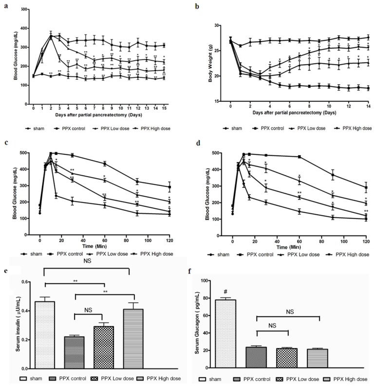
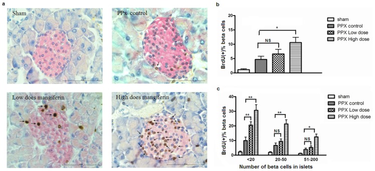
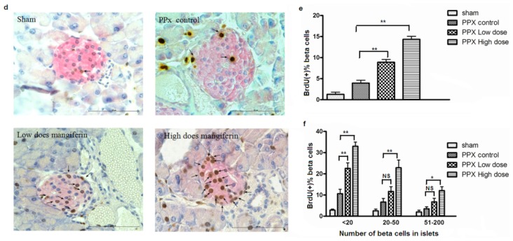
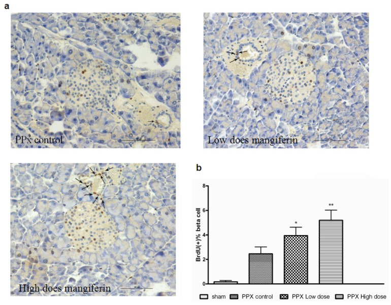
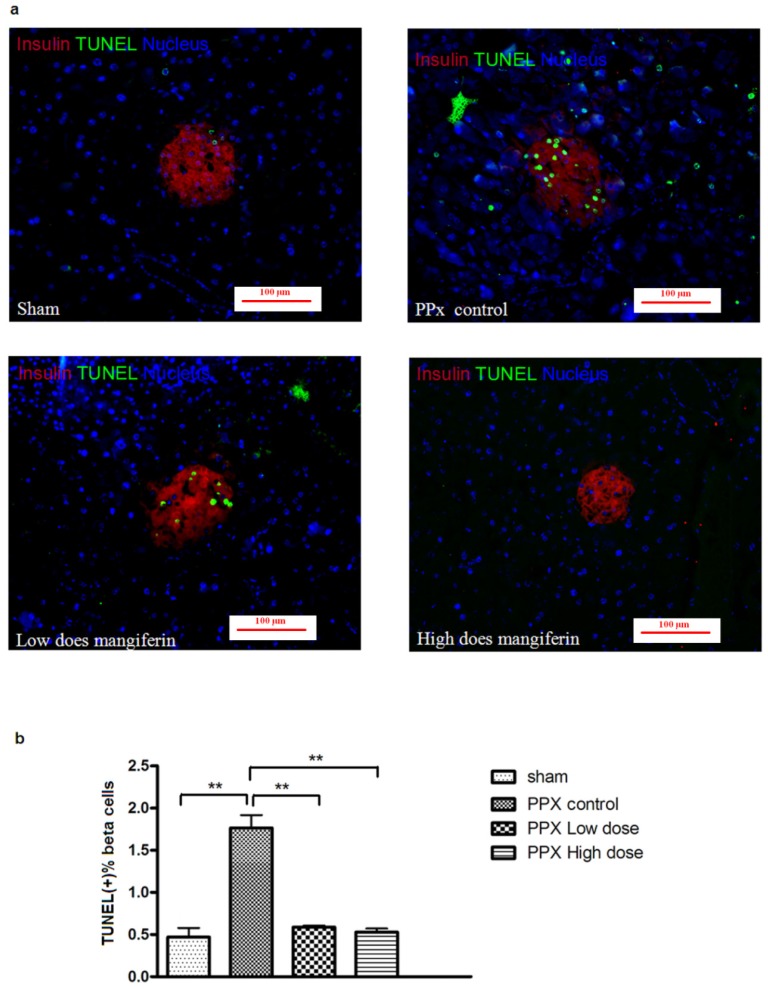
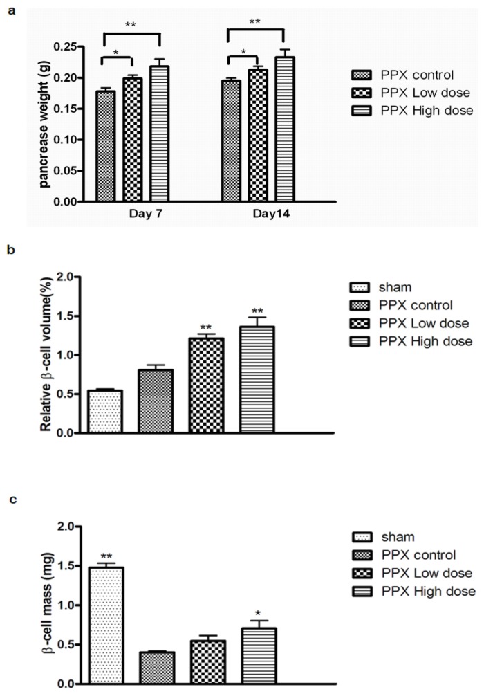
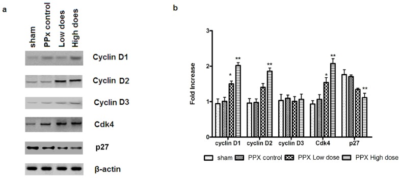
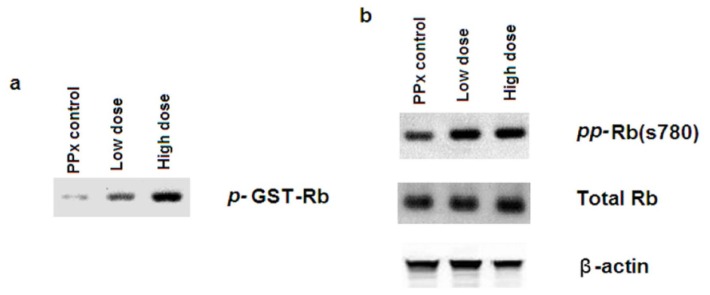
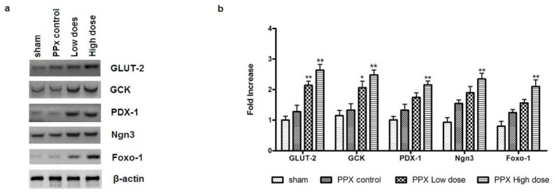
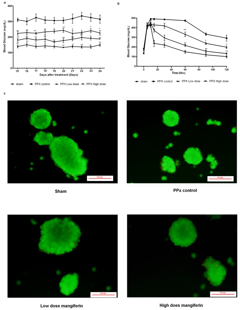
Mangiferin, a xanthonoid found in plants including mangoes and iris unguicularis, was suggested in previous studies to have anti-hyperglycemic function, though the underlying mechanisms are largely unknown. This study was designed to determine the therapeutic effect of mangiferin by the regeneration of β-cells in mice following 70% partial pancreatectomy (PPx), and to explore the mechanisms of mangiferin-induced β-cell proliferation. For this purpose, adult C57BL/6J mice after 7-14 days post-PPx, or a sham operation were subjected to mangiferin (30 and 90 mg/kg body weight) or control solvent injection. Mangiferin-treated mice exhibited an improved glycemia and glucose tolerance, increased serum insulin levels, enhanced β-cell hyperplasia, elevated β-cell proliferation and reduced β-cell apoptosis. Further dissection at the molecular level showed several key regulators of cell cycle, such as cyclin D1, D2 and cyclin-dependent kinase 4 (Cdk4) were significantly up-regulated in mangiferin-treated mice. In addition, critical genes related to β-cell regeneration, such as pancreatic and duodenal homeobox 1 (PDX-1), neurogenin 3 (Ngn3), glucose transporter 2 (GLUT-2), Forkhead box protein O1 (Foxo-1), and glucokinase (GCK), were found to be promoted by mangiferin at both the mRNA and protein expression level. Thus, mangiferin administration markedly facilitates β-cell proliferation and islet regeneration, likely by regulating essential genes in the cell cycle and the process of islet regeneration. These effects therefore suggest that mangiferin bears a therapeutic potential in preventing and/or treating the diabetes.


Figures

Figure 1.
Figure 1.
Mangiferin induces glucose metabolic changes in mice after pancreatectomy. (a) Comparisons of fasting blood glucose concentrations 14 days post surgery. Day 0 represented as the day mice received surgery, and all mice treated with different dosages of mangiferin from day 1; (b) The variation of body weight after a 14-day mangiferin treatment. Day 0 represented as the day mice received surgery, and all mice treated with different dosages of mangiferin from day 1; (c) Intravenous glucose tolerance test (IVGTT) on day 7 post surgery; (d) IVGTT on day 14 post surgery; (e) Fasting insulin and glucagon levels on day 14; and (f) Glucagon levels on day 14. n = 10 in each group. *, p < 0.05 and **, p < 0.01 vs. partial pancreatectomy (PPx) control at the same time point by two-way ANOVA for repeated measures. # p < 0.05 vs. all other groups at the same time point by one-way ANOVA. Data are means ± SEM. NS: not significant.
Figure 2.
Figure 2.
Mangiferin induces islet regeneration. (a) Representative pictures of immunohistochemistry staining of insulin-positive cells (red) and bromodeoxyuridine (BrdU)-labeled cells (brown) of different groups after 7 days treatment. Arrows point to the BrdU-labeled cells. Scale bar represents 100 μm; (b) BrdU-positive percentage of insulin-positive β-cells from different groups on day 7. At least 10 islets with more than 1000 β-cells were counted per mouse (n = 8). *, p < 0.05 and **, p < 0.01 vs. PPx control group at the same time point by one-way analysis of variance; (c) Assessment of the distribution of proliferated β-cells in different islet size on day 7. Data are means ± SEM of 3 independent experiments and statistical significance. *, p < 0.05 and **, p < 0.01 vs. PPx controls group by two-way analysis of variance; (d) Representative pictures of immunohistochemistry staining of insulin-positive cells (red) and bromodeoxyuridine (BrdU)-labeled cells (brown) of different groups after 14 days treatment. Arrows point to the BrdU-labeled cells. Scale bar represents 100 μm; (e) BrdU-positive percentage of insulin-positive β-cells from different groups on day 14. At least 10 islets with more than 1000 β-cells were counted per mouse (n = 8). **, p < 0.01 vs. PPx controls group by one-way ANOVA; and (f) Assessment of the distribution of proliferated β-cell in different islet size; n = 8 for each group and each time point. Data are means ± SEM of 3 independent experiments and statistical significance *, p < 0.05 and **, p < 0.01 vs. PPx controls group by two-way analysis of variance. NS: not significant.
Figure 2.
Figure 2.
Mangiferin induces islet regeneration. (a) Representative pictures of immunohistochemistry staining of insulin-positive cells (red) and bromodeoxyuridine (BrdU)-labeled cells (brown) of different groups after 7 days treatment. Arrows point to the BrdU-labeled cells. Scale bar represents 100 μm; (b) BrdU-positive percentage of insulin-positive β-cells from different groups on day 7. At least 10 islets with more than 1000 β-cells were counted per mouse (n = 8). *, p < 0.05 and **, p < 0.01 vs. PPx control group at the same time point by one-way analysis of variance; (c) Assessment of the distribution of proliferated β-cells in different islet size on day 7. Data are means ± SEM of 3 independent experiments and statistical significance. *, p < 0.05 and **, p < 0.01 vs. PPx controls group by two-way analysis of variance; (d) Representative pictures of immunohistochemistry staining of insulin-positive cells (red) and bromodeoxyuridine (BrdU)-labeled cells (brown) of different groups after 14 days treatment. Arrows point to the BrdU-labeled cells. Scale bar represents 100 μm; (e) BrdU-positive percentage of insulin-positive β-cells from different groups on day 14. At least 10 islets with more than 1000 β-cells were counted per mouse (n = 8). **, p < 0.01 vs. PPx controls group by one-way ANOVA; and (f) Assessment of the distribution of proliferated β-cell in different islet size; n = 8 for each group and each time point. Data are means ± SEM of 3 independent experiments and statistical significance *, p < 0.05 and **, p < 0.01 vs. PPx controls group by two-way analysis of variance. NS: not significant.
Figure 3.
Figure 3.
Mangiferin promotes proliferation of duct cells. (a) Representative pictures of immunohistochemistry staining of insulin-positive cells (red) and BrdU-labeled cells (brown). Arrows point to the BrdU-labeled duct cells. Scale bar represents 100 μm; and (b) BrdU-positive percentage of duct cells. n = 8 for each group.* p < 0.05 and **, p < 0.01 vs. PPx control group by one-way ANOVA; Data are means ± SEM. PPx, partial pancreatectomy.
Figure 4.
Figure 4.
Mangiferin inhibits β-cell apoptosis. (a) Representative pictures of immunofluorescent staining of insulin (red) and TUNEL (green) of mangiferin treated or PPx mice, on day 14. At least 10 islets with more than 1000 β-cells were counted per mouse. All graphs show means ± SEM from at least 2 independent experiments. Scale bar represents 100 μm; and (b) TUNEL-positive percentage of different groups on day 14. At least 10 islets with more than 1000 β-cells were counted per mouse; n = 8 for each group and each time point. All graphs show means ± SEM from three independent experiments. **, p < 0.01 vs. PPx controls group by one-way ANOVA.
Figure 5.
Figure 5.
Mangiferin induces β-cell hyperplasia. (a) Comparisons of remnant pancreas weight; (b) Relative β-cell volume by point counting; and (c) β-cell mass calculated by relative β-cell volume and total weight of remnant pancreas. Two or three slides (200 μm apart) from the broadest pancreatic sections were analyzed for β-cell mass measurement. (n > 6 for each group). All graphs show means ± SEM from three independent experiments. (*, p < 0.05 and **, p < 0.01 compared with PPx controls group).
Figure 6.
Figure 6.
Mangiferin modulates the mRNA and protein level of β-cell cycle regulators. Immunoblotting (a) and real time reverse transcription polymerase chain reaction (RT-PCR) (b) analyses of cyclin D1, cyclin D2, cyclin D3, Cdk4, p27 (n = 10 for each group, *, p < 0.05, and **, p <0.01, vs. PPx controls group by two-way analysis of variance).
Figure 7.
Figure 7.
Mangiferin up-regulates Cdk4 enzymatic activity. (a) In vitro Cdk4 kinase activity in islets from mangiferin-treated and untreated control mice. Islets from remnant pancreas of 50 mice were pooled, with recombinant GST-Rb; and (b) Immunoblotting for hyperphosphorylated Rb at Ser780 (upper panel). Total Rb (middel panel) and β-actin (bottom panel) in islet lysates were showed. The data represent one out of three independent experiments that gave similar results.
Figure 8.
Figure 8.
Expression change of important proteins for β-cell regeneration and function by mangiferin. (a) Representative immunoblots for islets isolated from remnant pancreas, by indicated antibodies. n = 6, for PPx control mice; n = 8, for mangiferin-treated mice. Protein expression of GLUT-2, GCK, PDX-1, Ngn3, and Foxo-1 were examined in cell lysates. Protein bands shown are representative from more than 3 independent experiments with similar results; and (b) Real time RT-PCR analyses of GLUT-2, GCK, PDX-1, Ngn3, Foxo-1. n = 10 for each group, All data are means ± SEM. * p < 0.05, and ** p < 0.01 vs. PPx control by two-way analysis of variance. The data represent one out of three independent experiments that gave similar results.
Figure 9.
Figure 9.
Mangiferin-induced irreversible islet β-cell regeneration (a) Comparisons of fasting blood glucose concentrations at the 10th day after mangiferin treatment; (b) IVGTT on day 10 after 14-day treatment of mangiferin or DMSO; and (c) Islet viability was preserved after 10 days feeding. FDA was represented as viable cells with green fluorescence, while nonviable cells were stained with PI and exhibited as red fluorescence. Scale bar represents 100 μm. All data are means ± SEM. * p < 0.05, and ** p < 0.01 vs. PPx control by two-way analysis of variance.

Similar articles

Cited by

References

    1. Dean P.G., Kudva Y.C., Stegall M.D. Long-term benefits of pancreas transplantation. Curr. Opin. Organ Tranplant. 2008;13:85–90. - PubMed
    1. Shapiro A.J., Ricordi C., Hering B.J., Auchincloss H., Lindblad R., Robertson R.P., Secchi A., Brendel M.D., Berney T., Brennan D.C. International trial of the Edmonton protocol for islet transplantation. N. Engl. J. Med. 2006;355:1318–1330. - PubMed
    1. Bonner-Weir S., Sharma A. Are there pancreatic progenitor cells from which new islets form after birth? Nat. Clin. Pract. 2006;2:240–224. - PubMed
    1. Palmer J.P., McCulloch D.K. Prediction and prevention of IDDM—1991. Diabetes. 1991;40:943–947. - PubMed
    1. Mauricio D., Mandrup-Poulsen T. Apoptosis and the pathogenesis of IDDM: A question of life and death. Diabetes. 1998;47:1537–1543. - PubMed

Publication types

MeSH terms

LinkOut - more resources