. 2014 Aug;60(2):116-28.
doi: 10.1016/j.molimm.2014.04.007. Epub 2014 May 20.

γδ T cells recognize the insulin B:9-23 peptide antigen when it is dimerized through thiol oxidation

Affiliations

γδ T cells recognize the insulin B:9-23 peptide antigen when it is dimerized through thiol oxidation

M Kemal Aydintug et al. Mol Immunol. 2014 Aug.

Abstract

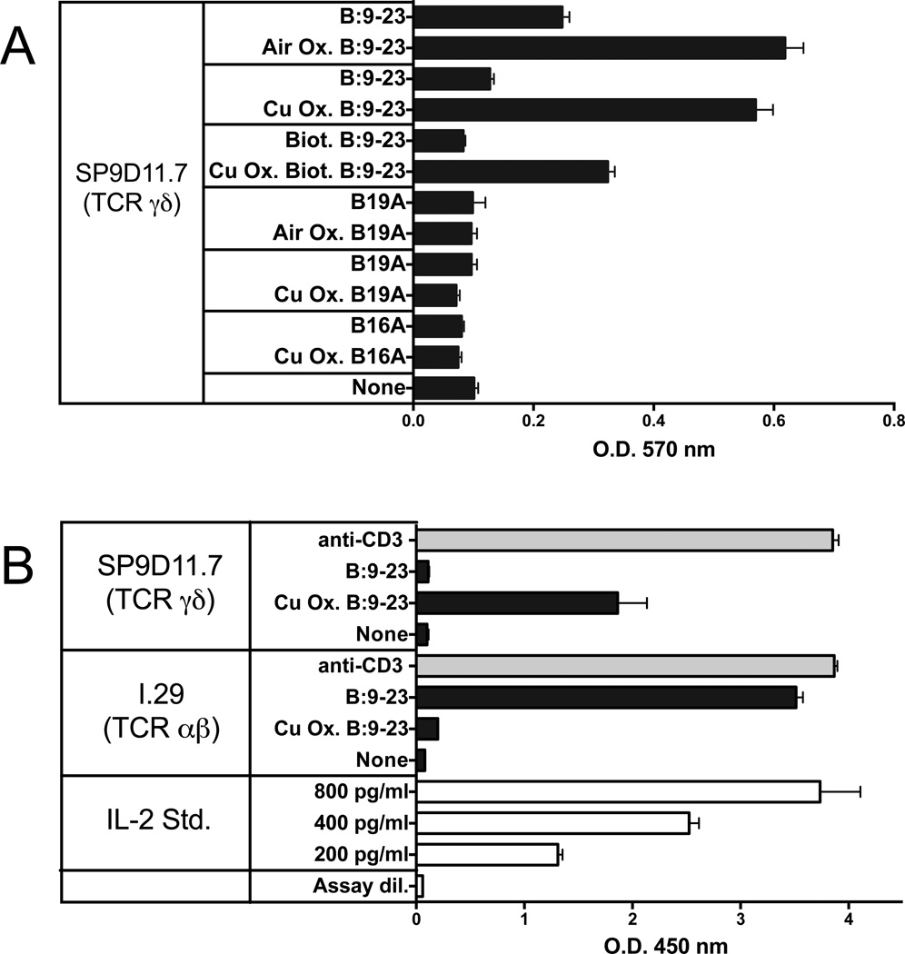
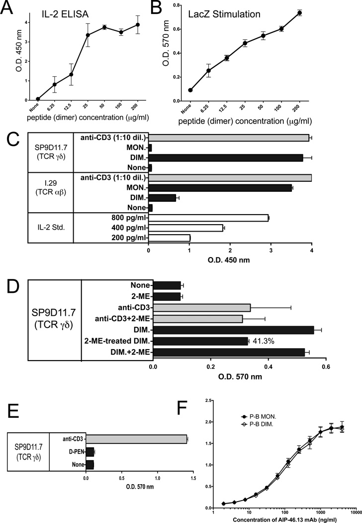
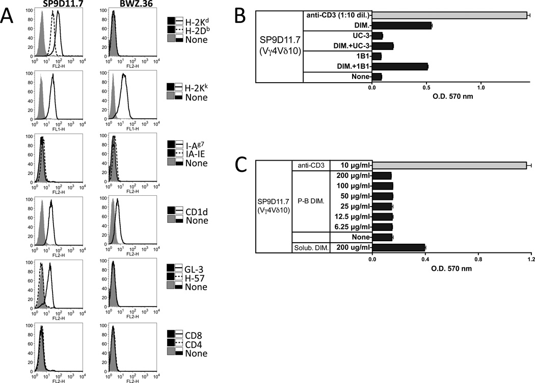
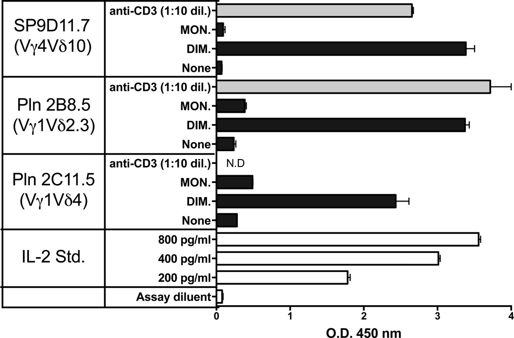
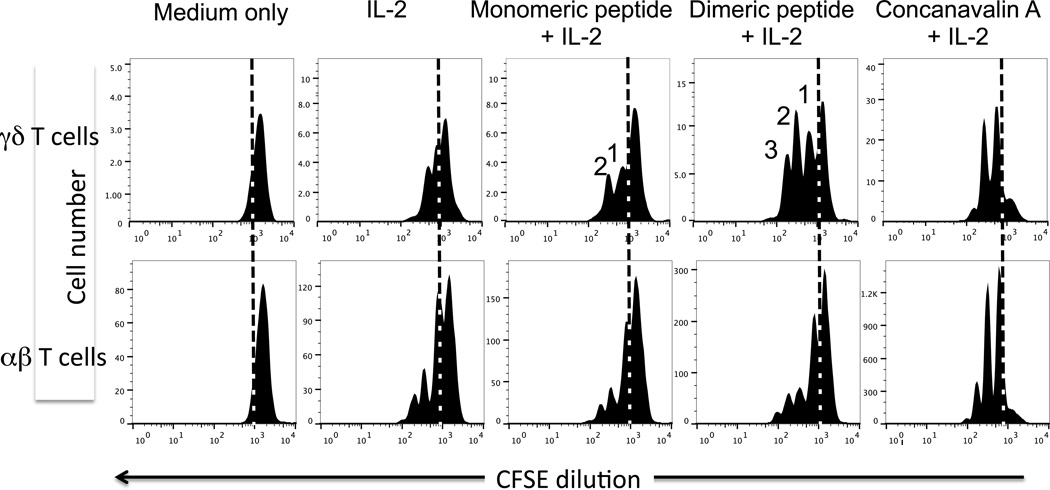
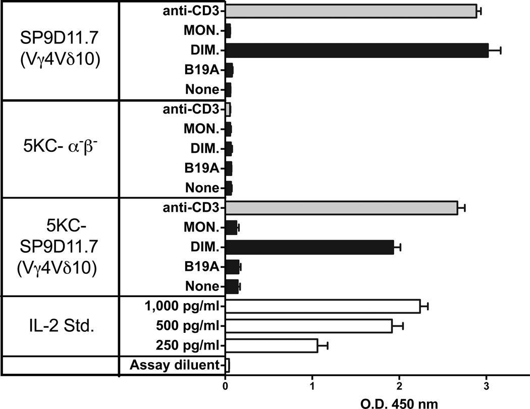
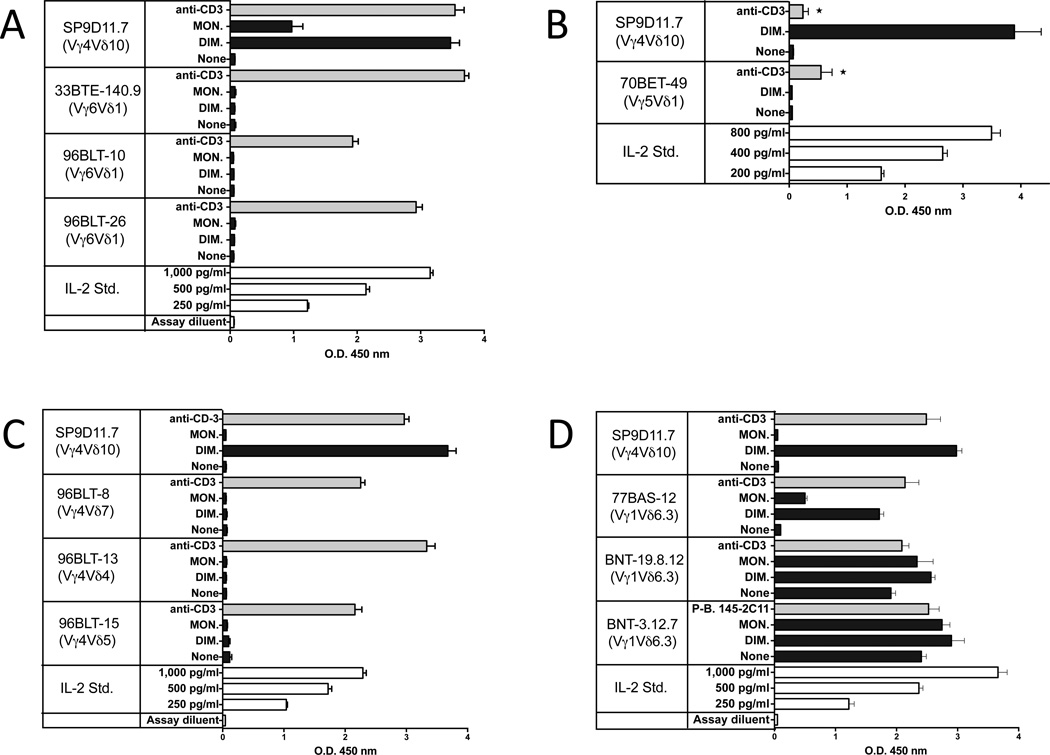
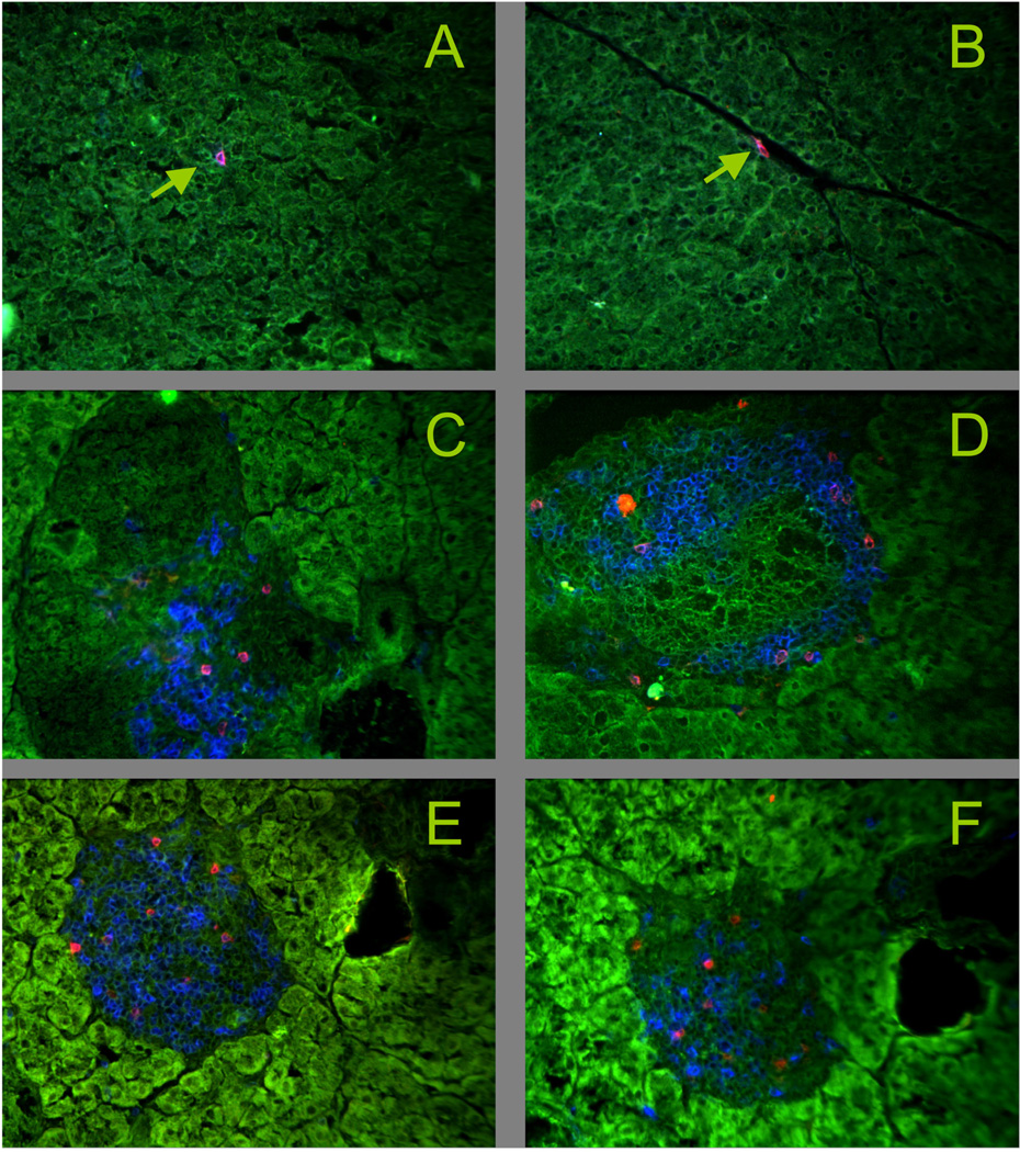
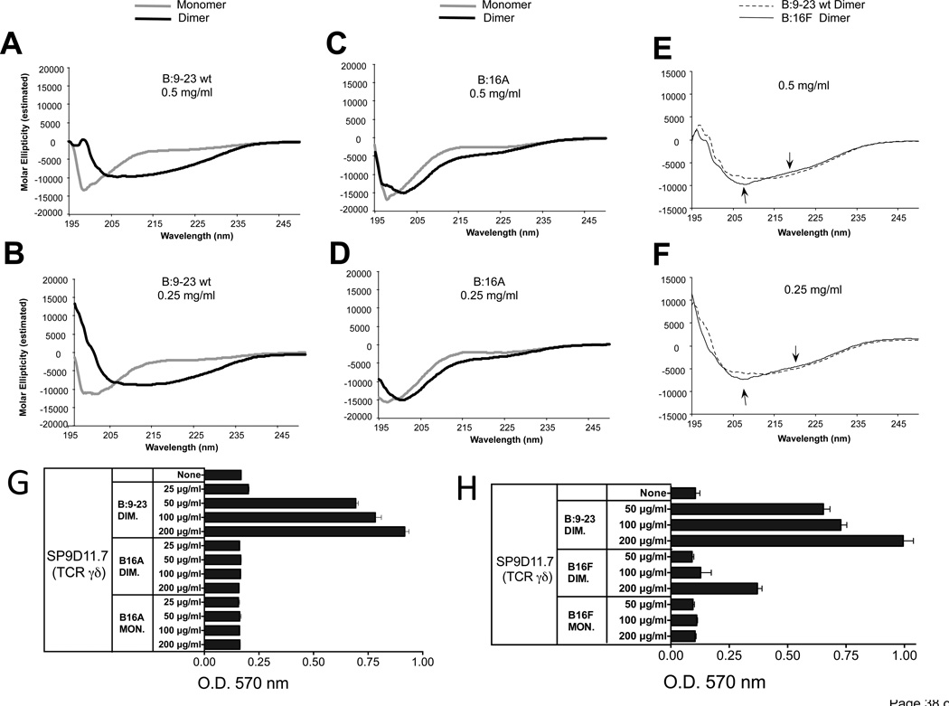
The insulin peptide B:9-23 is a natural antigen in the non-obese diabetic (NOD) mouse model of type 1 diabetes (T1D). In addition to αβ T cells and B cells, γδ T cells recognize the peptide and infiltrate the pancreatic islets where the peptide is produced within β cells. The peptide contains a cysteine in position 19 (Cys19), which is required for the γδ but not the αβ T cell response, and a tyrosine in position 16 (Tyr16), which is required for both. A peptide-specific mAb, tested along with the T cells, required neither of the two amino acids to bind the B:9-23 peptide. We found that γδ T cells require Cys19 because they recognize the peptide antigen in an oxidized state, in which the Cys19 thiols of two peptide molecules form a disulfide bond, creating a soluble homo-dimer. In contrast, αβ T cells recognize the peptide antigen as a reduced monomer, in complex with the MHCII molecule I-A(g7). Unlike the unstructured monomeric B:9-23 peptide, the γδ-stimulatory homo-dimer adopts a distinct secondary structure in solution, which differs from the secondary structure of the corresponding portion of the native insulin molecule. Tyr16 is required for this adopted structure of the dimerized insulin peptide as well as for the γδ response to it. This observation is consistent with the notion that γδ T cell recognition depends on the secondary structure of the dimerized insulin B:9-23 antigen.

Keywords: Autoimmune diabetes; Autoreactivity; Gamma delta T cells; Insulin; Oxidation; T Cell Receptor.


Figures

Figure 1
Figure 1. Oxidation of the B:9-23 peptide antigen enhances stimulation of antigen-specific γδ T cell hybridomas while reducing stimulation of an antigen-specific αβ T cell hybridoma
Panel A Responses of hybridoma SP9D11 to untreated and oxidized peptide antigens 3×10⁴ hybridoma cells were cultured overnight either alone (none) or with untreated or oxidized (Air Ox., oxidized by exposure to ambient air, Cu Ox., oxidized with copper (II) chloride) peptide antigens at 100 µg/ml. Cellular responses were measured in triplicate using the LacZ stimulation assay. Bars show mean response values +/− SE. Panel B Comparison of the responses of the γδ hybridoma SP9D11 and the αβ T cell hybridoma I.29 to untreated and oxidized B:9-23 peptide antigens Culture conditions and peptide stimulation were as described for panel A, except for the addition to all cultures of 1×10⁵ fixed APCs. Cellular responsiveness was determined by stimulation with plate-bound anti-CD3ε mAbs. Cellular responses were measured in triplicate using ELISA for IL-2. Bars show mean response values +/− SE.
Figure 2
Figure 2. The γδ T cell hybridoma SP9D11 specifically recognizes oxidized dimers of the B:9-23 peptide antigen whereas the αβ T cell hybridoma I.29 recognizes monomers
Panels A, B Response of SP9D11 to titrated amounts of dimeric B:9-23 peptide measured via IL-2 ELISA and LacZ assays, respectively. Culture conditions and response measurements were as described in Fig.1. Panel C SP9D11 γδ T cells selectively respond to dimers of the B:9-23-peptide, whereas I.29 αβ T cells respond to monomers. Culture conditions, peptide stimulation and response measurements as described in Fig.1, panel B, except that HPLC-purified monomeric B:9-23 peptide (MON.) and DMSO-oxidized dimeric B:9-23 peptide (DIM.) were used as peptide antigens. Cellular responsiveness was determined by stimulation with plate-bound anti CD3ε mAbs. Supernatants of cultures with anti CD3ε were diluted 10× prior to response measurements. Bars show mean response values +/− SE. Panel D SP9D11 Treatment of the dimeric B:9-23-peptide with 2-ME reduces its stimulatory activity. Culture conditions, peptide stimulation and response measurements as described in Fig.2, panel C. None: no stimulation. 2-ME: 2-ME added to the cultures to a final concentration of 0.25 mM. anti-CD3: soluble anti CD3ε mAb added. Anti-CD3+2-ME: soluble anti CD3ε mAb added as well as 0.25 mM 2-ME. DIM: dimeric B:9-23 peptide added at 100 µg/ml. 2-ME-treated DIM: dimeric B:9-23 peptide pretreated with 5mM 2-ME, then added to stimulation cultures to a final peptide concentration of 100 µg/ml and a final 2-ME concentration of 0.25 mM. DIM+2-ME: dimeric B:9-23 peptide added at 100 µg/ml and 2-ME added at 0.25 mM. Cellular responses were measured in triplicate using the LacZ stimulation assay. Bars show mean response values +/− SE. Panel E SP9D11 γδ T cells fail to respond to the penicillamin adduct of B:9-23 Culture conditions, stimulation and response measurements were as described in Fig.1, panel A. The B:9-23-penicillamin adduct was added at 100 µg/ml. Cellular responsiveness was determined by stimulation with plate-bound anti-CD3ε mAbs. Responses were measured in triplicate using the LacZ stimulation assay. Bars show mean response values +/− SE. Panel F Monoclonal antibody AIP-46.13 recognizes monomeric and dimeric B:9-23 peptide (binding assay) High protein binding ELISA plates were coated with purified monomeric or dimeric B:9-23 peptide at 3 µg peptide/ml. Titrated amounts of AIP-46.13 mAb were added and plate-bound antibody detected by ELISA. Curves show mean absorbance values of triplicate determinations +/− SE.
Figure 3
Figure 3. No roles for MHC II or CD1d in the peptide response of the B:9-23-reactive γδ T cell hybridoma SP9D11
Panel A Cell surface expression of TCR, MHC and CD1d molecules by SP9D11.7 cells SP9D11.7 hybridoma cells and the fusion line BWZ.36 were stained with mAbs specific for MHC I and II molecules, CD1d, TCR-δ, TCR-β, CD4 and CD8, and analyzed cytofluorimetrically. Panel B Anti CD1d mAb 1B1 fails to inhibit the peptide response of SP9D11.7 cells Culture conditions, stimulation and response measurements were as described in Fig.1, panel A. Antibodies specific for TCR-Vγ4 (mAb UC3) or CD1d (mAb 1B1) were added to some cultures at a concentration of 10 µg/ml. Cellular responsiveness was determined by stimulation with plate-bound anti-CD3ε mAbs. Supernatants of cultures stimulated with plate-bound anti-CD3ε were diluted 10× prior to response measurements. Responses were measured in triplicate using the LacZ stimulation assay. Bars show mean response values +/− SE. Panel C Plate-bound insulin peptide does not substantially contribute to the stimulation of SP9D11 hybridoma cells Prior to the stimulation cultures, dimeric insulin peptide dissolved in tissue culture medium was added to the wells of a tissue culture plate at the indicated concentrations, and plates were incubated overnight at 37°C. After washing the wells to remove unbound peptide, stimulation cultures were set up. Culture conditions, stimulation and response measurements were as described in Fig.1, panel A. Cellular responsiveness was determined by stimulation with plate-bound anti-CD3ε mAbs. Responses were measured in triplicate using the LacZ stimulation assay. Bars show mean response values +/− SE.
Figure 4
Figure 4. APC-independent responses of γδ T cell hybridomas expressing diverse TCRs to the oxidized dimeric B:9-23 antigen
Culture conditions and peptide stimulation were as described for Fig. 2, panel C. Cellular responsiveness was determined by stimulation with plate-bound anti CD3ε mAbs. Supernatants of cultures with anti-CD3ε were diluted 10× prior to response measurements. Responses were measured in triplicate using an IL-2 ELISA. Bars show mean response values +/− SE.
Figure 5
Figure 5. Proliferation of freshly isolated γδ T cells from NOD spleen in response to stimulation with the oxidized dimeric B:9-23 antigen
Nylon wool non-adherent lymphocytes from the spleens of 10 wks old NOD females were labeled in vitro with CFSE, then cultured at 5 × 10⁶ cells/ml for two days in medium with 10 units/ml of murine IL-2 plus the peptide indicated at 300 µg/ml. The mitogen Concanavalin A (5 µg/ml) plus IL-2 was used as a positive control. Following a two day culture period, cells were stained with mAbs against either the γδ or αβ TCR. After gating on blasts based on forward/side scatter properties, γδ and αβ T cells were identified and assessed for CFSE levels by fluorescence intensity. Cells left of the dashed vertical line in each histogram have divided, and peaks denoting the expected CFSE levels after 1, 2, or 3 cell divisions among the γδ T cells are indicated.
Figure 6
Figure 6. The γδ T cell response to the oxidized dimeric B:9-23 antigen is TCR-dependent
Hybridoma SP9D11.7 and transfectoma 5KC-SP9D11.7 expressing the SP9D11.7 γδ TCR show similar peptide responses whereas whereas non-transfected cells (5KC-α-β-) are non-responsive. Culture conditions and peptide stimulation were as described for Fig. 2, panel C. Cellular responsiveness was determined by stimulation with plate-bound anti-CD3ε mAbs. Responses were measured in triplicate using ELISA for IL-2. Bars show mean response values +/− SE.
Figure 7
Figure 7. Limitations in the TCR repertoire of γδ T cells responsive to the oxidized dimeric B:9-23 antigen
Panel A Hybridomas expressing Vγ6Vδ1 fail to respond to dimeric B:9-23 Panel B A hybridoma expressing Vγ5Vδ1 fails to respond to dimeric B:9-23 Panel C Several hybridomas expressing Vγ4 fail to respond to dimeric B:9-23 Panel D High spontaneous reactivity makes it difficult to detect B:9-23 reactivity in most Vγ1+ hybridomas, but hybridoma 77BAS-12 (Vγ1+Vδ6.3+) is a responder. For all panels, culture conditions and peptide stimulation were as described for Fig.2, panel C. Cellular responsiveness was determined by stimulation with plate-bound anti-CD3ε mAbs, except for panel B (asterisks). Here, soluble anti-CD3ε mAbs were used (10 µg/ml), which provide a weaker stimulus. Responses were measured in triplicate using ELISA for IL-2. Bars show mean response values +/− SE.
Figure 8
Figure 8. Vγ1+ γδ T cells infiltrate the islets of Langerhans in NOD mice
Snap frozen pancreas tissue was acetone dehydrated and stained with mAbs. Green: tissue autofluorescence. Panels A, B Control: C57BL/6, blue: CD3ε, red TCR-δ, arrows: individual γδ T cells, not in the islets Panels C–F NOD, C: 16 wks of age, blue: CD3ε, red: TCR-δ; D: 8 wks of age, blue: CD3ε, red: TCR-Vγ1; E: 12 wks of age, blue: CD3ε, red: TCR-Vγ1; F: 12 wks of age, blue: CD8α, red: TCR-Vγ1
Figure 9
Figure 9. As an oxidized dimer, the B:9-23 peptide antigen adopts a distinct secondary structure, which requires Tyr16
Panels A, B CD spectra of monomeric and oxidized dimeric B:9-23 peptides (wild-type) Panels C, D CD spectra of monomeric and oxidized dimeric B16A peptides Panel E, F Comparison of CD spectra of oxidized dimeric wild-type and B16F peptides CD spectroscopy was performed on HPLC-purified monomeric and dimeric B:9-23 wild-type (wt) (panels A and B) and amino-acid substituted peptides (panels C–F), at two peptide concentrations. Dimerization induces a shift in the circular dichroism of the wt peptide consistent with a change from a random structure of the monomer to a beta-pleated sheet structure of the dimer (panels A, B). In contrast, dimerization does not substantially change the circular dichroism of the B:16A substituted peptides (panels C, D). The dimeric B16F substituted peptide does shift in circular dichroism, but slightly less than the dimeric wt peptide (see arrows in panels E, F). Panel G The B16A substituted peptide fails to stimulate a response of γδ hybridoma SP9D11 Culture conditions and response measurements using the LacZ assay were as described in Fig.1. Panel H The B16F substituted peptide elicits a smaller response of γδ hybridoma SP9D11 than the wt peptide Culture conditions and response measurements using the LacZ assay were as described in Fig.1.

Similar articles

Cited by

References

    1. Rast JP, Anderson M, Strong SJ, Luer C, Litman RT, Litman GW. α, β, γ and δ T cell antigen receptor genes arose early in vertebrate phylogeny. Immunity. 1997;6:1–11. - PubMed
    1. Bonneville M, O'Brien RL, Born WK. Gammadelta T cell effector functions: a blend of innate programming and acquired plasticity. Nature Reviews Immunology. 2010;10:467–478. - PubMed
    1. Vantourout P, Hayday A. Six-of-the-best: unique contributions of gammadelta T cells to immunology. Nature Reviews Immunology. 2013;13:88–100. - PMC - PubMed
    1. Mukasa A, Lahn M, Pflum EK, Born W, O'Brien RL. Evidence that the same gd T cells respond during infection-induced and autoimmune inflammation. J Immunol. 1997;159:5787–5794. - PubMed
    1. Simonian PL, Wehrmann F, Roark CL, Born WK, O'Brien RL, Fontenot AP. gammadelta T cells protect against lung fibrosis via IL-22. Journal of Experimental Medicine. 2010;207:2239–2253. - PMC - PubMed

Publication types