

Knockout of PARG110 confers resistance to cGMP-induced toxicity in mammalian photoreceptors
- PMID: 24853412
- PMCID: PMC4047865
- DOI: 10.1038/cddis.2014.208
Knockout of PARG110 confers resistance to cGMP-induced toxicity in mammalian photoreceptors
Abstract
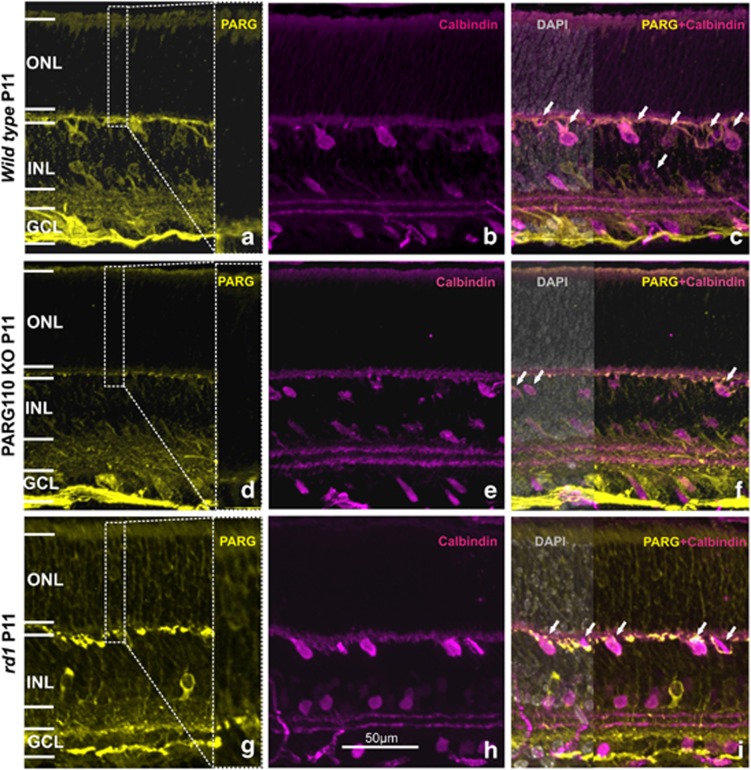
Hereditary retinal degeneration (RD) relates to a heterogeneous group of blinding human diseases in which the light sensitive neurons of the retina, the photoreceptors, die. RD is currently untreatable and the underlying cellular mechanisms remain poorly understood. However, the activity of the enzyme poly-ADP-ribose polymerase-1 (PARP1) and excessive generation of poly-ADP-ribose (PAR) polymers in photoreceptor nuclei have been shown to be causally involved in RD. The activity of PARP1 is to a large extent governed by its functional antagonist, poly-ADP-glycohydrolase (PARG), which thus also may have a role in RD. To investigate this, we analyzed PARG expression in the retina of wild-type (wt) mice and in the rd1 mouse model for human RD, and detected increased PARG protein in a subset of degenerating rd1 photoreceptors. Knockout (KO) animals lacking the 110 kDa nuclear PARG isoform were furthermore analyzed, and their retinal morphology and function were indistinguishable from wild-type animals. Organotypic wt retinal explants can be experimentally treated to induce rd1-like photoreceptor death, but PARG110 KO retinal explants were unexpectedly highly resistant to such treatment. The resistance was associated with decreased PAR accumulation and low PARP activity, indicating that PARG110 may positively regulate PARP1, an event that therefore is absent in PARG110 KO tissue. Our study demonstrates a causal involvement of PARG110 in the process of photoreceptor degeneration. Contrasting its anticipated role as a functional antagonist, absence of PARG110 correlated with low PARP activity, suggesting that PARG110 and PARP1 act in a positive feedback loop, which is especially active under pathologic conditions. This in turn highlights both PARG110 and PARP1 as potential targets for neuroprotective treatments for RD.
Figures


Similar articles
-
PARP1 gene knock-out increases resistance to retinal degeneration without affecting retinal function.PLoS One. 2010 Nov 23;5(11):e15495. doi: 10.1371/journal.pone.0015495. PLoS One. 2010. PMID: 21124852 Free PMC article.
-
Expression of poly(ADP-ribose) glycohydrolase in wild-type and PARG-110 knock-out retina.Adv Exp Med Biol. 2014;801:463-9. doi: 10.1007/978-1-4614-3209-8_59. Adv Exp Med Biol. 2014. PMID: 24664732
-
Excessive activation of poly(ADP-ribose) polymerase contributes to inherited photoreceptor degeneration in the retinal degeneration 1 mouse.J Neurosci. 2007 Sep 19;27(38):10311-9. doi: 10.1523/JNEUROSCI.1514-07.2007. J Neurosci. 2007. PMID: 17881537 Free PMC article.
-
Neurotoxicity of cGMP in the vertebrate retina: from the initial research on rd mutant mice to zebrafish genetic approaches.J Neurogenet. 2017 Sep;31(3):88-101. doi: 10.1080/01677063.2017.1358268. Epub 2017 Aug 16. J Neurogenet. 2017. PMID: 28812418 Review.
-
Poly(ADP-ribosylation) and genomic stability.Biochem Cell Biol. 2005 Jun;83(3):263-9. doi: 10.1139/o05-039. Biochem Cell Biol. 2005. PMID: 15959554 Review.
Cited by
-
Deletion of myosin VI causes slow retinal optic neuropathy and age-related macular degeneration (AMD)-relevant retinal phenotype.Cell Mol Life Sci. 2015 Oct;72(20):3953-69. doi: 10.1007/s00018-015-1913-3. Epub 2015 May 6. Cell Mol Life Sci. 2015. PMID: 25939269 Free PMC article.
-
Efficacy of PARP inhibition in Pde6a mutant mouse models for retinitis pigmentosa depends on the quality and composition of individual human mutations.Cell Death Discov. 2016 Jul 4;2:16040. doi: 10.1038/cddiscovery.2016.40. eCollection 2016. Cell Death Discov. 2016. PMID: 27551530 Free PMC article.
-
Poly ADP ribosylation and extracellular vesicle activity in rod photoreceptor degeneration.Sci Rep. 2019 Mar 6;9(1):3758. doi: 10.1038/s41598-019-40215-3. Sci Rep. 2019. PMID: 30842506 Free PMC article.
-
DNA methylation and differential gene regulation in photoreceptor cell death.Cell Death Dis. 2014 Dec 4;5(12):e1558. doi: 10.1038/cddis.2014.512. Cell Death Dis. 2014. PMID: 25476906 Free PMC article.
-
The role of epigenetic changes in the pathology and treatment of inherited retinal diseases.Front Cell Dev Biol. 2023 Aug 4;11:1224078. doi: 10.3389/fcell.2023.1224078. eCollection 2023. Front Cell Dev Biol. 2023. PMID: 37601102 Free PMC article. Review.
References
-
- Sancho-Pelluz J, Arango-Gonzalez B, Kustermann S, Romero FJ, van Veen T, Zrenner E, et al. Photoreceptor cell death mechanisms in inherited retinal degeneration. Mol Neurobiol. 2008;38:253–269. - PubMed
-
- Trifunovic D, Sahaboglu A, Kaur J, Mencl S, Zrenner E, Ueffing M, et al. Neuroprotective strategies for the treatment of inherited photoreceptor degeneration. Curr Mol Med. 2012;12:598–612. - PubMed
Publication types
MeSH terms
Substances
LinkOut - more resources
Full Text Sources
Other Literature Sources
Research Materials
Miscellaneous
