. 2014 May 20;106(10):2166-74.
doi: 10.1016/j.bpj.2014.04.013.

None of the rotor residues of F1-ATPase are essential for torque generation

Affiliations

None of the rotor residues of F1-ATPase are essential for torque generation

Ryohei Chiwata et al. Biophys J. .

Abstract

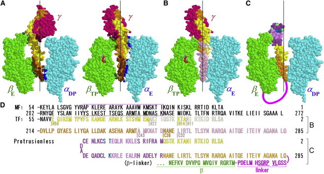
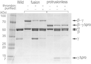
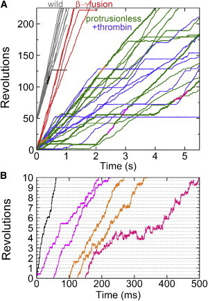
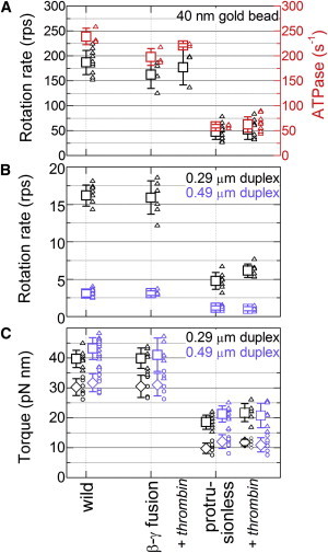
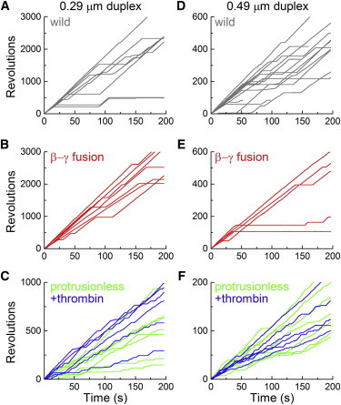
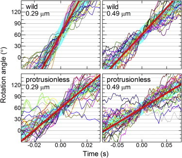
F1-ATPase is a powerful rotary molecular motor that can rotate an object several hundred times as large as the motor itself against the viscous friction of water. Forced reverse rotation has been shown to lead to ATP synthesis, implying that the mechanical work against the motor's high torque can be converted into the chemical energy of ATP. The minimal composition of the motor protein is α3β3γ subunits, where the central rotor subunit γ turns inside a stator cylinder made of alternately arranged α3β3 subunits using the energy derived from ATP hydrolysis. The rotor consists of an axle, a coiled coil of the amino- and carboxyl-terminal α-helices of γ, which deeply penetrates the stator cylinder, and a globular protrusion that juts out from the stator. Previous work has shown that, for a thermophilic F1, significant portions of the axle can be truncated and the motor still rotates a submicron sized bead duplex, indicating generation of up to half the wild-type (WT) torque. Here, we inquire if any specific interactions between the stator and the rest of the rotor are needed for the generation of a sizable torque. We truncated the protruding portion of the rotor and replaced part of the remaining axle residues such that every residue of the rotor has been deleted or replaced in this or previous truncation mutants. This protrusionless construct showed an unloaded rotary speed about a quarter of the WT, and generated one-third to one-half of the WT torque. No residue-specific interactions are needed for this much performance. F1 is so designed that the basic rotor-stator interactions for torque generation and control of catalysis rely solely upon the shape and size of the rotor at very low resolution. Additional tailored interactions augment the torque to allow ATP synthesis under physiological conditions.


Figures

Figure 1
Figure 1
Design of rotor mutations based on a crystal structure of MF1 (9). (A) Rotor-stator arrangement. The γ rotor (dark pink) and an opposing pair of a β subunit (green) and an α subunit (cyan) are shown for two views differing by 120°. The α and β subunits are designated according to the nucleotide bound in the original structure (8): TP, ATP (analog); DP, ADP; E, empty. The C-terminal α-helix of γ is painted orange, and N-terminal helix yellow. Dark-colored atoms in β and α are those within 0.5 nm of an atom in γ (excluding hydrogen), and dark atoms in γ are within 0.5 nm of β or α. The bound nucleotide is in CPK color. Black vertical lines show a putative rotation axis (10). (B) Positions of previous rotor truncations as diagrammed in D. (C) The protrusionless mutant. The exogenous hairpin is borrowed from Rop (dark violet) and seryl-tRNA synthetase (light violet) with extra four residues (blue) as diagramed in D. The hairpin structure shown is part of the MF1 structure enclosed in the violet rectangle in D (actual structure and position of the modified γ are unknown). Sea-green spheres show approximate positions of the three cysteines. Thick magenta line represents the linker between β and γ. The other two γs must be outside the stator cylinder. (D) N- and C-terminal sequences of the γ subunit of MF1 and TF1, showing the positions of truncations/replacements and the linker (thrombin site underlined) sequence for C. The sequence for TF1 starts from 2, because we count from the first Met, which is processed in the WT and this protrusionless construct but not in some mutants (18). To see this figure in color, go online.
Figure 2
Figure 2
Confirmation of mutation by SDS-PAGE. Samples were run in a 10–20% gradient gel, and stained with Coomasie Brilliant Blue R-250. The β-γ fusion mutant and the protrusionless mutant were each run in three lanes, where – – indicates purified subcomplex before thrombin treatment, + – after thrombin treatment for 10 min (reaction mixture), and + + the subcomplex after further purification by size-exclusion chromatography. γΔpro, the γ subunit of the protrusionless mutant.
Figure 3
Figure 3
Time courses of 40-nm gold bead rotation recorded at 4000 or 8000 frames s−1. (A) Overall time courses. Portions shown in a different color are magnified in B with the same color. (B) Magnified time courses corresponding to portions in A shown in the same color. Horizontal solid lines are spaced every one revolution, and gray dotted lines every 120°. To see this figure in color, go online.
Figure 4
Figure 4
Summary of rotation and ATP hydrolysis activities at 2 mM MgATP. Small symbols are individual data and large symbols their average with error bars showing SD. (A) Time-averaged rotary speed for 40-nm gold beads (black), and hydrolysis activity in the presence of LDAO (red). (B) Time-averaged rotary speed of duplexes of polystyrene beads of indicated diameters. (C) Torque estimated from time-averaged rotary speeds of polystyrene bead duplexes (diamonds). Torque values estimated from the instantaneous rotary speed in consecutive 120° steps (Fig. 6) are also shown (squares). To see this figure in color, go online.
Figure 5
Figure 5
Time courses of polystyrene bead rotation at 2 mM MgATP. (AC) Rotation of 0.29-μm bead duplexes attached to the WT (A), the β-γ fusion (B), and the protrusionless mutant (C). Note the difference in the vertical scales. (DF) 0.49-μm bead duplexes. Time-averaged rotary speeds were estimated on uninterrupted portions (see text for the protrusionless mutant) and are plotted in Fig. 4B as small dots. To see this figure in color, go online.
Figure 6
Figure 6
Torque estimation from 120° portions of a rotation record. Thin colored curves show 30 consecutive 120° steps overlaid on top of each other, thick cyan lines representing their average. Individual 120° steps in a continuous record were shifted vertically by a multiple of 120° to obtain the overlap. Time zero for each step was assigned by eye to the data point closest to 60°. Straight red lines indicate linear fit to the cyan curve between 30° and 90°. The slope of the red line, the angular velocity ω in rad s−1, gives the torque N (Eq. 1). To see this figure in color, go online.

References

    1. Noji H., Yasuda R., Kinosita K., Jr. Direct observation of the rotation of F1-ATPase. Nature. 1997;386:299–302. - PubMed
    1. Yoshida M., Muneyuki E., Hisabori T. ATP synthase—a marvellous rotary engine of the cell. Nat. Rev. Mol. Cell Biol. 2001;2:669–677. - PubMed
    1. Boyer P.D. A research journey with ATP synthase. J. Biol. Chem. 2002;277:39045–39061. - PubMed
    1. Senior A.E., Nadanaciva S., Weber J. The molecular mechanism of ATP synthesis by F1F0-ATP synthase. Biochim. Biophys. Acta. 2002;1553:188–211. - PubMed
    1. Kinosita K., Jr., Adachi K., Itoh H. Rotation of F1-ATPase: how an ATP-driven molecular machine may work. Annu. Rev. Biophys. Biomol. Struct. 2004;33:245–268. - PubMed

Publication types

Substances

LinkOut - more resources