. 2014 Dec;25(12):2717-29.
doi: 10.1681/ASN.2013121329. Epub 2014 May 22.

MicroRNA-24 antagonism prevents renal ischemia reperfusion injury

Affiliations

MicroRNA-24 antagonism prevents renal ischemia reperfusion injury

Johan M Lorenzen et al. J Am Soc Nephrol. 2014 Dec.

Abstract

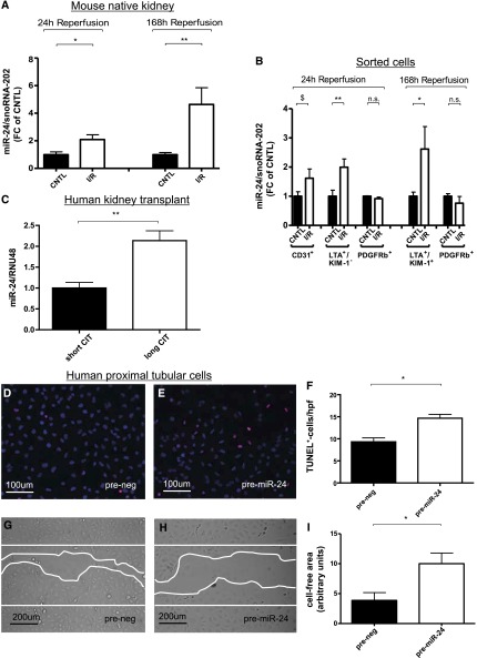
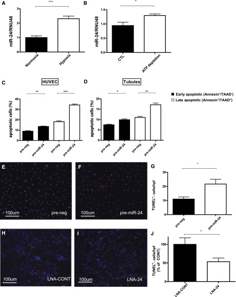
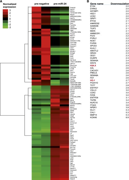
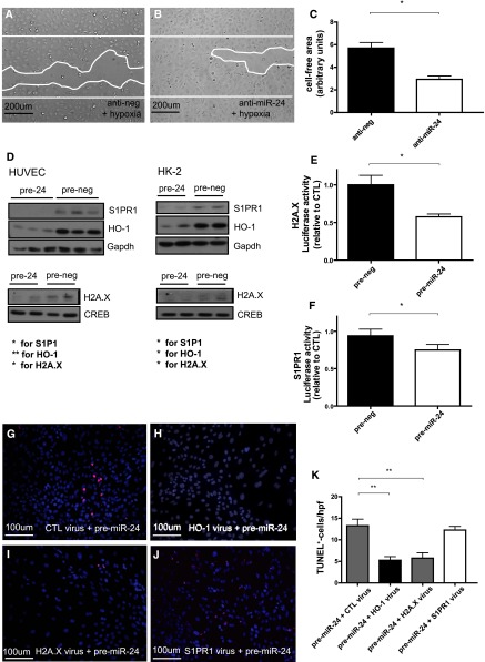
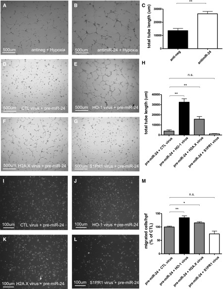
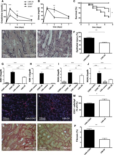
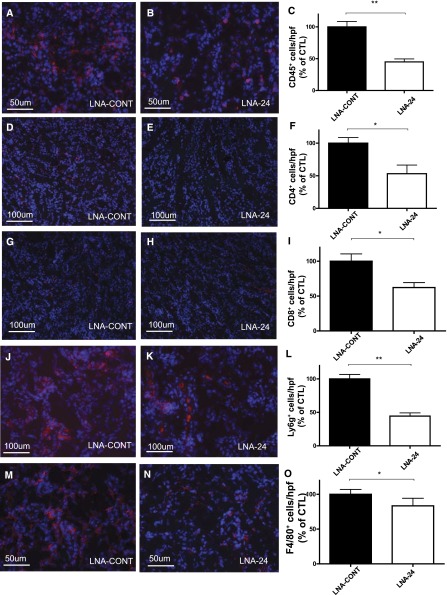
Ischemia-reperfusion (I/R) injury of the kidney is a major cause of AKI. MicroRNAs (miRs) are powerful regulators of various diseases. We investigated the role of apoptosis-associated miR-24 in renal I/R injury. miR-24 was upregulated in the kidney after I/R injury of mice and in patients after kidney transplantation. Cell-sorting experiments revealed a specific miR-24 enrichment in renal endothelial and tubular epithelial cells after I/R induction. In vitro, anoxia/hypoxia induced an enrichment of miR-24 in endothelial and tubular epithelial cells. Transient overexpression of miR-24 alone induced apoptosis and altered functional parameters in these cells, whereas silencing of miR-24 ameliorated apoptotic responses and rescued functional parameters in hypoxic conditions. miR-24 effects were mediated through regulation of H2A histone family, member X, and heme oxygenase 1, which were experimentally validated as direct miR-24 targets through luciferase reporter assays. In vitro, adenoviral overexpression of miR-24 targets lacking miR-24 binding sites along with miR-24 precursors rescued various functional parameters in endothelial and tubular epithelial cells. In vivo, silencing of miR-24 in mice before I/R injury resulted in a significant improvement in survival and kidney function, a reduction of apoptosis, improved histologic tubular epithelial injury, and less infiltration of inflammatory cells. miR-24 also regulated heme oxygenase 1 and H2A histone family, member X, in vivo. Overall, these results indicate miR-24 promotes renal ischemic injury by stimulating apoptosis in endothelial and tubular epithelial cell. Therefore, miR-24 inhibition may be a promising future therapeutic option in the treatment of patients with ischemic AKI.

Keywords: acute renal failure; apoptosis; endothelium; heme oxygenase; ischemia-reperfusion; proximal tubule.


Figures

Figure 1.
Figure 1.
Expression and function of miR-24 in the kidney and distinct renal cell populations. The expression of miR-24 in mouse kidneys is depicted at 24 and 168 hours (A) after unilateral I/R injury (n=7 each). Expression of miR-24 in sorted cells after digestion of postischemic mouse kidney at reperfusion for 24 and 168 hours is shown (B). Levels of miR-24 were compared with snoRNA-202 as control. MiR-24 expression normalized to RNU-48 in biopsy specimens from kidney transplant recipients with long compared with short CIT (n=5 in each group) CD31+, endothelial cells; LTA+/KIM-1, uninjured proximal tubular epithelial cells; LTA+/KIM-1+, injured proximal tubular epithelial cells; PDGFRb+, pericytes. (C). TUNEL staining in cultured HK-2 cells after prenegative control (D) and pre–miR-24 oligonucleotide (E) transfection and quantification of results (F) (n=6 experiments). Scratch migration analysis in normoxia in HK-2 cells after prenegative control (G) and pre–miR-24 oligonucleotide (H) transfection and quantification of results (I) (n=6 experiments). $P=0.08; **P<0.01; *P<0.05. CNTL, contralateral control kidney; hpf, high-power field.
Figure 2.
Figure 2.
Expression of miR-24 in distinct kidney cells and role in apoptosis regulation. Expression of miR-24 normalized to RNU-48 in hypoxic compared with normoxic control human umbilical vein endothelial cells (HUVECs) (A) and HK-2 cells exposed to ATP depletion (B) is shown. Percentage of early apoptotic (Annexin+/7AAD) as well as late apoptotic (Annexin+/7AAD+) HUVECs (C) and proximal tubular epithelial cells (D) in FACS analysis is shown. TUNEL staining in cultured endothelial cells after prenegative control (E) and pre–miR-24 oligonucleotide (F) transfection and quantification of results (G) (n=6 experiments). TUNEL+ cells in outer medulla in mice after ischemia and reperfusion for 24 hours treated with control LNA (LNA-CONT; H) and LNA-24 (I) and quantification of results (J). ***P<0.0001; **P<0.01; *P<0.05. CTL = control.
Figure 3.
Figure 3.
Cluster analysis of miR-24 targets in tubular epithelial cells overexpressing miR-24. Affymetrix gene array and cluster analysis in tubular epithelial cells transfected with prenegative control and pre–miR-24 oligonucleotide (A) is shown. Bioinformatically predicted targets of miR-24 (as obtained from Targetscan) were cross-checked with the results of the array (B). Downregulated genes (fold regulation) of the array, subsequently merged with predicted targets of miR-24 (Targetscan), are shown in Figure 3B. Targets further analyzed (H2A.X and HO-1) are highlighted in red (B).
Figure 4.
Figure 4.
Validation of miR-24 targets and adenoviral rescue assays. Protective role of anti–miR-24 treatment in tubular epithelial cells and miR-24 targets involved in miR-24s action in renal epithelial cells and endothelial cells. Scratch migration analysis in hypoxic HK-2 cells after antinegative control (A) and anti–miR-24 oligonucleotide (B) transfection and quantification of results (C) (n=6 experiments). Western blot analysis in HUVECs and HK-2 cells of cytosolic S1PR1 and HO-1 normalized to glyceraldehyde 3-phosphate dehydrogenase (Gapdh) and nuclear H2A.× normalized to cAMP response element-binding protein after transfection with prenegative control and pre–miR-24 oligonucleotides. Results of luciferase gene reporter assays concerning H2A.X (E) and S1PR1 (F). TUNEL stainings in miR-24–overexpressing tubular epithelial cells after transduction with adenoviral constructs lacking miR-24 binding sites for control virus (CTL; G), HO-1 virus (H), H2A.X virus (I), S1PR1 virus (J), and quantification of results (K). **P<0.01; *P<0.05. hpf, high power field.
Figure 5.
Figure 5.
Adenoviral rescue assays in miR-24 overexpressing endothelial cells. Tube formation capacity (total tube length in micrometers) in hypoxic HUVECs transfected with antinegative control oligonucleotides (A), anti–miR-24 oligonucleotides (B), and quantification of results (C). Tube formation capacity in miR-24–overexpressing endothelial cells after transduction with adenoviral constructs lacking miR-24 binding sites for control virus (CTL; D), HO-1 virus (E), H2A.X virus (F), S1PR1 virus (G), and quantification of results (H). Migration capacity in Boyden chamber assays in miR-24–overexpressing endothelial cells after transduction with adenoviral constructs lacking miR-24 binding sites for control virus (CTL; I), HO-1 virus (J), H2A.X virus (K), S1PR1 virus (L), and quantification of results (M). **P<0.01; *P<0.05.
Figure 6.
Figure 6.
In vivo effect of miR-24 antagonism in murine I/R injury. Protective rescue of renal I/R injury following anti–miR-24 therapy. Renal function parameters (serum creatinine [A] and urea [B]) as well as Kaplan–Meier curve survival analysis (C) in mice treated with an LNA-modified anti-miR targeting miR-24 (LNA-24) and a mismatch control LNA (CONT) 24 hours before induction of I/R injury, as well as sham-operated animals. Bilateral renal I/R injury was performed for 27 minutes. Observation period from days 0 to 7; n=20 per treatment group, n=4 in the sham group. Differences in urea levels at day 7 are underestimated because of loss of uremic mice in the control group. Histologic degree of epithelial injury after ischemia and reperfusion for 24 hours in mice receiving LNA-MM (D) and LNA-24 (E) and unilateral clamping of renal pedicles, as well as quantification of results (F, n=7 each). KIM-1 as well as NGAL mRNA levels in postischemic unilaterally clamped kidneys after reperfusion for 24 hours (G and I) and 168 hours (H and J); CONT contra, contralateral kidney of mice with LNA-CONT; CONT IR, clamped kidney of mice with LNA-CONT; LNA contra, contralateral kidney of mice with LNA-24; LNA IR, clamped kidney of mice with LNA-24. n=7 mice in each group and time point. Capillary rarefaction (CD31-staining) analysis in mice treated with control LNA (LNA-CONT) (K) and LNA-24 (L) and quantification of results (M) (n=7 per group) at reperfusion for 24 hours after clamping. Fibrosis development (sirius red staining) analysis in mice treated with control LNA (LNA-CONT) (N) and LNA-24 (O) and quantification of results at reperfusion for 168 hours (P) (n=7 each). *P<0.05; **P<0.01; ***P<0.0001.
Figure 7.
Figure 7.
Infiltration of inflammatory cells following miR-24 antagonism in vivo. Immunofluorescence stainings and quantification of representative cryosections (4 μm) in outer medulla of mice treated with control LNA (LNA-CONT) and LNA-24 concerning CD45+ (A–C), CD4+ (D–F), CD8+ (G–I), Ly6g+- neutrophils (J–L), and F4/80+ macrophages (M–O) at reperfusion time of 168 hours. Specific immunofluorescence stainings in red and DAPI in blue. **P<0.01; *P<0.05.

References

    1. Kelly KJ: Acute renal failure: Much more than a kidney disease. Semin Nephrol 26: 105–113, 2006 - PubMed
    1. Bon D, Chatauret N, Giraud S, Thuillier R, Favreau F, Hauet T: New strategies to optimize kidney recovery and preservation in transplantation. Nat Rev Nephrol 8: 339–347, 2012 - PubMed
    1. Weight SC, Bell PR, Nicholson ML: Renal ischaemia—reperfusion injury. Br J Surg 83: 162–170, 1996 - PubMed
    1. Bonventre JV, Yang L: Cellular pathophysiology of ischemic acute kidney injury. J Clin Invest 121: 4210–4221, 2011 - PMC - PubMed
    1. Lorenzen JM, Haller H, Thum T: MicroRNAs as mediators and therapeutic targets in chronic kidney disease. Nat Rev Nephrol 7: 286–294, 2011 - PubMed

Publication types

MeSH terms

LinkOut - more resources