

In vivo needle-based electromechanical reshaping of pinnae: New Zealand White rabbit model
- PMID: 24854476
- PMCID: PMC4123460
- DOI: 10.1001/jamafacial.2014.85
In vivo needle-based electromechanical reshaping of pinnae: New Zealand White rabbit model
Abstract
Importance: Electromechanical reshaping (EMR) is a low-cost, needle-based, and simple means to shape cartilage tissue without the use of scalpels, sutures, or heat that can potentially be used in an outpatient setting to perform otoplasty.
Objectives: To demonstrate that EMR can alter the shape of intact pinnae in an in vivo animal model and to show that the amount of shape change and the limited cell injury are proportional to the dosimetry.
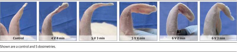
Design, setting, and specimens: In an academic research setting, intact ears of 18 New Zealand white rabbits underwent EMR using 6 different dosimetry parameters (4 V for 5 minutes, 4 V for 4 minutes, 5 V for 3 minutes, 5 V for 4 minutes, 6 V for 2 minutes, and 6 V for 3 minutes). A custom acrylic jig with 2 rows of platinum needle electrodes was used to bend ears at the middle of the pinna and to perform EMR. Treatment was repeated twice per pinna, in proximal and distal locations. Control pinnae were not subjected to current application when being bent and perforated within the jig. Pinnae were splinted for 3 months along the region of the bend using soft silicon sheeting and a cotton bolster.
Main outcomes and measures: The ears were harvested the day after splints were removed and before euthanasia. Photographs of ears were obtained, and bend angles were measured. Tissue was sectioned for histologic examination and confocal microscopy to assess changes to microscopic structure and cellular viability.
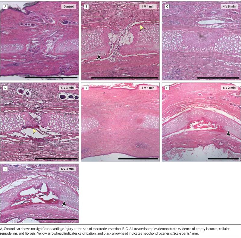
Results: Treated pinnae were bent more and retained shape better than control pinnae. The mean (SD) bend angles in the 7 dosimetry groups were 55° (35°) for the control, 60° (15°) for 4 V for 4 minutes, 118° (15°) for 4 V for 5 minutes, 88° (26°) for 5 V for 3 minutes, 80° (17°) for 5 V for 4 minutes, 117° (21°) for 6 V for 2 minutes, and 125° (18°) for 6 V for 3 minutes. Shape change was proportional to electrical charge transfer, which increased with voltage and application time. Hematoxylin-eosin staining of the pinnae identified localized areas of cell injury and fibrosis in the cartilage and in the surrounding soft tissue where the needle electrodes were inserted. This circumferential zone of injury (range, 1.5-2.5 mm) corresponded to dead cells on cell viability assay, and the diameter of this region increased with total electrical charge transfer to a maximum of 2.5 mm at 6 V for 3 minutes.
Conclusions and relevance: Electromechanical reshaping produced shape change in intact pinnae of rabbits in this expanded in vivo study. A short application of 4 to 6 V can achieve adequate reshaping of the pinnae. Tissue injury around the electrodes increases with the amount of total current transferred into the tissue and is modest in spatial distribution. This study is a critical step toward evaluation of EMR in clinical trials.
Level of evidence: NA.
Figures


Similar articles
-
Optimal Electromechanical Reshaping of the Auricular Ear and Long-term Outcomes in an In Vivo Rabbit Model.JAMA Facial Plast Surg. 2016 Jul 1;18(4):277-84. doi: 10.1001/jamafacial.2016.0166. JAMA Facial Plast Surg. 2016. PMID: 27101542 Free PMC article.
-
In vivo electromechanical reshaping of ear cartilage in a rabbit model: a minimally invasive approach for otoplasty.JAMA Facial Plast Surg. 2013 Jan;15(1):34-8. doi: 10.1001/2013.jamafacial.2. JAMA Facial Plast Surg. 2013. PMID: 23117484 Free PMC article.
-
Long-term in vivo electromechanical reshaping for auricular reconstruction in the New Zealand white rabbit model.Laryngoscope. 2015 Sep;125(9):2058-66. doi: 10.1002/lary.25237. Epub 2015 Mar 16. Laryngoscope. 2015. PMID: 25779479 Free PMC article.
-
Handheld-Level Electromechanical Cartilage Reshaping Device.Facial Plast Surg. 2015 Jun;31(3):295-300. doi: 10.1055/s-0035-1555623. Epub 2015 Jun 30. Facial Plast Surg. 2015. PMID: 26126226
-
Needle electrode-based electromechanical reshaping of cartilage.Ann Biomed Eng. 2010 Nov;38(11):3389-97. doi: 10.1007/s10439-010-0088-1. Epub 2010 Jul 8. Ann Biomed Eng. 2010. PMID: 20614240 Free PMC article.
Cited by
-
Potential-Driven Electrochemical Clearing of Ex Vivo Alkaline Corneal Injuries.Transl Vis Sci Technol. 2022 Jan 3;11(1):32. doi: 10.1167/tvst.11.1.32. Transl Vis Sci Technol. 2022. PMID: 35061010 Free PMC article.
-
Association of Electrochemical Therapy With Optical, Mechanical, and Acoustic Impedance Properties of Porcine Skin.JAMA Facial Plast Surg. 2017 Dec 1;19(6):502-509. doi: 10.1001/jamafacial.2017.0341. JAMA Facial Plast Surg. 2017. PMID: 28654968 Free PMC article.
-
[Preliminary study on microdissection needle-assisted ear cartilage reshaping in vivo rabbit models].Zhongguo Xiu Fu Chong Jian Wai Ke Za Zhi. 2019 May 15;33(5):601-605. doi: 10.7507/1002-1892.201807032. Zhongguo Xiu Fu Chong Jian Wai Ke Za Zhi. 2019. PMID: 31090355 Free PMC article. Chinese.
-
Exploring feedback-controlled versus open-circuit electrochemical lipolysis in ex vivo and in vivo porcine fat: A feasibility study.Lasers Surg Med. 2022 Jan;54(1):157-169. doi: 10.1002/lsm.23466. Epub 2021 Aug 19. Lasers Surg Med. 2022. PMID: 34412154 Free PMC article.
-
Monitoring of Biological Changes in Electromechanical Reshaping of Cartilage Using Imaging Modalities.Biomed Res Int. 2016;2016:7089017. doi: 10.1155/2016/7089017. Epub 2016 Dec 8. Biomed Res Int. 2016. PMID: 28053987 Free PMC article.
References
-
- Weerda H. Surgery of the Auricle: Tumors, Trauma, Defects, and Abnormalities. Thieme; New York, NY: 2007.
-
- Park C, Yoo YS, Hong ST. An update on auricular reconstruction: three major auricular malformations of microtia, prominent ear and cryptotia. Curr Opin Otolaryngol Head Neck Surg. 2010;18(6):544–549. - PubMed
-
- Janz BA, Cole P, Hollier LH, Jr, Stal S. Plast Reconstr Surg. 1. suppl. Vol. 124. 27e-37e: 2009. Treatment of prominent and constricted ear anomalies. doi:10.1097/PRS.0b013e3181aa0e9d. - PubMed
-
- van Wijk MP, Breugem CC, Kon M. Non-surgical correction of congenital deformities of the auricle: a systematic review of the literature. J Plast Reconstr Aesthet Surg. 2009;62(6):727–736. - PubMed
-
- Ragab A. Carbon dioxide laser–assisted cartilage reshaping otoplasty: a new technique for prominent ears. Laryngoscope. 2010;120(7):1312–1318. - PubMed
Publication types
MeSH terms
Grants and funding
LinkOut - more resources
Full Text Sources
Other Literature Sources
Research Materials