. 2014 May 23:4:5050.
doi: 10.1038/srep05050.

Transposon-mediated targeted and specific knockdown of maternally expressed transcripts in the ascidian Ciona intestinalis

Affiliations

Transposon-mediated targeted and specific knockdown of maternally expressed transcripts in the ascidian Ciona intestinalis

Takako Iitsuka et al. Sci Rep. .

Abstract

Maternal mRNAs play crucial roles during early embryogenesis of ascidians, but their functions are largely unknown. In this study, we developed a new method to specifically knockdown maternal mRNAs in Ciona intestinalis using transposon-mediated transgenesis. We found that GFP expression is epigenetically silenced in Ciona intestinalis oocytes and eggs, and this epigenetic silencing of GFP was used to develop the knockdown method. When the 5' upstream promoter and 5' untranslated region (UTR) of a maternal gene are used to drive GFP in eggs, the maternal gene is specifically knocked down together with GFP. The 5' UTR of the maternal gene is the major element that determines the target gene silencing. Zygotic transcription of the target gene is unaffected, suggesting that the observed phenotypes specifically reflect the maternal function of the gene. This new method can provide breakthroughs in studying the functions of maternal mRNAs.


Figures
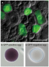
Figure 1
Figure 1. Maternal expression of GFP is epigenetically silenced in Ciona.
(a) A typical GFP expression pattern in the ovary. An ovary of an enhancer detection line EJ[MiTSAdTPOG]78, which entrapped an enhancer responsible for expression in oocytes. Only a few oocytes express GFP. Bar, 100 μm. (b, c) Expression of GFP mRNA in unfertilized eggs of a maternal GFP line, as revealed by whole-mount in situ hybridization (WISH). Dark blue staining suggests the presence of GFP mRNA. (b) An egg that had GFP fluorescence. (c) An egg that lacked GFP fluorescence.
Figure 2
Figure 2. Morphological defects seen in pem > GFP lines.
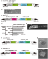
(a) The transposon vector used to knockdown Ci-pem. Black arrowheads indicate inverted repeats (ITR) of Minos. UTR, untranslated region; NLS, nuclear localization signal sequence; Ter, transcription termination sequence. (b) GFP expression in unfertilized eggs of pem > GFP line 1. The egg in the upper right corner emitted GFP fluorescence, while the egg in the lower left corner did not. Bar, 100 μm. (c) A larva derived from sperm of pem > GFP line 4 and a wild-type egg. Bar, 100 μm. (d) A larva derived from an egg of pem > GFP line 4 and wild-type sperm. No, notochord. (e–i) Differentiation of major tissues in abnormal larvae derived from eggs of pem > GFP lines. (e) Epidermis (green). (f) Muscle (green). (g) Notochord (green). (h) Neural tissues (red). (i) Endoderm (En).
Figure 3
Figure 3. Knockdown of Ci-pem.
(a) Abnormal larvae of pem > GFP lines were derived from GFP-negative eggs. One normal and one abnormal larvae derived from the same individual of a pem > GFP line are shown. In the normal larva, GFP fluorescence was detected throughout the body, suggesting that the fluorescence was derived from maternal GFP expression. Such maternal GFP fluorescence was not detected in the abnormal larva, although zygotic GFP expression derived from the Ci-TnI > GFP cassette was evident. (b–f) Ci-pem maternal mRNA was decreased in GFP-negative eggs of pem > GFP lines, as revealed by WISH. Arrows indicate the position of the signal. (b) An egg derived from a wild-type animal. (c) An egg of pem > GFP line 1 that had GFP fluorescence. (d) An egg of line 1 that lacked GFP fluorescence. (e) An egg of pem > GFP line 2. All of the eggs were GFP negative. (f) An egg of pem > GFP line 9. All of the eggs were GFP-positive. (g) Relative expression levels of Ci-pem in eggs of pem > GFP lines, as revealed by quantitative RT-PCR. n = 2 for every line. P values were calculated using the two-tailed Student's t test. (h) A larva derived from an egg of pem > GFP line 2. (i) A larva derived from an egg of pem > GFP line 2 that had been microinjected with in vitro-synthesized Ci-pem mRNA.
Figure 4
Figure 4. The 5′ UTR is essential for maternal gene silencing of the target gene.
(a) The transposon vector from which the Ci-pem 5′ UTR was omitted. (b) A larva derived from a transgenic line created by the vector shown in (a). Bar, 100 μm. (c) The transposon vector in which the 5′ UTR of Ci-pem was substituted with the 5′ UTR of Ci-Nut. (d) Relative expression levels of Ci-pem and Ci-Nut in eggs of Nut5′UTR lines, as revealed by quantitative RT-PCR. n = 3. Note that three samples were derived from different Nut5′UTR lines. P values were calculated using two-tailed Student's t test. (e) Typical morphology of larvae derived from eggs of Nut5′UTR lines. (f) The transposon vector in which the Kaede reporter was fused to the Ci-pem promoter and 5′ UTR. (g) A larva derived from an egg of the pem > Kaede line showed a typical morphology associated with Ci-pem knockdown. (h) The transposon vector in which the DsRed-based marker cassette was utilized. (i) A larva derived from a transgenic line created with the vector in (h) showed the typical morphology associated with Ci-pem knockdown.
Figure 5
Figure 5. Targeted knockdown of maternal transcripts.
(a) The transposon vector for Ci-mT knockdown. (b) The transposon vector for Ci-Nut knockdown. (c) Relative expression levels of Ci-mT and Ci-Nut in Ci-mT− or Ci-Nut−knockdown eggs, as revealed by quantitative RT-PCR. n = 2. P values were calculated using the two-tailed Student's t test. (d) Morphology of Ci-mT−knockdown larva derived from eggs of Tg[MiCiTnIGCimTG]1. (e) Morphology of maternal Ci-Nut−knocked down larva derived from eggs of Tg[MiCiNutG]3. (f) Relative expression levels of Ci-Nut in tailbud embryos, as revealed by quantitative RT-PCR. n = 2. Control, embryos derived from wild-type eggs fertilized with sperm from Tg[MiCiNutG]3. Ci-Nut > GFP, embryos derived from Ci-Nut−knockdown eggs fertilized with wild-type sperm. P values were calculated using the two-tailed Student's t test. (g) Zygotic Ci-Nut expression was not affected by knockdown of maternal Ci-Nut, as revealed by WISH. Upper, Ci-Nut expression in a wild-type egg (left), a late gastrula-stage embryo (middle), and a late tailbud-stage embryo (right). Bottom, Ci-Nut expression in an egg (left), a late gastrula-stage embryo (middle), and a late tailbud-stage embryo (right) derived from maternal Ci-Nut−knockdown eggs. In embryos from Ci-Nut−knockdown eggs, the maternal Ci-Nut transcript was not detected, whereas zygotic expression of Ci-Nut was observed in the neural tissue.

Similar articles

Cited by

References

    1. Conklin E. G. Organ forming substances in the eggs of ascidians. Biol. Bull. 8, 205–230 (1905).
    1. Nishida H. Specification of embryonic axis and mosaic development in ascidians. Dev. Dyn. 233, 1177–1193 (2005). - PubMed
    1. Nishida H. & Sawada K. macho-1 encodes a localized mRNA in ascidian eggs that specifies muscle fate during embryogenesis. Nature 409, 679–680 (2001). - PubMed
    1. Nishiyama A. & Fujiwara S. RNA interference by expressing short hairpin RNA in the Ciona intestinalis embryo. Dev. Growth Differ. 50, 521–529 (2008). - PubMed
    1. Satou Y., Imai K. S. & Satoh N. Action of morpholinos in Ciona embryos. Genesis 30, 103–106 (2001). - PubMed

Publication types

MeSH terms