. 2014 Nov;20(21-22):3031-40.
doi: 10.1089/ten.TEA.2014.0101. Epub 2014 Jun 30.

Positive selection for bone morphogenetic protein receptor type-IB promotes differentiation and specification of human adipose-derived stromal cells toward an osteogenic lineage

Affiliations

Positive selection for bone morphogenetic protein receptor type-IB promotes differentiation and specification of human adipose-derived stromal cells toward an osteogenic lineage

Adrian McArdle et al. Tissue Eng Part A. 2014 Nov.

Abstract

Background: Adipose tissue represents an abundant and easily accessible source of multipotent cells that may serve as an excellent building block for tissue engineering. However, adipose-derived stromal cells (ASCs) are a heterogeneous group and subpopulations may be identified with enhanced osteogenic potential.

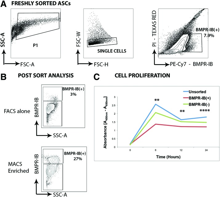
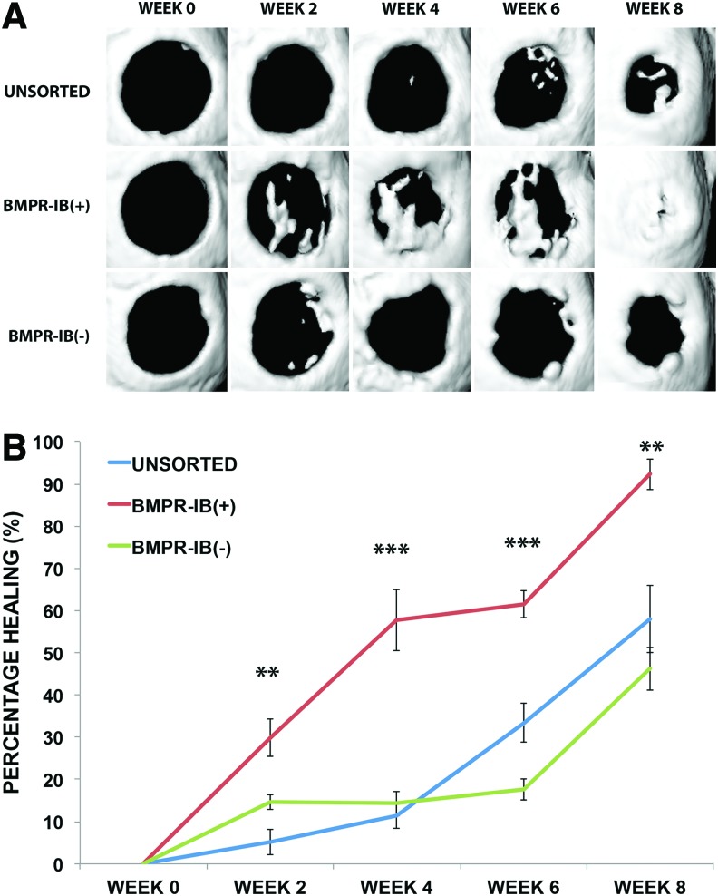
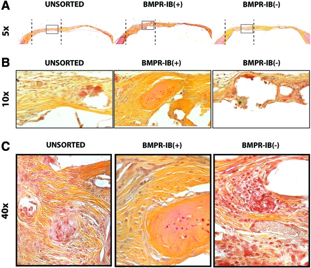
Methods: Human ASC subpopulations were prospectively isolated based on expression of bone morphogenetic protein receptor type-IB (BMPR-IB). Unsorted, BMPR-IB(+), and BMPR-IB(-) cells were analyzed for their osteogenic capacity through histological staining and gene expression. To evaluate their in vivo osteogenic potential, critical-sized calvarial defects were created in immunocompromised mice and treated with unsorted, BMPR-IB(+), or BMPR-IB(-) cells. Healing was assessed using microcomputed tomography and pentachrome staining of specimens at 8 weeks.

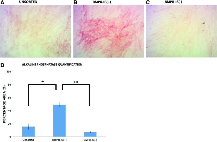
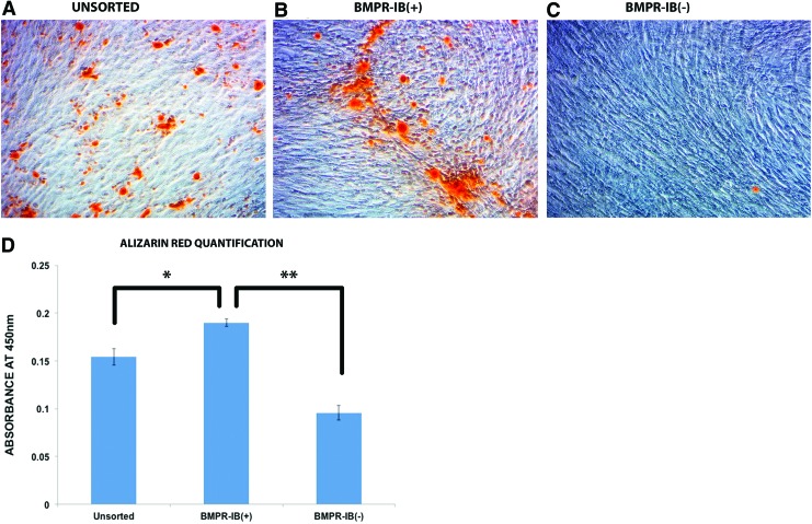
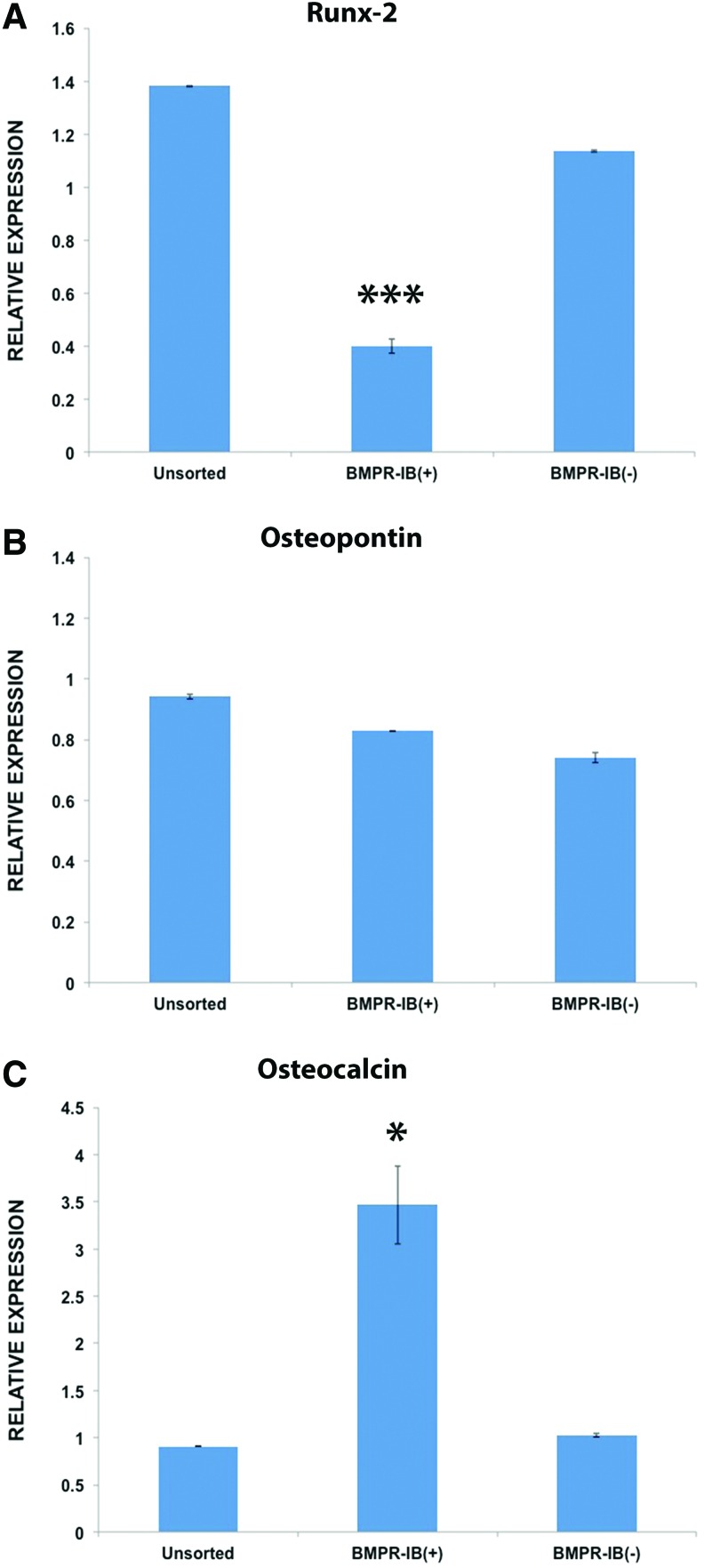
Results: Increased osteogenic differentiation was noted in the BMPR-IB(+) subpopulation, as demonstrated by alkaline phosphatase staining at day 7 and extracellular matrix mineralization with Alizarin red staining at day 14. This was also associated with increased expression for osteocalcin, a late marker of osteogenesis. Radiographic analysis demonstrated significantly enhanced healing of critical-sized calvarial defects treated with BMPR-IB(+) ASCs compared with unsorted or BMPR-IB(-) cells. This was confirmed through pentachrome staining, which revealed more robust bone regeneration in the BMPR-IB(+) group.

Conclusion: BMPR-IB(+) human ASCs have an enhanced ability to form bone both in vitro and in vivo. These data suggest that positive selection for BMPR-IB(+) and manipulation of the BMP pathway in these cells may yield a highly osteogenic subpopulation of cells for bone tissue engineering.


Figures

<b>FIG. 1.</b>
FIG. 1.
Cell sorting of ASCs for expression of BMPR-IB. Gating scheme for BMPR-IB expression in ASCs. Preliminary cell selection based on size and complexity to exclude debris (P1 gate). Doublets were excluded through selective gating to yield single cells. (A) FACS analysis of freshly isolated ASCs stained for BMPR-IB-PE-Cy7 demonstrates that 7.9% of the population is BMPR-IB(+). (B) FACS analysis of ASCs for BMPR-IB demonstrates that 27% of cells isolated after MACS enrichment are BMPR-IB(+) (bottom panel) compared with 3% using FACS alone (top panel). (C) XTT cell proliferation assay revealed BMPR-IB(+) cells to have the lowest proliferation rate over 24 h compared with unsorted and BMPR-IB(−) cells (**p<0.01, ****p<0.0001, analysis of variance). ASCs, adipose-derived stromal cells; BMPR-IB, bone morphogenetic protein receptor type-IB; FACS, fluorescence-activated cell sorting; FSC-A, forward scatter area; SSC-A, side scatter area; FSC-H, forward scatter height; FSC-W, forward scatter width. Color images available online at www.liebertpub.com/tea
<b>FIG. 2.</b>
FIG. 2.
Alkaline phosphatase staining of ASC subpopulations. Alkaline phosphatase staining of (A) unsorted, (B) BMPR-IB(+), and (C) BMPR-IB(−) ASCs after 7 days of osteogenic differentiation. (D) Quantification of staining demonstrates significantly increased staining in the BMPR-IB(+) group compared with unsorted ASCs (*p<0.05) and BMPR-IB(−) ASCs (**p<0.01). Color images available online at www.liebertpub.com/tea
<b>FIG. 3.</b>
FIG. 3.
Alizarin red staining of ASC subpopulations. Alizarin red staining of ASCs after 14 days of osteogenic differentiation for (A) unsorted, (B) BMPR-IB(+), and (C) BMPR-IB(−) ASCs. (D) Quantification of staining demonstrates significantly greater extracellular matrix mineralization from BMPR-IB(+) ASCs compared with unsorted (*p<0.05) and BMPR-IB(−) ASCs (**p<0.01). Color images available online at www.liebertpub.com/tea
<b>FIG. 4.</b>
FIG. 4.
Osteogenic gene expression in ASC subpopulations. Gene expression for markers of osteogenic differentiation in unsorted, BMPR-IB(+), and BMPR-IB(−) ASCs. (A). Levels of Runx2 were significantly lower in the BMPR-IB(+) group compared with unsorted and BMPR-IB(−) ASCs (***p<0.001). (B) No significant differences were noted in osteopontin expression between any of the groups (p>0.05). (C) Significantly greater osteocalcin expression was noted in BMPR-IB(+) ASCs relative to unsorted and BMPR-IB(−) ASCs (*p<0.05). Color images available online at www.liebertpub.com/tea
<b>FIG. 5.</b>
FIG. 5.
Healing of critical-sized calvarial defects with different ASC subpopulations. (A) Three-dimensional micro-CT reconstructions were performed for calvarial defects repaired with unsorted, BMPR-IB(+), or BMPR-IB(−) ASCs. (B) Quantification of healing at 8 weeks demonstrated significantly greater bone regeneration with BMPR-IB(+) ASCs (92%) compared with unsorted and BMPR-IB(−) ASCs (58% and 46%, respectively, **p<0.01). Significant differences in healing were also seen at 2 weeks (**p<0.01), 4 weeks (***p<0.001), and 6 weeks (***p<0.001). micro-CT, microcomputed tomography. Color images available online at www.liebertpub.com/tea
<b>FIG. 6.</b>
FIG. 6.
Histological staining of bone regenerate. (A) Movat's pentachrome staining of bone regenerate in defects repaired with unsorted, BMPR-IB(+), or BMPR-IB(−) ASCs at 5× magnification using bright field microscopy. Note more robust bone formation in BMPR-IB(+) group compared with BMPR-IB(−) group. The dotted line represents the extent of the defect area. The area within the black rectangle is shown on higher magnification using (B) 10× and (C) 40× bright field microscopy of defect area. Color images available online at www.liebertpub.com/tea

Similar articles

Cited by

References

    1. Chapekar M.S.Tissue engineering: challenges and opportunities. J Biomed Mater Res 53,617, 2000 - PubMed
    1. McArdle A., Lo D.D., Hyun J.S., Senarath-Yapa K., Chung M.T., Wan D.C., et al. . Discussion: a report of the ASPS task force on regenerative medicine: opportunities for plastic surgery. Plast Reconstr Surg 131,400, 2013 - PubMed
    1. Zuk P.A., Zhu M., Ashjian P., De Ugarte D.A., Huang J.I., Mizuno H., et al. . Human adipose tissue is a source of multipotent stem cells. Mol Biol Cell 13,4279, 2002 - PMC - PubMed
    1. Gimble J.M., and Guilak F.Differentiation potential of adipose derived adult stem (ADAS) cells. Curr Top Dev Biol 58,137, 2003 - PubMed
    1. Cowan C.M., Shi Y.Y., Aalami O.O., Chou Y.F., Mari C., Thomas R., et al. . Adipose-derived adult stromal cells heal critical-size mouse calvarial defects. Nat Biotechnol 22,560, 2004 - PubMed

Publication types

Substances