Review
. 2014 Dec 5;9(12):2147-63.
doi: 10.2215/CJN.05920613. Epub 2014 May 22.

Distal convoluted tubule

Affiliations
Review

Distal convoluted tubule

Arohan R Subramanya et al. Clin J Am Soc Nephrol. .

Abstract

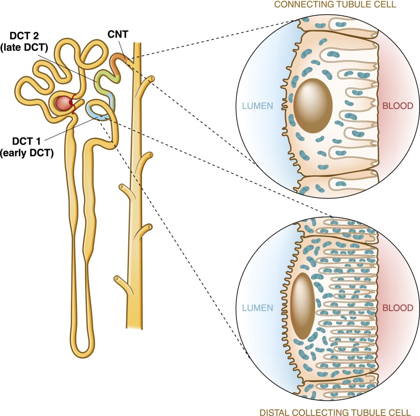
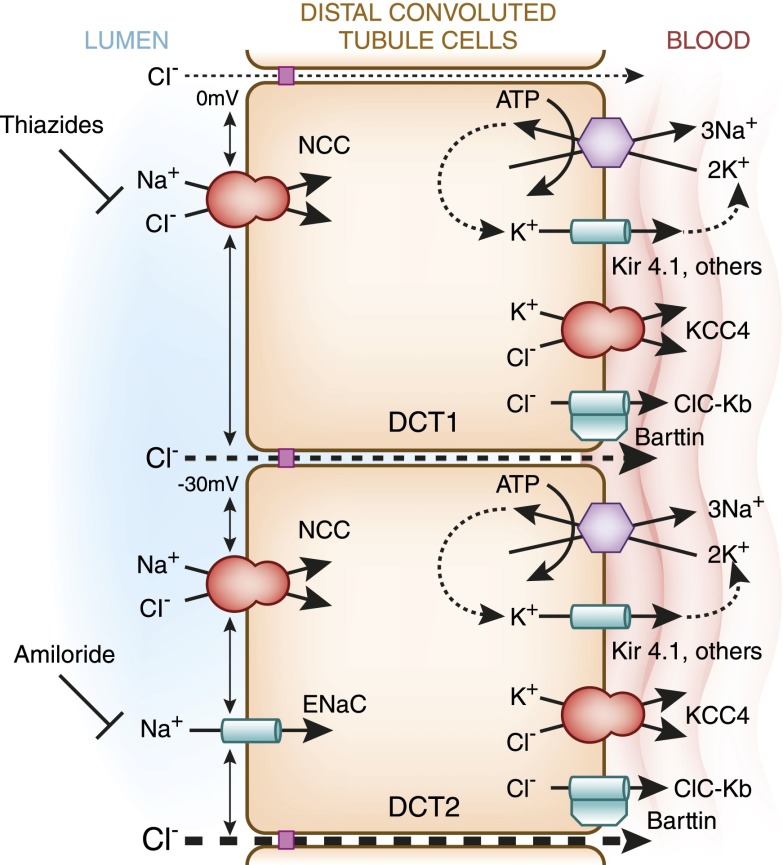
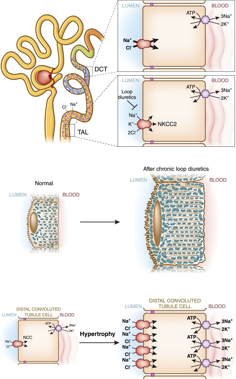
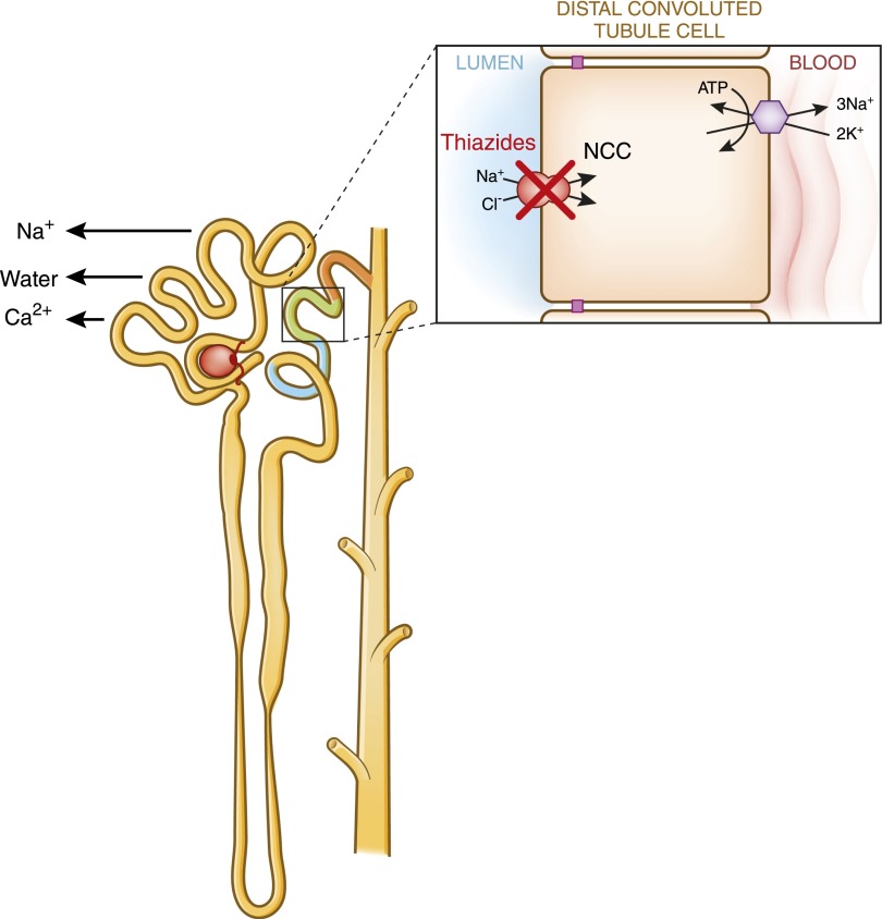
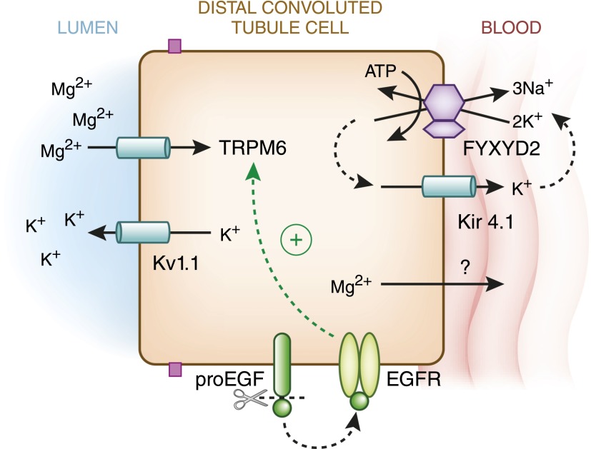
The distal convoluted tubule is the nephron segment that lies immediately downstream of the macula densa. Although short in length, the distal convoluted tubule plays a critical role in sodium, potassium, and divalent cation homeostasis. Recent genetic and physiologic studies have greatly expanded our understanding of how the distal convoluted tubule regulates these processes at the molecular level. This article provides an update on the distal convoluted tubule, highlighting concepts and pathophysiology relevant to clinical practice.

Keywords: Na transport; distal tubule; mineral metabolism; potassium channels; renal physiology.


Figures

Figure 1.
Figure 1.
A human nephron, showing the anatomic location of the distal tubule. The distal convoluted tubule (DCT) is divided into early and late segments, termed DCT1 and DCT2, respectively. The connecting tubule (CNT) is located immediately downstream of the DCT2. Insets compare DCT and CNT cell morphology. The cells of the DCT contain an extensive basolateral membrane system and are mitochondria-rich, indicating high transport activity of the Na+-K+-ATPase. CNT cells, in contrast, contain fewer mitochondria and basolateral membrane invaginations, suggesting that they are less metabolically active.
Figure 2.
Figure 2.
A model of NaCl reabsorption by cells of the early and late DCTs. Note that the transepithelial voltage is close to zero in the DCT1 and becomes progressively negative in the DCT2. In the early DCT, apical sodium reabsorption is exclusively mediated by thiazide-sensitive NaCl cotransporter (NCC), whereas in the late DCT, both NCC and amiloride-sensitive epithelial sodium channels (ENaCs) are present. The electrogenic transport of sodium by ENaC contributes to the lumen-negative transepithelial voltage. Basolateral sodium efflux is mediated by the Na+-K+-ATPase and aided by Kir4.1-mediated potassium leak currents. Chloride transport is carried out by the chloride channel ClC-Kb and potassium chloride cotransporter 4 (KCC4; SLC12A7).
Figure 3.
Figure 3.
Mechanism of diminished NaCl and Mg2+ reabsorption in EAST/SeSAME syndrome. EAST/SeSAME syndrome is caused by mutations of the basolateral K+ channel (KCNJ10; Kir4.1). This channel mediates leakage of potassium to the peritubular fluid, which provides a supply of potassium ions that drives the activity of the Na+-K+-ATPase, resulting in constant reabsorption of sodium across the basolateral membrane. In EAST/SeSAME syndrome, inactivating mutations of Kir4.1 impair a leak current, which probably reduces activity of the sodium/potassium pump. Patients with the syndrome also display a reduced basolateral membrane surface area, consistent with diminished Na+-K+-ATPase activity. The reduction in basolateral sodium transport reduces the gradient for NCC-mediated Na+ reabsorption and causes salt wasting. Luminal magnesium reabsorption is similarly reduced in these patients, possibly because of diminished basolateral magnesium transport. EAST/SeSAME, epilepsy, ataxia, sensorineural deafness, and tubulopathy/seizures, sensorineural deafness, ataxia, mantal retardation, and electrolyte imbalance.
Figure 4.
Figure 4.
Effect of chronic loop diuretic administration on DCT activity and morphology. (Upper panel) Sodium reabsorption normally mediated by the thick ascending limb Na-K-2Cl cotransporter (NKCC2) is blocked by loop diuretics, such as furosemide and bumetanide. This results in enhanced Na+ delivery to the DCT, which likely acts as a stimulus for DCT hypertrophy. (Lower panel) DCT hypertrophy manifests as an increase in mitochondrial size and basolateral membrane infoldings. The increase in NCC-mediated apical sodium transport coupled with enhanced activity of the sodium/potassium pump at the basolateral membrane results in a vectorial increase in sodium reabsorption and diuretic resistance.
Figure 5.
Figure 5.
SPAK and OSR1 phosphorylate and activate NCC. For NCC to be active, it must traffic to the plasma membrane from an intracellular storage pool. After it reaches the surface, it is in an inactive state until it is phosphorylated by two kinases, Ste20-like proline-alanine rich kinase (SPAK) and oxidative stress responsive kinase 1 (OSR1).
Figure 6.
Figure 6.
A model of WNK-SPAK/OSR1 regulation of NCC and its role in the pathogenesis of Familial Hyperkalemic Hypertension (FHHt). (A) In the baseline inactive state, WNK4 suppresses NCC trafficking to the plasma membrane, holding the cotransporter in an intracellular storage pool. The kinase active form of WNK1 can reverse this process. The Kelch-like 3/Cullin-3 (KLHL3/CUL3) E3 ubiquitin ligase complex constitutively degrades the WNKs. (B) FHHt-associated mutations in WNK4 reduce binding to KLHL3, increasing WNK4 abundance and triggering NCC activation through the WNK effector kinases SPAK and OSR1. Additionally, FHHt-causing mutations in WNK4 reduce its inhibitory effect on NCC traffic (represented by the hatched bar-headed line), which releases NCC from its intracellular compartment, increasing its trafficking to the cell surface. Thus, FHHt mutations in WNK4 convert it into an NCC stimulator. (C) WNK1 gene mutations increase kinase-active WNK1 expression, which overcomes constitutive degradation by KLHL3/CUL3. Because kinase active WNK1 can inhibit wild-type WNK4 and activate SPAK/OSR1, increased WNK1 expression stimulates NCC surface delivery and phosphorylation. (D) Mutations in KLHL3 either reduce binding of KLHL3/CUL3 to WNK1 and WNK4 or disconnect CUL3 from KLHL3; in either case, the CUL3 E3 ligase is unable to mark WNK signaling complexes for degradation. Increased WNK1 and WNK4 abundance stimulates NCC trafficking to the surface and triggers NCC phosphorylation. FHHt-causing mutations in CUL3 also likely reduce its activity to WNKs, although the mechanism by which this occurs remains unknown. WNK, With-No-Lysine [amino acid=K] kinase.
Figure 7.
Figure 7.
A working model for NCC regulation through the WNK-SPAK/OSR1 signaling cascade. To date, a number of hormones have been shown to stimulate NCC phosphorylation at residues that are directly phosphorylated by SPAK and OSR1. These include aldosterone, angiotensin II, insulin, and vasopressin. Tacrolimus also enhances NCC phosphorylation at these sites, resulting in thiazide-sensitive NaCl reabsorption. In all cases, the mechanism likely involves hormonal activation of WNKs, which in turn, activates SPAK/OSR1. In some cases, such as aldosterone (66) and angiotensin II (58,64,65), hormone-induced changes in WNK-SPAK/OSR1-dependent signaling are also associated with increased trafficking of NCC to the plasma membrane.
Figure 8.
Figure 8.
Model of potassium secretion in the DCT. K+ secretion in the distal nephron is voltage- and flow-dependent. In the low-flow state, “big”/“maxi”-K+ channels (BK) are closed, and potassium secretion exclusively occurs through the renal outer medullary potassium channel (ROMK). ROMK is expressed in the early DCT, but its activity there is relatively low because of the transepithelial voltage, which is near zero. In the late DCT, ENaC-mediated Na+ transport generates a driving force for ROMK-mediated K+ secretion. During a high-flow state, both ROMKs and BKs mediate K+ secretion. Increased sodium delivery and tubular fluid flow (for example, by infusing intravenous saline or Na bicarbonate or administering loop diuretics) increases ENaC activity, resulting in enhanced ROMK-mediated K+ secretion. Additionally, the increased flow triggers an intracellular signaling mechanism in the DCT that opens BK channels, facilitating K+ efflux into the tubular lumen.
Figure 9.
Figure 9.
Role of magnesium in ROMK potassium channel function. Potassium is the most abundant intracellular cation, creating a large chemical gradient that favors the outward flow of K+ through ROMK. (Left panel) Normally, magnesium binds to a cytosol-exposed site in ROMK to limit this outward flow. (Right panel) During hypomagnesemia, fewer Mg2+ ions can bind to this site, and K+ is secreted more freely. Thus, magnesium deficiency causes K+ wasting. This likely explains why magnesium repletion is required to efficiently restore potassium concentrations to normal during concomitant hypomagnesemia and hypokalemia.
Figure 10.
Figure 10.
Model of calcium reabsorption in the DCT. Apical calcium transport is mediated by transient receptor potential channel subfamily V member 5 (TRPV5) channels, which can be activated by the β-glucuronidase Klotho. Cytosolic calcium is immediately bound by calbindin-D28K, which shuttles calcium to the basolateral aspect of the DCT cell, where it can be transported out by the type 1 sodium calcium exchanger (NCX1) or calcium ATPases. These processes are tightly regulated by hormones, such as parathyroid hormone and 1,25-dihydroxyvitamin D (not shown).
Figure 11.
Figure 11.
Thiazide-induced hypocalciuria. Administration of thiazide diuretics reduces urinary calcium excretion and can sometimes cause hypercalcemia. The mechanism likely involves an increase in bulk calcium reabsorption with sodium and water in the proximal tubule. Thiazide-induced volume depletion might be the stimulus that triggers this process.
Figure 12.
Figure 12.
Model of magnesium reabsorption in the DCT. The magnesium channel transient receptor potential cation channel subfamily M member 6 (TRPM6) mediates luminal magnesium entry. At the luminal membrane, the K+ channel Kv1.1 extrudes K+ ions into the tubular lumen; this process probably generates an electrical driving force for Mg2+ entry through TRPM6. TRPM6 activity is stimulated by the magnesiotropic hormone EGF, which triggers an intracellular signaling cascade in the DCT after cleavage from pro-EGF and binding to basolateral EGF receptors (EGFRs). After transluminal entry, cytosolic Mg2+ is then transported out of the basolateral side of the DCT through unclear mechanisms, although cyclin M2 and SLC41A1 are candidate magnesium transport pathways that might mediate the process. Basolateral membrane voltage generated by the Na+-K+-ATPase is critical for Mg2+ exit, which is illustrated by Kir4.1 mutations in EAST/SeSAME syndrome that reduce pump activity by impaired recycling (Figure 3) or small γ-subunit of the Na+-K+-ATPase (FXYD2) mutations, which alter the pump’s affinity for Na+ and K+.

References

    1. Chabardès D, Gagnan-Brunette M, Imbert-Teboul M, Gontcharevskaia O, Montégut M, Clique A, Morel F: Adenylate cyclase responsiveness to hormones in various portions of the human nephron. J Clin Invest 65: 439–448, 1980 - PMC - PubMed
    1. Reilly RF, Ellison DH: Mammalian distal tubule: Physiology, pathophysiology, and molecular anatomy. Physiol Rev 80: 277–313, 2000 - PubMed
    1. Arriza JL, Weinberger C, Cerelli G, Glaser TM, Handelin BL, Housman DE, Evans RM: Cloning of human mineralocorticoid receptor complementary DNA: Structural and functional kinship with the glucocorticoid receptor. Science 237: 268–275, 1987 - PubMed
    1. Bostanjoglo M, Reeves WB, Reilly RF, Velázquez H, Robertson N, Litwack G, Morsing P, Dørup J, Bachmann S, Ellison DH: 11Beta-hydroxysteroid dehydrogenase, mineralocorticoid receptor, and thiazide-sensitive Na-Cl cotransporter expression by distal tubules. J Am Soc Nephrol 9: 1347–1358, 1998 - PubMed
    1. Meneton P, Loffing J, Warnock DG: Sodium and potassium handling by the aldosterone-sensitive distal nephron: The pivotal role of the distal and connecting tubule. Am J Physiol Renal Physiol 287: F593–F601, 2004 - PubMed

Publication types

MeSH terms