. 2014 May 8:8:459-74.
doi: 10.2147/DDDT.S58414. eCollection 2014.

Improving anticancer efficacy of (-)-epigallocatechin-3-gallate gold nanoparticles in murine B16F10 melanoma cells

Affiliations

Improving anticancer efficacy of (-)-epigallocatechin-3-gallate gold nanoparticles in murine B16F10 melanoma cells

Cheng-Cheung Chen et al. Drug Des Devel Ther. .

Abstract

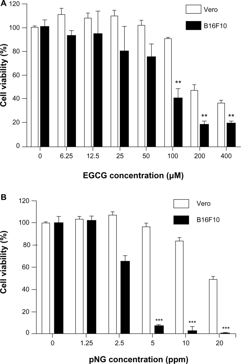
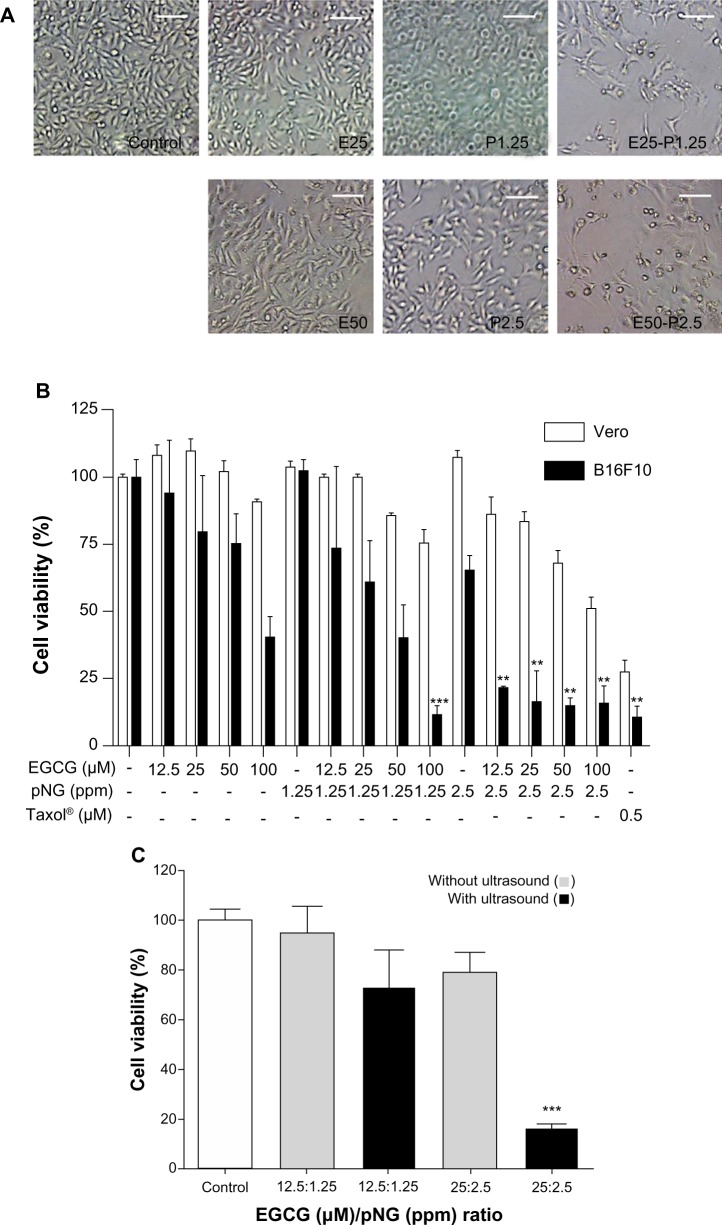
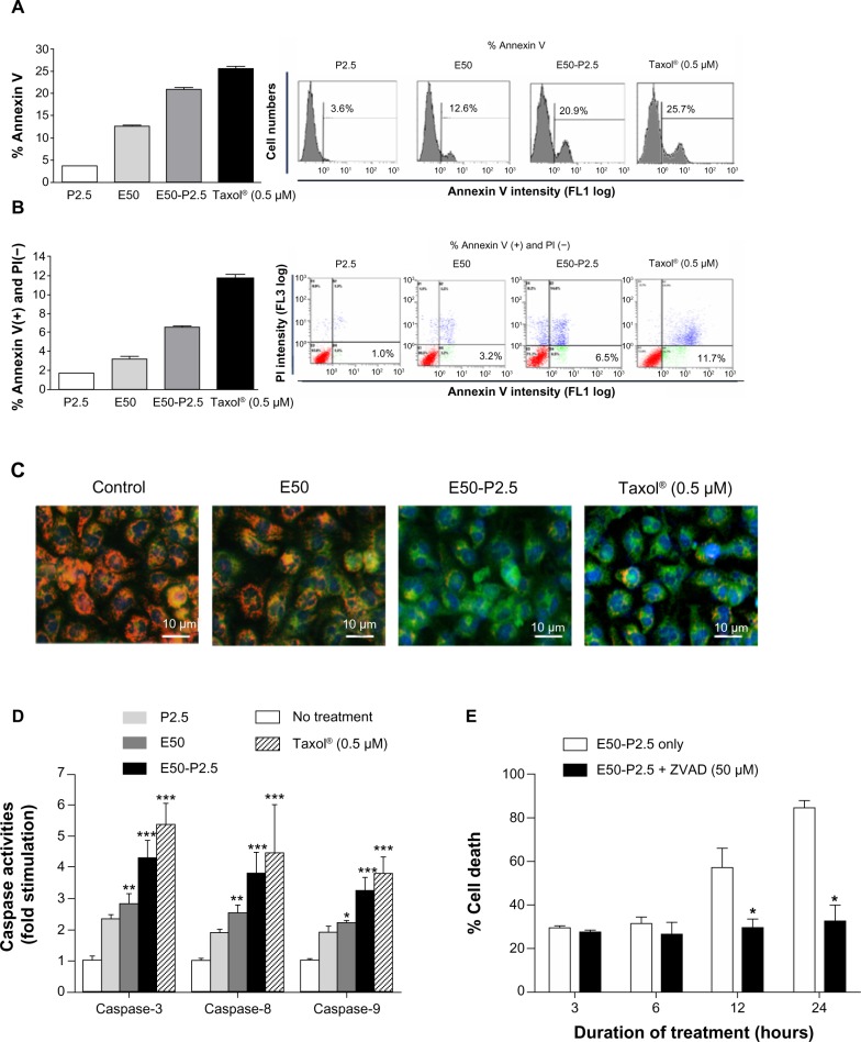
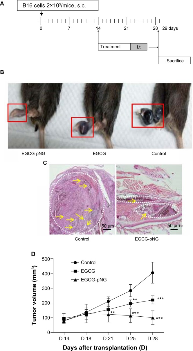
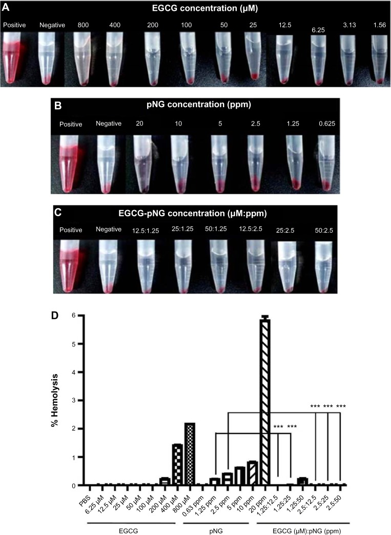
(-)-Epigallocatechin-3-gallate (EGCG), the major bioactive constituent in green tea, has been reported to effectively inhibit the formation and development of tumors. To maximize the effectiveness of EGCG, we attached it to nanogold particles (EGCG-pNG) in various ratios to examine in vitro cytotoxicity and in vivo anti-cancer activity. EGCG-pNG showed improved anti-cancer efficacy in B16F10 murine melanoma cells; the cytotoxic effect in the melanoma cells treated with EGCG-pNG was 4.91 times higher than those treated with EGCG. The enhancement is achieved through mitochondrial pathway-mediated apoptosis as determined by annexin V assay, JC-10 staining, and caspase-3, -8, -9 activity assay. Moreover, EGCG-pNG was 1.66 times more potent than EGCG for inhibition of tumor growth in a murine melanoma model. In the hemolysis assay, the pNG surface conjugated with EGCG is most likely the key factor that contributes to the decreased release of hemoglobin from human red blood cells.

Keywords: EGCG; anticancer; gold nanoparticles; melanoma.


Figures

Figure 1
Figure 1
The effects of EGCG and pNG on Vero and B16F10 cell proliferation. Notes: (A) Cells were treated with or without increasing concentrations of EGCG for 24 hours; (B) cells were treated with or without increasing concentrations of pNG for 24 hours. Data shown are means ± standard deviations for three samples. Data containing asterisks are significantly different from the control values at **P<0.01, ***P<0.001. Abbreviations: EGCG, (–)-epigallocatechin-3-gallate; pNG, physical nanogold.
Figure 2
Figure 2
Improving anti-proliferative activity of EGCG and pNG in vitro. Notes: (A) Morphologies of B16F10 cells treated with or without 25–50 μM EGCG and/or 1.25–2.5 ppm pNG for 24 hours (magnification ×40). Scale bar, 100 μm; (B) All the mixtures of EGCG and pNG underwent ultrasound pretreatment and showed significant 24-hour cytotoxicity compared with pNG or EGCG, respectively, in B16F10 but not Vero cells; (C) improved cytotoxicity of the EGCG and pNG mixtures was only achieved with ultrasound pretreatment; this was not observed in the mixture without ultrasound pretreatment. The cell viabilities were determined via WST-8 assay. Data shown are mean ± standard deviation for three samples. Data containing asterisks are significantly different from the control values at **P<0.01; ***P<0.001. Abbreviations: EGCG, (–)-epigallocatechin-3-gallate; pNG, physical nanogold; E25, EGCG 25 μM; E50, EGCG 50 μM; P1.25, pNG 1.25 ppm; P2.5, pNG 2.5 ppm; E25–P1.25, EGCG-pNG 25 μM:1.25 ppm; E50–P2.5, EGCG-pNG 50 μM:2.5 ppm.
Figure 3
Figure 3
Enhanced apoptosis induction of EGCG by pNG in B16F10 cells. Notes: (A) The fractions of annexin V-positive B16F10 cells were 3.59%±0.30%, 12.63%±0.61%, 20.93%±0.55%, and 25.7%±0.54%, after treatment with pNG 2.5 ppm, EGCG 50 μM, EGCG-pNG 50 μM:2.5 ppm, and Taxol® (Sigma-Aldrich; St Louis, MO, USA) 0.5 μM, respectively, at 24 hours; (B) the fractions of annexin V-positive and PI-negative B16F10 cells were 1.0% ± 0.17%, 3.2% ± 0.23%, 6.5% ± 0.23%, and 11.7% ± 0.41%, after treatment with pNG 2.5 ppm, EGCG 50 μM, EGCG-pNG 50 μM:2.5 ppm, and Taxol® 0.5 μM, respectively, at 24 hours; (C) cells treated with EGCG-pNG for 24 hours expressed more green fluorescence than those treated with EGCG (magnification ×200). Depolarized mitochondria are indicated by green fluorescence (JC-10 monomer), and polarized mitochondria are indicated by orange fluorescence (aggregated JC-10). Cell nuclei are indicated by blue fluorescence coupled with Hoechst 33342 staining. Scale bar, 10 μm; (D) cells treated with EGCG-pNG for 24 hours showed a significant increase of caspase-3, -8, and -9 activity compared with those treated with EGCG; (E) initiation of time-dependent apoptotic activation by EGCG-pNG treatment via the caspase pathway in B16F10 melanoma cells. Data shown are mean ± standard deviation for three samples. Data containing asterisks are significantly different from the control values at *P<0.05; **P<0.01; ***P<0.001. Abbreviations: PI, propidium iodide; EGCG, (–)-epigallocatechin-3-gallate; pNG, physical nanogold; P2.5, pNG 2.5 ppm; E50, EGCG 50 μM; E50-P2.5, EGCG-pNG 50 μM:2.5 ppm; ZVAD, Z-VAD-FMK, N-Benzyloxycarbonyl-Val-Ala-Asp (O-Me) fluoromethyl ketone.
Figure 4
Figure 4
Increased tumor growth inhibition of EGCG by pNG in vivo. Notes: (A) Melanoma murine model protocol. Mice were injected with PBS, EGCG, or EGCG-pNG intratumorally twice a week on day 14 after tumor implantation; (B) photographs of tumor appearance on day 29 after cell inoculation. Mice receiving EGCG-pNG showed greater reduction in tumor volume than those receiving EGCG; (C) histology (H&E staining, 40×) of tumors from mouse footpad 29 days after cell inoculation. The control group showed larger engorgement of tumor vessels (yellow arrows) than the EGCG-pNG group. The control group showed greater melanocyte infiltration (white dotted line circles) in the cross-sectional view than the EGCG-pNG group. Scale bar, 50 μm; (D) EGCG-pNG was more effective (1.66 times on day 29) than free EGCG at inhibiting tumor growth in the melanoma murine model. Tumor volumes were measured in two dimensions and calculated as width × length/2. Each data point represents the mean ± standard deviation of tumor volumes from six animals and they are significantly different from the control group at **P<0.01; ***P<0.001. Abbreviations: PBS, phosphate-buffered saline; EGCG, (–)-epigallocatechin-3-gallate; H&E, hematoxylin and eosin; i.t., intratumorally; pNG, physical nanogold; s.c., subcutaneously.
Figure 5
Figure 5
RBC hemolysis after treatment. Notes: Photographs of RBC hemolysis after treatment with (A) EGCG; (B) pNG; and (C) EGCG-pNG for 3 hours (the red hemoglobin in the supernatant indicates the damage to RBCs; dd-H2O and PBS were used as positive and negative controls, respectively); (D) hemolysis results reported as a percentage of the positive control condition. A concentration-dependent trend in hemolysis is as shown for 3 hours of EGCG or pNG exposure, respectively. EGCG-pNG showed significantly lower release of hemoglobin from cell lysis compared with pNG alone. Data shown are mean ± standard deviation for three samples. Data containing asterisks are significantly different from the control values at ***P<0.001. Abbreviations: dd-H2O, double-distilled water; EGCG, (–)-epigallocatechin-3-gallate; H&E, hematoxylin and eosin; PBS, phosphate-buffered saline; pNG, physical nanogold; RBC, red blood cell.

References

    1. Markovic SN, Erickson LA, Rao RD, et al. Melanoma Study Group of the Mayo Clinic Cancer Center Malignant melanoma in the 21st century, part 1: epidemiology, risk factors, screening, prevention, and diagnosis. Mayo Clin Proc. 2007;82(3):364–380. - PubMed
    1. Guevara-Canales JO, Gutiérrez-Morales MM, Sacsaquispe-Contreras SJ, Sánchez-Lihón J, Morales-Vadillo R. Malignant melanoma of the oral cavity. Review of the literature and experience in a Peruvian Population. Med Oral Patol Oral Cir Bucal. 2012;17(2):e206–e211. - PMC - PubMed
    1. Pusceddu S, Bajetta E, Carcangiu ML, Formisano B, Ducceschi M, Buzzoni R. A literature overview of primary cervical malignant melanoma: an exceedingly rare cancer. Crit Rev Oncol Hematol. 2012;81(2):185–195. - PubMed
    1. Vikey AK, Vikey D. Primary malignant melanoma, of head and neck: a comprehensive review of literature. Oral Oncol. 2012;48(5):399–403. - PubMed
    1. Bhatia S, Tykodi SS, Thompson JA. Treatment of metastatic melanoma: an overview. Oncology (Williston Park) 2009;23(6):488–496. - PMC - PubMed