

Case Reports
doi: 10.5826/dpc.0402a09.
eCollection 2014 Apr.
Non-choroidal yellow melanoma showing positive staining with Sudan Black consistent with the presence of lipofuscin: a case report
Affiliations
- PMID: 24855574
- PMCID: PMC4029254
- DOI: 10.5826/dpc.0402a09
Item in Clipboard
Case Reports
Non-choroidal yellow melanoma showing positive staining with Sudan Black consistent with the presence of lipofuscin: a case report
Dermatol Pract Concept.
.
Abstract
A case of a predominantly yellow primary superficial spreading melanoma arising on the back of a 44-year-old woman is presented. Possible causes of the clinical and dermatoscopic yellow color are discussed. Staining with the histochemical stain, Sudan Black, revealed a differential uptake compared to a closely matched control melanoma. We speculate that the clinical and dermatoscopic yellow color could be due to the presence of increased amounts of the pigment lipofuscin, which is known to produce subtle orange color in some choroidal melanomas.
Keywords: Sudan Black; dermatopathology; dermatoscopy; dermoscopy; hypomelanotic melanoma; lipofuscin; yellow melanoma.
Figures
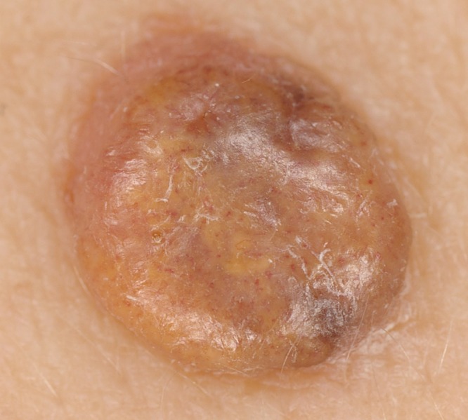
Close-up image (Pentax DS camera, Pentax Ricoh, Tokyo, Japan) of a nodular lesion over the right scapular area of a 44-year-old female patient. Irregular dominant yellow color is apparent. [Copyright: ©2014 Jegou Penouil et al.]
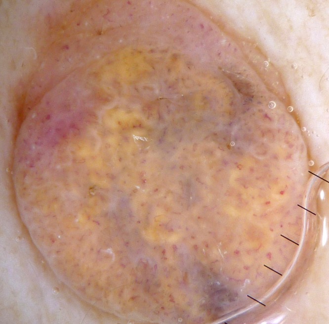
Dermatoscopy image (Heine delta 20 dermatoscope [Heine, Optotechnic, GmbH, Hersching, Germany] manually coupled to a Panasonic Lumix DMC ZX1 camera [Panasonic Corp., Kadoma, Japan]) of the lesion shown in Figure 1. The pattern is structureless, predominantly yellow, with an eccentric structureless pink area (upper pole of image) and evidence of light melanin pigmentation with some areas of structureless gray interspersed between the dominant yellow areas but absent at the upper pole of the image with resulting asymmetry. Polymorphous linear vessels (serpentine, looped and curved) are arranged randomly and densely over the surface of the lesion. There are a small number of dot vessels. [Copyright: ©2014 Jegou Penouil et al.]
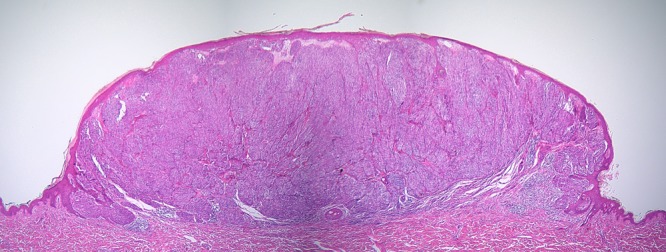
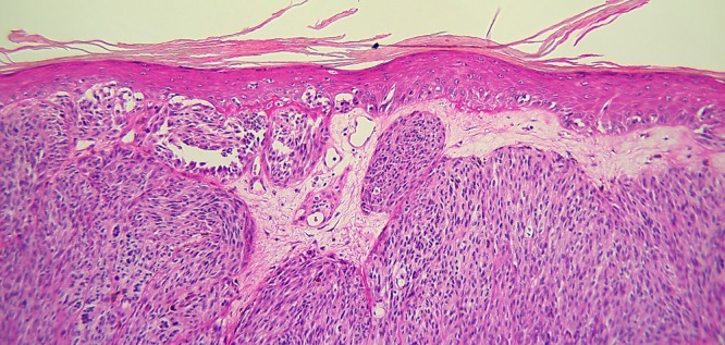
Dermatopathologic overview of the lesion shown in Figures 1 and 2. Significant nodular morphology is apparent although close examination of additional dermatopathology sections revealed that strict criteria for the classification as nodular subtype were not met. [Copyright: ©2014 Jegou Penouil et al.]
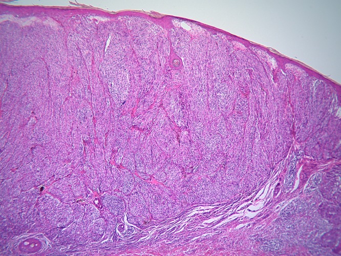
Medium high power dermatopathologic view of the lesion shown in Figure 3. Nesting is apparent at the base of the lesion. [Copyright: ©2014 Jegou Penouil et al.]
Higher power dermatopathologic view of the lesion shown in Figure 3 showing a proliferation of melanocytes at the dermoepidermal junction both as single cells and confluent nests with some clefting. A limited amount of pagetoid spread is seen. Sheets of spindle-shaped cells fill the dermis. [Copyright: ©2014 Jegou Penouil et al.]
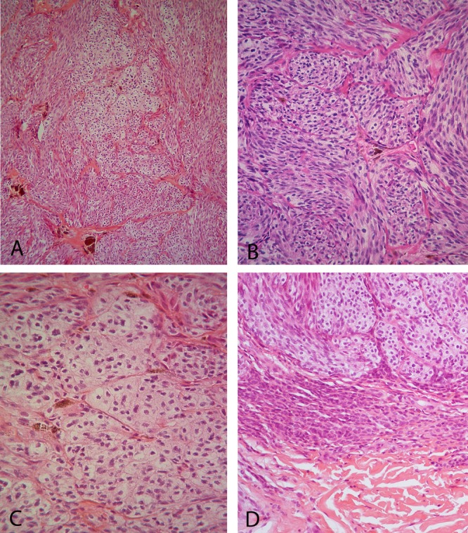
(A) Medium high power dermatopathologic view of the center of the lesion shown in Figure 3. Two distinct cell types are apparent, including spindle-shaped cells on one hand and cells with plump oval nuclei, prominent nucleoli and abundant clear cytoplasm (balloon cells) on the other. (B) High power view of spindle shaped cells and (C) balloon cells. (D) High power view of the base of the lesion shown in Figure 3. A sheet of mature nevomelanocytes can be seen beneath the nests of abnormal melanocytes, consistent with the contiguous presence of a nevus. [Copyright: ©2014 Jegou Penouil et al.]
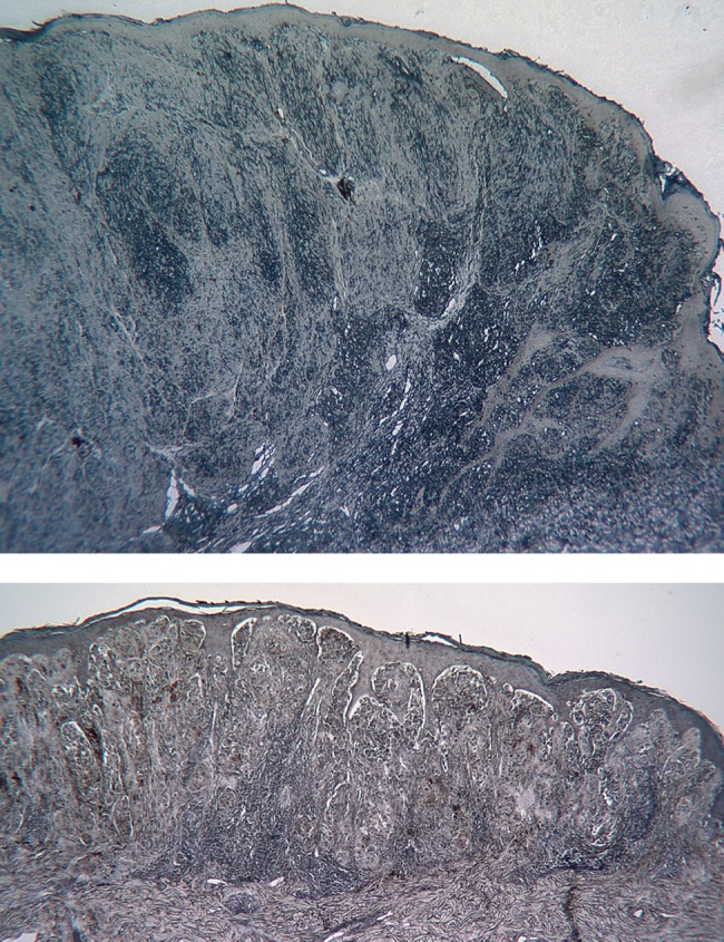
(Upper image) Dermatopathologic view of the lesion shown in Figure 3 stained with Sudan Black. (Lower image) Dermatopathologic image of a control melanoma of similar Breslow thickness and mitotic rate but with heavy melanin pigmentation. Both lesions had 5 micron thick sections stained with Sudan Black and the images were taken with the same exposure and white-balance settings. There has been no photo manipulation apart from cropping and identical resizing for publication. The case being reported here (upper image) is seen to stain differentially consistent with the possible presence of the pigment lipofuscin. [Copyright: ©2014 Jegou Penouil et al.]
Similar articles
-
Retinal and pigment epithelial alterations over choroidal malignant melanomas.Ann Ophthalmol. 1975 Apr;7(4):487-9, 491-2. Ann Ophthalmol. 1975. PMID: 50039
-
Role of In Vivo Reflectance Confocal Microscopy in the Analysis of Melanocytic Lesions.Acta Dermatovenerol Croat. 2018 Apr;26(1):64-67. Acta Dermatovenerol Croat. 2018. PMID: 29782304 Review.
-
Amelanotic/Hypomelanotic melanoma--is dermatoscopy useful for diagnosis?J Dtsch Dermatol Ges. 2003 May;1(5):369-73. doi: 10.1046/j.1610-0387.2003.02042.x. J Dtsch Dermatol Ges. 2003. PMID: 16285302
-
Dermatoscopy of amelanotic and hypomelanotic melanoma.J Dtsch Dermatol Ges. 2014 Jun;12(6):467-72. doi: 10.1111/ddg.12368. Epub 2014 May 14. J Dtsch Dermatol Ges. 2014. PMID: 24825465 Review.
-
Autofluorescence of orange pigment overlying small choroidal melanoma.Retina. 2007 Oct;27(8):1107-11. doi: 10.1097/IAE.0b013e31814934ef. Retina. 2007. PMID: 18040254
Cited by
-
Lipofuscin, lipofuscin-like pigments and autofluorescence.Eur J Histochem. 2015 Feb 6;59(1):2485. doi: 10.4081/ejh.2015.2485. Eur J Histochem. 2015. PMID: 25820564 Free PMC article. Review.
-
Cell autofluorescence and lipofuscin.Front Med (Lausanne). 2015 Feb 19;2:6. doi: 10.3389/fmed.2015.00006. eCollection 2015. Front Med (Lausanne). 2015. PMID: 25745630 Free PMC article. No abstract available.
References
-
- Rosendahl C, Cameron A, McColl I, Wilkinson D. Dermatoscopy in routine practice—“Chaos and Clues.”. Aust Fam Physician. 2012 Jul;41(7):482–7. - PubMed
-
- Chamberlain AJ, Fritschi L, Kelly JW. Nodular melanoma: patients’ perceptions of presenting features and implications for earlier detection. J Am Acad Dermatol. 2003 May;48(5):694–701. - PubMed
-
- Moloney FJ, Menzies SW. Key points in the dermoscopic diagnosis of hypomelanotic melanoma and nodular melanoma. J Dermatol. 2011 Jan;38(1):10–5. - PubMed
-
- Kittler H. Dermatoscopy: introduction of a new algorithmic method based on pattern analysis for diagnosis of pigmented skin lesions. Dermatopathology: Practical & Conceptual. 2007;13:3.
Publication types
LinkOut - more resources
Full Text Sources
Other Literature Sources