. 2014 Jul 4;289(27):19019-30.
doi: 10.1074/jbc.M114.570382. Epub 2014 May 22.

Novel zinc-binding site in the E2 domain regulates amyloid precursor-like protein 1 (APLP1) oligomerization

Affiliations

Novel zinc-binding site in the E2 domain regulates amyloid precursor-like protein 1 (APLP1) oligomerization

Magnus C Mayer et al. J Biol Chem. .

Abstract

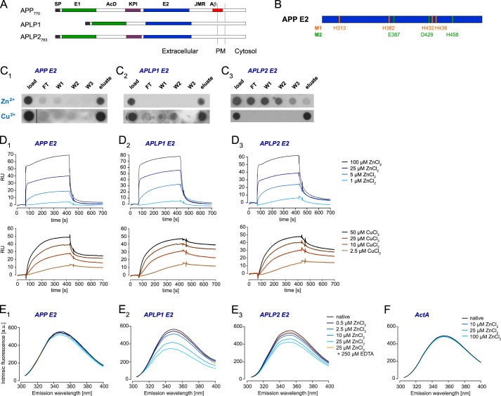
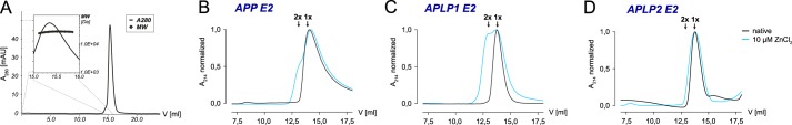
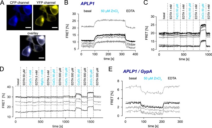
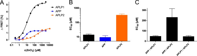
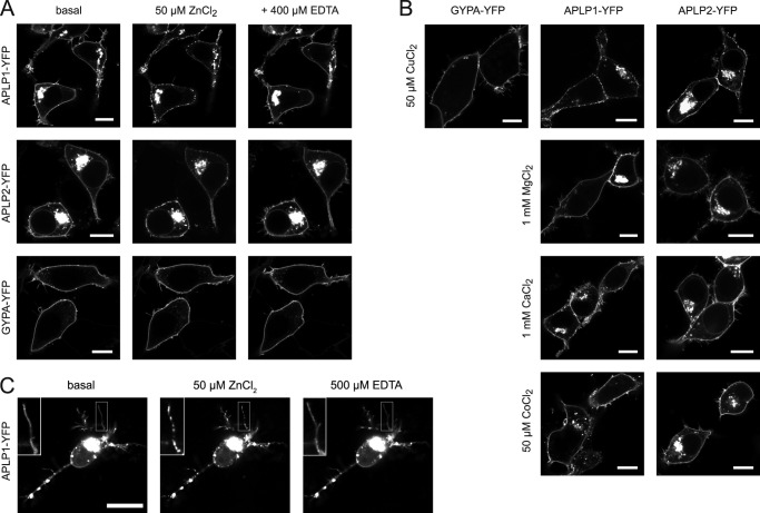
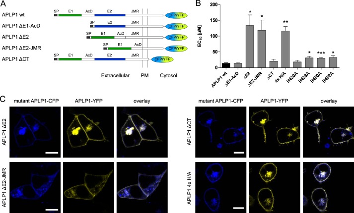
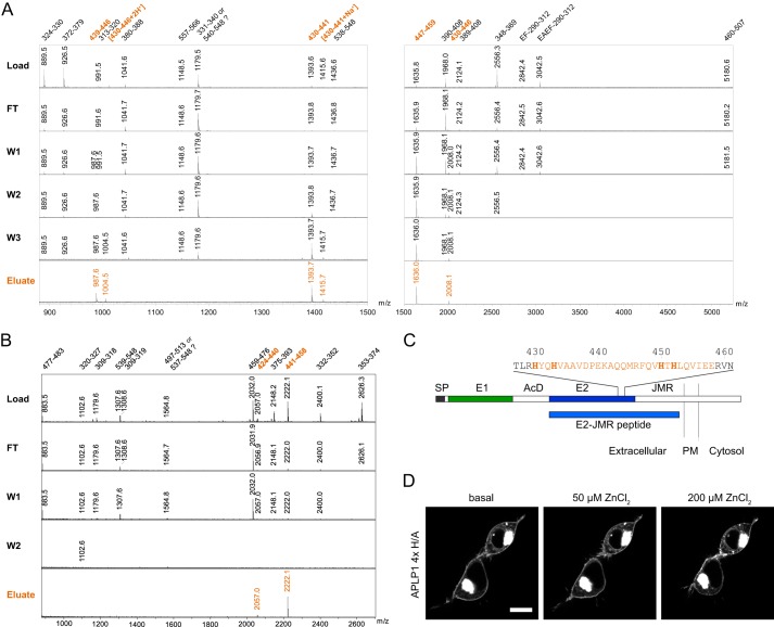
The amyloid precursor protein (APP) and the APP-like proteins 1 and 2 (APLP1 and APLP2) are a family of multidomain transmembrane proteins possessing homo- and heterotypic contact sites in their ectodomains. We previously reported that divalent metal ions dictate the conformation of the extracellular APP E2 domain (Dahms, S. O., Könnig, I., Roeser, D., Gührs, K.-H., Mayer, M. C., Kaden, D., Multhaup, G., and Than, M. E. (2012) J. Mol. Biol. 416, 438-452), but unresolved is the nature and functional importance of metal ion binding to APLP1 and APLP2. We found here that zinc ions bound to APP and APLP1 E2 domains and mediated their oligomerization, whereas the APLP2 E2 domain interacted more weakly with zinc possessing a less surface-exposed zinc-binding site, and stayed monomeric. Copper ions bound to E2 domains of all three proteins. Fluorescence resonance energy transfer (FRET) analyses examined the effect of metal ion binding to APP and APLPs in the cellular context in real time. Zinc ions specifically induced APP and APLP1 oligomerization and forced APLP1 into multimeric clusters at the plasma membrane consistent with zinc concentrations in the blood and brain. The observed effects were mediated by a novel zinc-binding site within the APLP1 E2 domain as APLP1 deletion mutants revealed. Based upon its cellular localization and its dominant response to zinc ions, APLP1 is mainly affected by extracellular zinc among the APP family proteins. We conclude that zinc binding and APP/APLP oligomerization are intimately linked, and we propose that this represents a novel mechanism for regulating APP/APLP protein function at the molecular level.

Keywords: Amyloid Precursor Protein (APP); Fluorescence Resonance Energy Transfer (FRET); Mass Spectrometry (MS); Metalloprotein; Zinc.


Figures

FIGURE 1.
FIGURE 1.
Zinc and copper binding properties of APP, APLP1, and APLP2 E2 domains. A, schematic representation of the amyloid precursor protein family: SP, signal peptide; E1 and E2, two conserved regions of the ectodomain; AcD, acidic stretch linker region between E1 and E2; JMR, juxtamembrane region; Aβ, amyloid β sequence; PM, plasma membrane. B, metal-binding residues of metal-binding sites 1 and 2 (M1 and M2) observed in crystals of the APP E2 domain (30). C, recombinant E2 domains were loaded onto an IMAC column charged with immobilized zinc or copper ions; flow-through (FT), washes (W1–3), and elution volumes were analyzed by dot-blotting. D, overlays of reference-subtracted SPR titrations for ZnCl2 or CuCl2 binding (1–100 μm ZnCl2 or 2.5–50 μm CuCl2 at 30 μl/min) to amine-coupled E2 domains (∼3300 RU each). E, corrected intrinsic fluorescence titrations for ZnCl2 binding (0–25 μm) to E2 domains in solution (0.5 μm) in the absence or presence of 250 μm EDTA. F, intrinsic fluorescence repeated (0–100 μm ZnCl2 binding) with ActA control peptide. C–F, blots and spectra shown are representatives of at least three independent experiments.
FIGURE 2.
FIGURE 2.
Oligomerization of recombinant APP, APLP1, and APLP2 E2 domains. A, gel filtration chromatography coupled to static light scattering (SEC-SLS) to analyze APLP1 E2 (line, absorbance at 280 nm; symbols, SLS-based molecular weight) as a function of retention volume. B–D, SECs for E2 domains in the absence (black lines) and presence (cyan lines) of 10 μm zinc; retention volumes at which monomers (1×) and dimers (2×) elute are indicated. A–D, chromatograms shown are representative of at least three independent experiments.
FIGURE 3.
FIGURE 3.
Live cell FRET analysis of APP and APLP oligomerization in the presence of metal ions. HEK293 cells were transiently transfected with APLP1-CFP and APLP1-YFP (A–D) or APLP1-CFP and GypA-YFP (E). A, representative CFP, YFP, and overlay images of cells used for FRET measurements, monitored without binning. Scale bar, 10 μm. B, real time analysis of FRET signal changes upon perfusion with 50 μm ZnCl2 (90–330 s) followed by 500 μm EDTA; displayed are single-cell curves (gray) and averaged FRET signals (black) of SE measurements of five cells. C and D, single-cell curves of SE. FRET signals reveal a specific increase only for 50 μm Zn2+ and 50 μm Cu2+ but not for 50 μm Fe2+, 50 μm Fe3+ (C), 1 mm Ca2+, 1 mm Mg2+, or 50 μm Co2+ (D); a minimal increase of less than 1% was observed for 150 μm Co2+ (D); cells were incubated with metal ions and washed in continuous cycles using a perfusion device. E, GypA-YFP/APLP1-CFP pair shows a slight decrease of the baseline FRET signal in the presence of zinc between APLP1 and GypA most likely due to increased homophilic oligomerization of APLP1; displayed are single-cell curves (gray) and averaged FRET signals (black) of SE measurements of five cells. A–E, measurements presented are representative of at least three independent transfections.
FIGURE 4.
FIGURE 4.
Concentration-dependent FRET analysis of zinc-mediated oligomerization of APP, APLP1, and APLP2. A, increased FRET efficiencies as a function of increasing zinc concentrations reveal sigmoidal concentration responses for homophilic APP, APLP1, and APLP2 FRET pairs, each fitted with a logistic function. B and C, EC50 values for FRET increases after zinc treatment (n = 4–6 experiments with 6–12 cells per experiment) with FRET homoligomer (B) or hetero-oligomer pairs (C; APP-CFP/APLP1-YFP, APP-CFP/APLP2-YFP, or APLP2-CFP/APLP1-YFP).
FIGURE 5.
FIGURE 5.
Plasma membrane distribution of APLPs in the presence of metal ions. Plasmids encoding for APLP1, APLP2, and GypA with C-terminal YFP tags were transiently transfected in HEK293 cells (A and B) or rat hippocampal neurons (C) and imaged 1 day after transfection by cLSM. Representative images from at least three independent transfections were taken before and 2 min after addition of divalent metal ions to the medium (50 μm ZnCl2, 50 μm CuCl2, 1 mm MgCl2, 1 mm CaCl2, or 50 μm CoCl2) and 2 min after a further addition of EDTA. C, inset, upper left, is a magnification of the boxed neurite illustrating the punctate appearance of APLP1 clusters in the presence of zinc ions. Scale bar, 10 μm.
FIGURE 6.
FIGURE 6.
FRET analysis of zinc-mediated oligomerization of APLP1 mutants. A, schematic representation of APLP1 deletion mutants: SP, signal peptide; E1 and E2, two conserved regions of the ectodomain; AcD, acidic stretch linker region between E1 and E2; JMR, juxtamembrane region; PM, plasma membrane. B, HEK293 cells were transfected with APLP1 deletion mutants (A) or full-length site-directed APLP1 mutants (4× H/A represents the quadruple point mutation H430A/H433A/H450A/H452A); FRET concentration-response curves for zinc treatment were recorded, and EC50 values were determined (n = 4–6 experiments with 5–12 cells per experiment). Asterisks indicate significant differences to APLP1 WT as follows: *, p < 0.05; **, p < 0.01; ***, p < 0.001. C, plasmids encoding for wild-type APLP1 fused to YFP and APLP1-CFP mutant constructs were transiently transfected in HEK293 cells; representative of at least three independent transfections, the cells were imaged by cLSM 1 day post-transfection. Scale bar, 10 μm.
FIGURE 7.
FIGURE 7.
Identification of the zinc-binding site in APLP1 E2. Recombinant APLP1 E2-JMR was digested with trypsin (A) or GluC (B) while bound to zinc-chelated Sepharose. Load, flow-through (FT), wash (W1–3), and elution volumes were analyzed by MALDI-MS. For trypsin analysis, EDTA elutions were performed using two different acetonitrile concentrations to cover a broad molecular mass range for peptide recovery. Mass spectrum for W3 digested with GluC is not shown because no APLP1 peptides were found in this fraction. Peaks are labeled with the respective peptide mass. Numbers are referring to the amino acid residues in the APLP1 sequence. A, trypsin digestion revealed six zinc-binding peptides harboring amino acids 430–459 of full-length APLP1. The nontryptic peptide with m/z = 987.6 was sequenced and found to result from an internal cleavage between Asp-438 and Pro-439. This internal cleavage is frequently observed in MALDI-MS due to the exceptional susceptibility of the amide linkage C-terminal to an Asp and N-terminal to a Pro residue (67, 68). The peptide with m/z = 3042.5 was sequenced and found to contain an additional EA sequence at its N terminus before the EF linker. This was previously observed in heterologous protein production and described as a result of incomplete processing by P. pastoris signal peptidases (57). B, GluC digestion yielded two zinc-binding peptides comprising amino acids 424–458. C, schematic representation of APLP1 with the E2 sequence from residues 427 to 461; histidines are shown in bold, and the identified zinc-binding region is highlighted; SP, signal peptide; E1 and E2, two conserved regions of the ectodomain; AcD, acidic stretch linker region between E1 and E2; JMR, juxtamembrane region; PM, plasma membrane. D, representative cLSM images of APLP1-YFP 4×H/A (H430A/H433A/H450A/H452A)-transfected HEK293 cells from three independent transfections taken before and after addition of zinc solution to final concentrations of 50 μm ZnCl2 or 200 μm ZnCl2. Scale bar, 10 μm.

References

    1. Coulson E. J., Paliga K., Beyreuther K., Masters C. L. (2000) What the evolution of the amyloid protein precursor supergene family tells us about its function. Neurochem. Int. 36, 175–184 - PubMed
    1. Hardy J., Selkoe D. J. (2002) The amyloid hypothesis of Alzheimer's disease: progress and problems on the road to therapeutics. Science 297, 353–356 - PubMed
    1. Eggert S., Paliga K., Soba P., Evin G., Masters C. L., Weidemann A., Beyreuther K. (2004) The proteolytic processing of the amyloid precursor protein gene family members APLP-1 and APLP-2 involves α-, β-, γ-, and ϵ-like cleavages: modulation of APLP-1 processing by N-glycosylation. J. Biol. Chem. 279, 18146–18156 - PubMed
    1. Li Q., Südhof T. C. (2004) Cleavage of amyloid-β precursor protein and amyloid-β precursor-like protein by BACE 1. J. Biol. Chem. 279, 10542–10550 - PubMed
    1. Kuhn P.-H., Koroniak K., Hogl S., Colombo A., Zeitschel U., Willem M., Volbracht C., Schepers U., Imhof A., Hoffmeister A., Haass C., Rossner S., Bräse S., Lichtenthaler S. F. (2012) Secretome protein enrichment identifies physiological BACE1 protease substrates in neurons. EMBO J. 31, 3157–3168 - PMC - PubMed

Publication types