. 2014 May 22;157(5):1073-87.
doi: 10.1016/j.cell.2014.03.047.

Deconstructing the peptide-MHC specificity of T cell recognition

Affiliations

Deconstructing the peptide-MHC specificity of T cell recognition

Michael E Birnbaum et al. Cell. .

Abstract

In order to survey a universe of major histocompatibility complex (MHC)-presented peptide antigens whose numbers greatly exceed the diversity of the T cell repertoire, T cell receptors (TCRs) are thought to be cross-reactive. However, the nature and extent of TCR cross-reactivity has not been conclusively measured experimentally. We developed a system to identify MHC-presented peptide ligands by combining TCR selection of highly diverse yeast-displayed peptide-MHC libraries with deep sequencing. Although we identified hundreds of peptides reactive with each of five different mouse and human TCRs, the selected peptides possessed TCR recognition motifs that bore a close resemblance to their known antigens. This structural conservation of the TCR interaction surface allowed us to exploit deep-sequencing information to computationally identify activating microbial and self-ligands for human autoimmune TCRs. The mechanistic basis of TCR cross-reactivity described here enables effective surveillance of diverse self and foreign antigens without necessitating degenerate recognition of nonhomologous peptides.


Figures

Figure 1
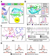
Figure 1. Library design and selection of I-Ek, a murine class II MHC molecule
(A) Schematic of the murine class II MHC I-Ek displayed on yeast, as β1α1 ‘mini’ MHC with peptide covalently linked to MHC N-terminus. (B) Mutations required for correct folding of the β1α1 ‘mini’ I-Ek (top). Mutations were derived from error prone mutagenesis and selection (purple) and rational design (red). Staining with 2B4 and 226 tetramers demonstrate function of error prone-only construct (1st gen MHC) as well as error prone + designed mutant construct (2nd gen MHC) (bottom). (C) Design of the peptide library displayed by I-Ek. Design is based upon the structure of 2B4 bound to MCC-I-Ek (left). Residues from P(-2) to P10 are randomized, with limited diversity at P(-2), P10, and the P1/P9 anchors (right). (D) 500 nM TCR tetramer staining of three clones selected for binding to 2B4 TCR compared to MCC (wild-type). TCR contact residues are colored red. See also Figure S1.
Figure 2
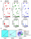
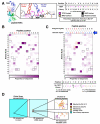
Figure 2. Deep sequencing of peptide selections on I-Ek converges on one dominant epitope for 2B4 TCR recognition
(A) Plots for amino acid prevalence at the three primary TCR contact positions (P3 (cyan), P5 (magenta), and P8 (orange)) show the peptide library enriches from even representation of all amino acids in the pre-selection library to a WT-like motif at each position. A secondary preference can be seen at P5 and P8 in round 3 but is outcompeted by round 4. (B) Sequence enrichment of 250 most abundant peptides show a convergence from a broad array of sequences to a few related clones. Area in grey represents all clones other than the most prevalent 250. (C) Comparison of total number of peptides and prevalence of 10 most abundant peptides for each round of selection. See also Figure S2.
Figure 3
Figure 3. Three different MCC/I-Ek reactive TCRs require a WT-like recognition motif in the peptide antigens
(A) Heatmaps of amino acid preference by position for 2B4 (left, red) 5cc7 (center, green) and 226 (right, blue) TCRs after three rounds of selection. The sequence for MCC is represented via outlined boxes. TCR contact residues are labeled red on x axis. (B) Covariation analysis of TCR contact positions P5 (x axis) and P8 (y axis) show distinct coupling of amino acid preferences. (C) Minimum distance clustering of all TCR sequences selected above background show sequences for all TCRs form one large cluster with MCC (black circle, not represented in library but added for reference). Sequence cluster placed in a representation of whole-library sequence space (left: 1x magnification, center: 1000x magnification) for reference. See also Figure S3.
Figure 4
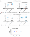
Figure 4. Relationships between affinity and activity of peptides selected for binding to IEk-reactive TCRs
(A) EC50s of IL-2 release and CD69 upregulation for 2B4 T cells stimulated with peptides selected with 2B4 TCR, plus MCC (red) (left), or peptides selected with 226 or 5cc7 TCRs (right). Sequences with close homology to MCC at P3, P5, and P8 are represented in blue. Sequences that do not share 3/3 TCR contacts with MCC are in black. (B) EC50s as in A for 5cc7 T cells with peptides selected with 5cc7 (left) or 226/2B4 (right) TCRs. (C) Correlation between TCR-pMHC affinity and peptide signaling potency. Each data point represents one peptide. See also Figure S4.
Figure 5
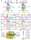
Figure 5. Peptides distantly related to MCC show highly similar mechanism of recognition and linkages to the cognate antigen
Crystal structures of TCR-pMHC complexes for 2B4-2A-I-Ek and 2B4-MCC-I-Ek (PDB ID: 3QIB) (A) and 5cc7-5c1-I-Ek and 226-MCC-I-Ek (PDB ID: 3QIU) (B) compared. TCR contacts are shown in magenta (top, noted with triangles). There is very little change in overall binding geometry despite significant variation of peptide sequence. The TCRs accommodate differences in peptide sequence primarily through differences in CDR3β (bottom). (C) TCR CDR loop footprints for 2B4 recognizing MCC and 2A peptides, 226 recognizing MCC and MCC K99E peptides, and 5cc7 recognizing 5c1 and 5c2 peptides show very little deviation. (D) Relationship between MCC and 2A peptides revealed through intermediate selected peptide sequences. See also Table S1 and Figure S5.
Figure 6
Figure 6. Design and selection of HLA-DR15 based libraries for myelin basic protein (MBP)-reactive human TCRs
(A) HLA-DR15 library design based upon structure of Ob.1A12-MBP-HLA-DR15 complex. Residues P(-4)-P10 are fully randomized, except for the P1 and P4 anchors (in black). TCR contacts are colored magenta. (B) Heatmap of amino acid preference by position for Ob.1A12 TCR. The sequence for MBP is represented via outlined boxes. TCR contacts are labeled red on the x axis. (C) Design and selection results of library that suppresses central ‘HF’ TCR recognition motif at P2-P3 of peptide. Resulting register shift is shown in blue on x axis. (D) Sequence clustering shows distinct, related clusters of selected peptides. Sequence cluster placed in a representation of whole-library sequence space (left: 1x magnification, center: 1000x magnification) for reference. See also Figure S6.
Figure 7
Figure 7. Discovery of naturally occurring TCR ligands through deep sequencing and substitution matrix-based homology search
(A) Schematic for ligand search strategy, in which a positional substitution matrix is generated from deep sequencing data and then used to find naturally occurring peptides that are represented within the matrix. (B) Functional characterization of a selection of naturally occurring peptides with predicted activity. Activity is tested via proliferation of T cells when exposed to peptide. Heatmaps are normalized to 10μM dose of MBP peptide for each T cell clone. See also Tables S2 and S3 and Figure S7.

Comment in

References

    1. Adams JJ, Narayanan S, Liu B, Birnbaum ME, Kruse AC, Bowerman NA, Chen W, Levin AM, Connolly JM, Zhu C, et al. T Cell Receptor Signaling Is Limited by Docking Geometry to Peptide-Major Histocompatibility Complex. Immunity. 2011;35:681–693. - PMC - PubMed
    1. Adams PD, Afonine PV, Bunkoczi G, Chen VB, Davis IW, Echols N, Headd JJ, Hung LW, Kapral GJ, Grosse-Kunstleve RW, et al. PHENIX: a comprehensive Python-based system for macromolecular structure solution. Acta Crystallogr D Biol Crystallogr. 2010;66:213–221. - PMC - PubMed
    1. Basu D, Horvath S, Matsumoto I, Fremont DH, Allen PM. Molecular basis for recognition of an arthritic peptide and a foreign epitope on distinct MHC molecules by a single TCR. J Immunol. 2000;164:5788–5796. - PubMed
    1. Benoist C, Mathis D. Autoimmunity provoked by infection: how good is the case for T cell epitope mimicry? Nat Immunol. 2001;2:797–801. - PubMed
    1. Birnbaum ME, Dong S, Garcia KC. Diversity-oriented approaches for interrogating T-cell receptor repertoire, ligand recognition, and function. Immunol Rev. 2012;250:82–101. - PMC - PubMed

Publication types