. 2014 Jun;20(6):950-9.
doi: 10.3201/eid2006.131438.

Rapid spread and diversification of respiratory syncytial virus genotype ON1, Kenya

Rapid spread and diversification of respiratory syncytial virus genotype ON1, Kenya

Charles N Agoti et al. Emerg Infect Dis. 2014 Jun.

Abstract

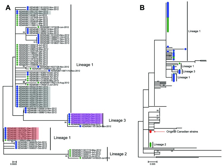
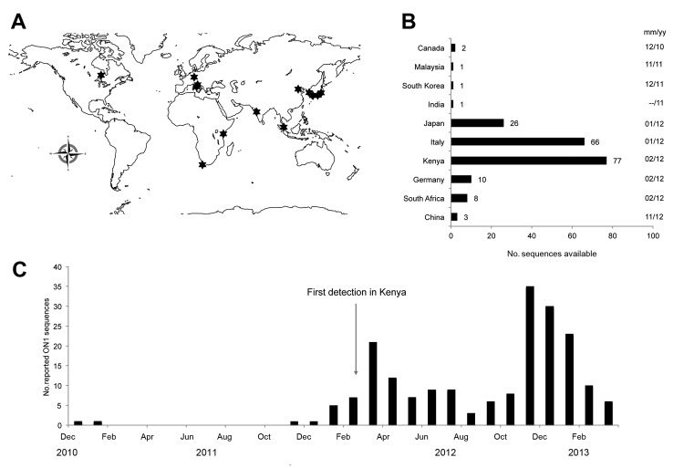
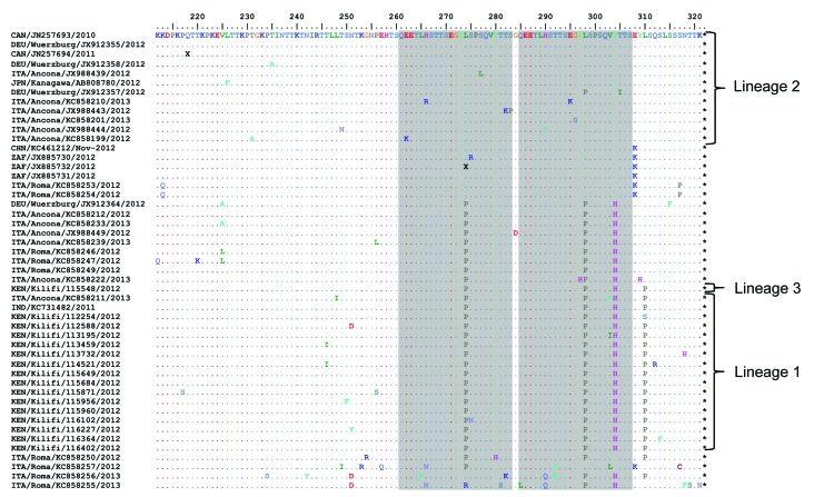
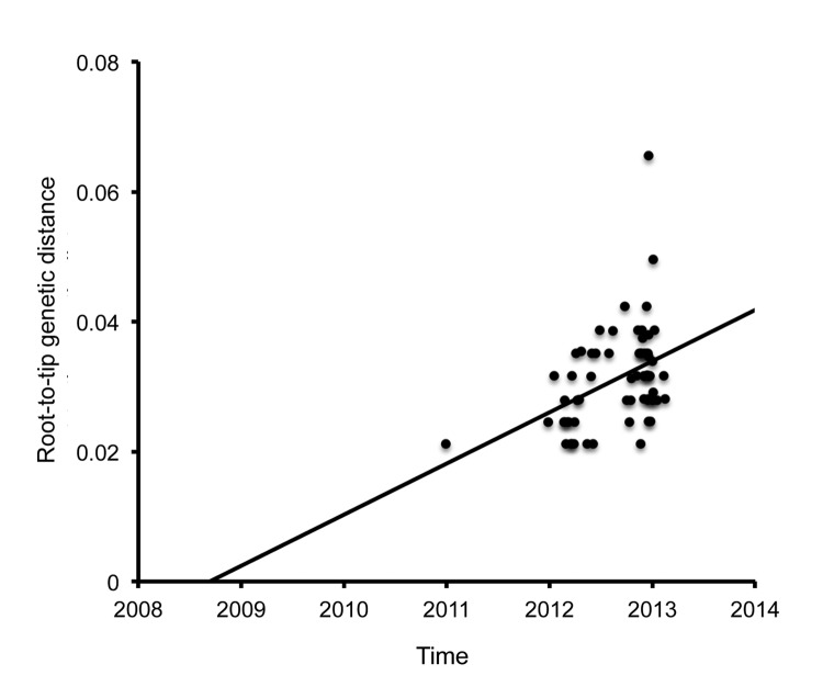
Respiratory syncytial virus genotype ON1, which is characterized by a 72-nt duplication in the attachment protein gene, has been detected in >10 countries since first identified in Ontario, Canada, in 2010. We describe 2 waves of genotype ON1 infections among children admitted to a rural hospital in Kenya during 2012. Phylogenetic analysis of attachment protein gene sequences showed multiple introductions of genotype ON1; variants distinct from the original Canadian viruses predominated in both infection waves. The genotype ON1 dominated over the other group A genotypes during the second wave, and some first wave ON1 variants reappeared in the second wave. An analysis of global genotype ON1 sequences determined that this genotype has become considerably diversified and has acquired signature coding mutations within immunogenic regions, and its most recent common ancestor dates to ≈2008-2009. Surveillance of genotype ON1 contributes to an understanding of the mechanisms of rapid emergence of respiratory viruses.

Keywords: G glycoprotein; G protein gene; Kenya; ON1; RSV; attachment protein; epidemics; genotype ON1; humans; phylogenetic analysis; pneumonia; respiratory syncytial virus; respiratory viruses; surveillance; viruses.


Figures

Figure 1
Figure 1
A) Number of persons positive for respiratory syncytial virus (RSV) genotype ON1 during 2 infection waves, Kilifi, Kenya, 2012. Blue line indicates cases of RSV group A infection; red line indicates cases of RSV group B infection; bars indicate number of ON1 case-patients admitted to Kilifi District Hospital during January 1–December 31, 2012. The first ON1 infection wave (green shading) overlapped with a 2011–12 RSV epidemic, and the second ON1 infection wave (blue shading) overlapped with a 2012–13 RSV epidemic (see arrows above graph). B) Lifespans of the main ON1 lineages observed at Kilifi. Numbers inside arrows indicate the number of sequences for the lineage named at the far left.
Figure 2
Figure 2
Phylogeny of respiratory syncytial virus genotype ON1 viruses detected globally and from Kilifi, Kenya. A) Maximum-likelihood, nucleotide-based phylogenetic tree showing the evolutionary relationships of the 77 Kilifi ON1 viruses across the sequenced portion (702 nt long) of the attachment (G) protein gene. The taxon nomenclature on the tree is as follows: A 3-letter code representing country of isolation/(location within country of isolation, if provided)/GenBank accession number (or identification for Kilifi sequences)/date of isolation. Kilifi viruses identified during the first infection wave are preceded by a green diamond; those identified during the second infection wave are preceded by a blue circle. Highlighted names (i.e., red, gray, and purple, indicating viruses by taxa) had sequences identical to those for viruses from the first ON1 infection wave. B) Maximum-likelihood, nucleotide-based phylogenetic tree showing the evolutionary backbone structure of 118 global ON1 viruses sequences, together with the 77 Kilifi ON1 viruses sequences across the sequenced portion (333 nt long) of the G third C-terminus region. The positions of the Kilifi viruses from the first infection wave are indicated by a green diamond, those from the second wave are indicated by a blue circle; the red circle indicates the position of the original Canadian ON1 viruses. On both trees, only bootstrap support values >60 are shown on the branches. Scale bars indicate nucleotide substitutions per site.
Figure 3
Figure 3
A) Geographic locations (indicated by stars) where respiratory syncytial virus genotype ON1 sequences had been detected and reported as of September 8, 2013. B) Number of ON1 sequences present in GenBank as of September 8, 2013, including sequences submitted for the viruses detected in Kilifi, Kenya, during 2012. The month and year that the first ON1 virus was reported for each country is given on the far right. C) Monthly reported detections of the ON1 viruses with sequences from the combined worldwide dataset. The month of first ON1 detection at Kilifi is indicated by an arrow. Note that the data presented in this figure have not been systematically collected to represent the geographic and temporal distribution of the ON1 genotype. Instead, the data are derived from sequence submissions to GenBank, with inherent sampling bias, that do not necessarily reflect the total number of cases from the different locations; the submissions do, however, indicate the rapid spread of the genotype.
Figure 4
Figure 4
Alignment of unique deduced amino acid sequences from the combined dataset of sequences from the C-terminal third of the attachment protein of respiratory syncytial virus genotype ON1. The sequences are compared with the sequence for the earliest ON1 variant (from Ontario, Canada). The duplicated parent and the resulting regions are in gray.
Figure 5
Figure 5
Most recent common ancestor analysis of the 65 respiratory syncytial virus genotype ON1 viruses in GenBank and the sequences for ON1 viruses detected in Kilifi, Kenya, during 2012. The analysis was done by root-to-tip regression of the genetic distances from the maximum-likelihood tree.

References

    1. Collins PL, Melero JA. Progress in understanding and controlling respiratory syncytial virus: still crazy after all these years. Virus Res. 2011;162:80–99. 10.1016/j.virusres.2011.09.020 - DOI - PMC - PubMed
    1. Cane PA, Matthews DA, Pringle CR. Analysis of respiratory syncytial virus strain variation in successive epidemics in one city. J Clin Microbiol. 1994;32:1–4 . - PMC - PubMed
    1. Cane PA, Pringle CR. Evolution of subgroup A respiratory syncytial virus: evidence for progressive accumulation of amino acid changes in the attachment protein. J Virol. 1995;69:2918–25 . - PMC - PubMed
    1. Melero JA, Garcia-Barreno B, Martinez I, Pringle CR, Cane PA. Antigenic structure, evolution and immunobiology of human respiratory syncytial virus attachment (G) protein. J Gen Virol. 1997;78:2411–8 . - PubMed
    1. Smith GJ, Vijaykrishna D, Bahl J, Lycett SJ, Worobey M, Pybus OG, et al. Origins and evolutionary genomics of the 2009 swine-origin H1N1 influenza A epidemic. Nature. 2009;459:1122–5. 10.1038/nature08182 - DOI - PubMed

Publication types

MeSH terms

Substances