Review
. 2014 Jun;45 Suppl 2(0 2):S8-S15.
doi: 10.1016/j.injury.2014.04.003.

Biological perspectives of delayed fracture healing

Affiliations
Review

Biological perspectives of delayed fracture healing

K D Hankenson et al. Injury. 2014 Jun.

Abstract

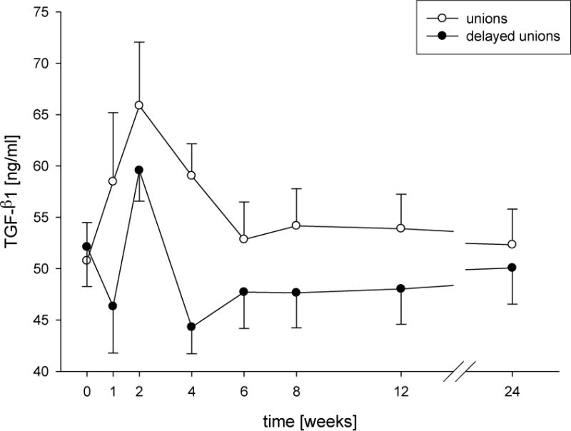
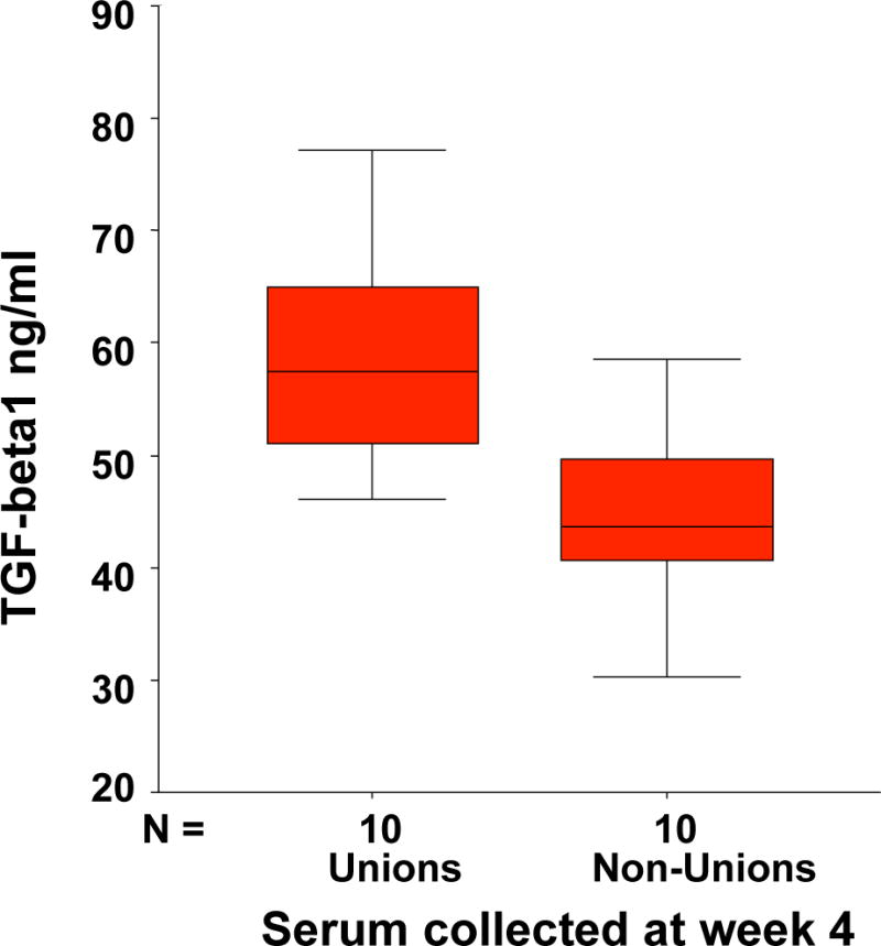
Fracture healing is a complex biological process that requires interaction among a series of different cell types. Maintaining the appropriate temporal progression and spatial pattern is essential to achieve robust healing. We can temporally assess the biological phases via gene expression, protein analysis, histologically, or non-invasively using biomarkers as well as imaging techniques. However, determining what leads to normal versus abnormal healing is more challenging. Since the ultimate outcome of fracture healing is to restore the original functions of bone, assessment of fracture healing should include not only monitoring the restoration of structure and mechanical function, but also an evaluation of the restoration of normal bone biology. Currently few non-invasive measures of biological factors of healing exist; however, recent studies that have correlated non-invasive measures with fracture healing outcome in humans have shown that serum TGFbeta1 levels appear to be an indicator of healing versus non-healing. In the future, developing additional measures to assess biological healing will improve the reliability and permit us to assess stages of fracture healing. Additionally, new functional imaging technologies could prove useful for better understanding both normal fracture healing and predicting dysfunctional healing in human patients.

Keywords: Bone healing; Mediators; Non-union; TGFbeta1; Vascularity.


Figures

Figure 1
Figure 1. Tibial shaft fracture in a mouse at days 2, 5, 10, 15, 20, and 30 post-fracture (DPF)
Top row are histology sections stained with Fast Green FCF and Safranin-O, and represent a series of 2X images that have been stitched together to show the entire tibia. Rows 2 and 3 are micro-CT images of the callus; the middle row is a three-dimensional reconstruction of the callus and the bottom row is a coronal section of the specimen. The 2 and 5 DPF time points represent the early inflammatory and marrow response stage. 10 DPF represents the early intermediate phase dominated by chondrogenesis. 15 DPF demonstrates the transition to the late intermediate stage marked by primary bone formation observed at 20 DPF. Finally, secondary bone formation and remodeling can be observed at 30 DPF.
Figure 2
Figure 2. The process of fracture healing in non-stabilized fractures of the mouse tibia
Safranin-O/fast green (A,F,K,P,Q,U,V) and trichrome (C,H,M,R,S,W,X) were used to staining cartilage red and bone blue respectively. Transcripts of Col2 (B,G,L), Col10 (L), and osteocalcin (D,I,N) were detected by in situ hybridization, pseudocolored red, yellow, and green and used to visualize chondrocytes, hypertrophic chondrocytes, and osteoblasts respectively. Macrophages were detected via immunohistochemistry using antibody F4/80 (E), and endothelial cells were stained blach using immunohistochemistry with an antibody against PECAM (J,O, also known as CD31). Osteoclasts were stained red using tartrate-resistant acid phosphatase (T,Y). (A) At day 3 post fracture no cartilage or (B) Col2/Col10 transcripts were detected. (C) No bone or (D) osteocalcin was present. (E) At this time, macrophages have infiltrated the fracture site. (F) At 5 days, immature cartilage (G) expressing Col2 but not Col10 was observed in the periosteum. (H) A small amount of new bone (I) and osteocalcin expression was also apparent in the periosteal reaction, (J) which is highly vascularized. (K,l) At 7 days, cartilage is beginning to mature, and Col10 transcripts are evident. (M,N) More new bone and osteocalcin expression are also evident. (O) Vascular invasion was observed around hypertrophic chondrocytes. (P,Q) At day 14 a large amount of cartilage and (R,S) bone were formed. (T) Multiple osteoclasts were observed at the front of endochondral ossification. (U,V) By day 21 after injury, cartilage has been replaced by bone. (W,X) Fractures have healed by bony bridging. (Y) Osteoclasts on the surface of trabecular bone continue to remodel the newly formed bone. Scale bar: A-D, G-I, L-O, Q,S,V, and X=250mm; E,T, and Y=60mm, F,K,P,U,R=1mm.
Figure 3
Figure 3. Changes in whole-callus gene expression reflect the rapidly changing temporal biology of fracture healing
RNA was harvested from murine calluses following fracture and assessed for markers of inflammation (IL1-beta), chondrogenesis (type 2 collagen), and bone formation (osteocalcin).
Figure 4
Figure 4. TGF-b1 serum levels presented as absolute concentrations
Time course of concentrations of TGF-b1 (mean + SEM) from patients after surgery for long-bone shaft fractures with normal (unions) or delayed consolidation (delayed unions). Significant changes compared to the reference value (48-week unions; 72-week delayed unions) are indicated by asterisks (*P < 0.05, **P < 0.01). # Indicates significant differences a certain point between the union and delayed union group (#P < 0.05, ##P < 0.01). Clamps illustrate significant time related changes within both groups.
Figure 5
Figure 5. Decreases in serum TGF-b1 levels reflect a failure to heal
Based on a number of 15 patients in each group the distribution of the values in the group of non unions and unions could be seen. At a level of under 45 ng/cc TGFß-1 only one patient of 15 would have healed in the next months and only one patient over 45 ng/cc of 15 developed a non union.

References

    1. Einhorn TA. The cell and molecular biology of fracture healing. Clin Orthop. 1998;(355 Suppl):S7–21. - PubMed
    1. Colnot C, Zhang X, Knothe Tate ML. Current insights on the regenerative potential of the periosteum: molecular, cellular, and endogenous engineering approaches. J Orthop Res. 2012;30(12):1869–78. - PMC - PubMed
    1. Bahney CS, et al. Stem Cell Derived Endochondral Cartilage Stimulates Bone Healing by Tissue Transformation. J Bone Miner Res. 2013 - PMC - PubMed
    1. Little DG, Ramachandran M, Schindeler A. The anabolic and catabolic responses in bone repair. J Bone Joint Surg Br. 2007;89(4):425–33. - PubMed
    1. Colnot C, Huang S, Helms J. Analyzing the cellular contribution of bone marrow to fracture healing using bone marrow transplantation in mice. Biochem Biophys Res Commun. 2006;350(3):557–61. - PubMed

Publication types