Review
. 2014:e258-66.
doi: 10.14694/EdBook_AM.2014.34.e258.

Patient-derived xenograft models in gynecologic malignancies

Affiliations
Review

Patient-derived xenograft models in gynecologic malignancies

Clare L Scott et al. Am Soc Clin Oncol Educ Book. 2014.

Abstract

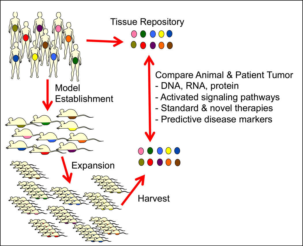
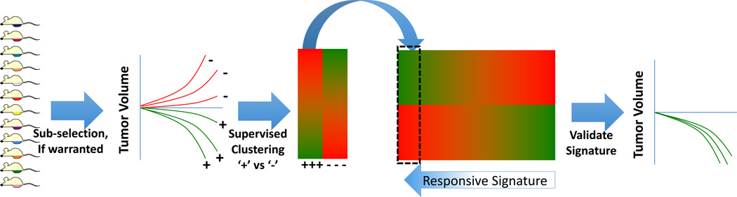
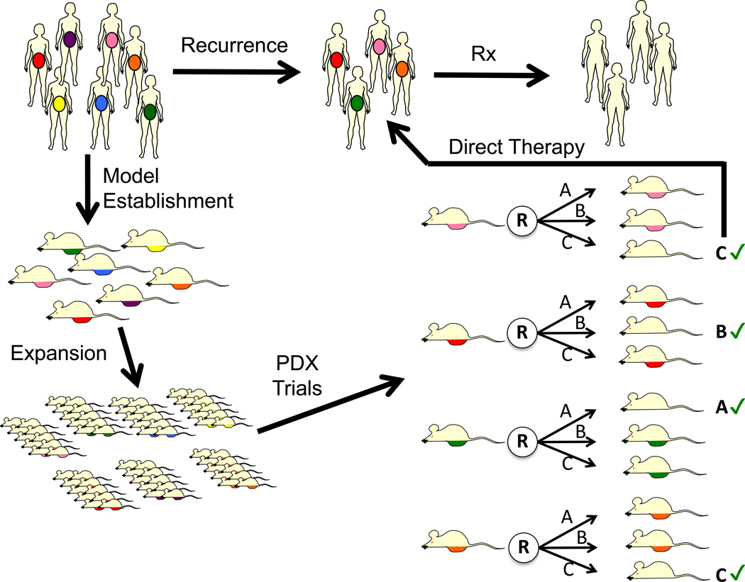
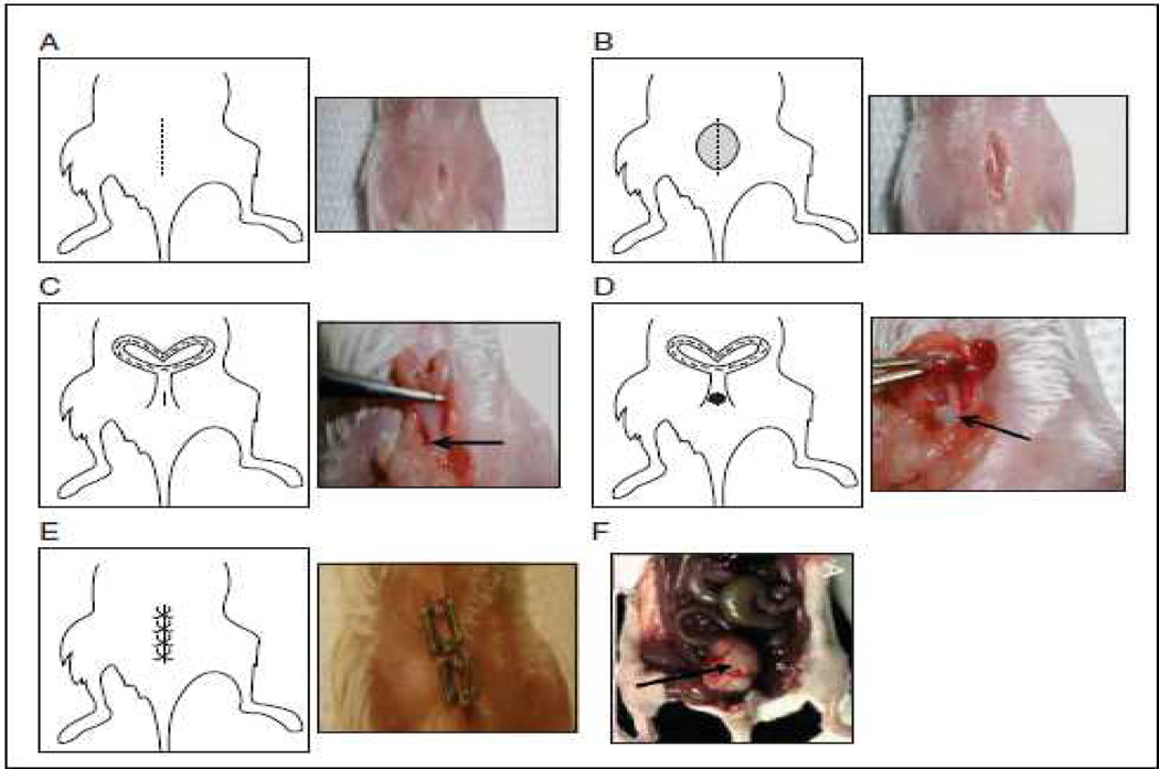
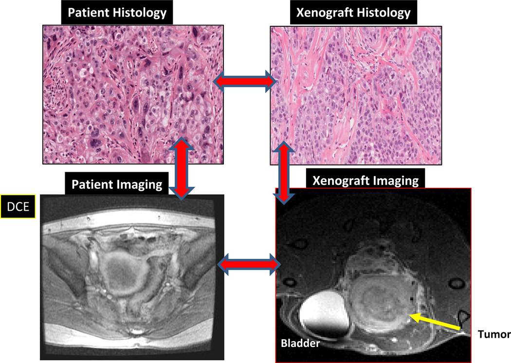
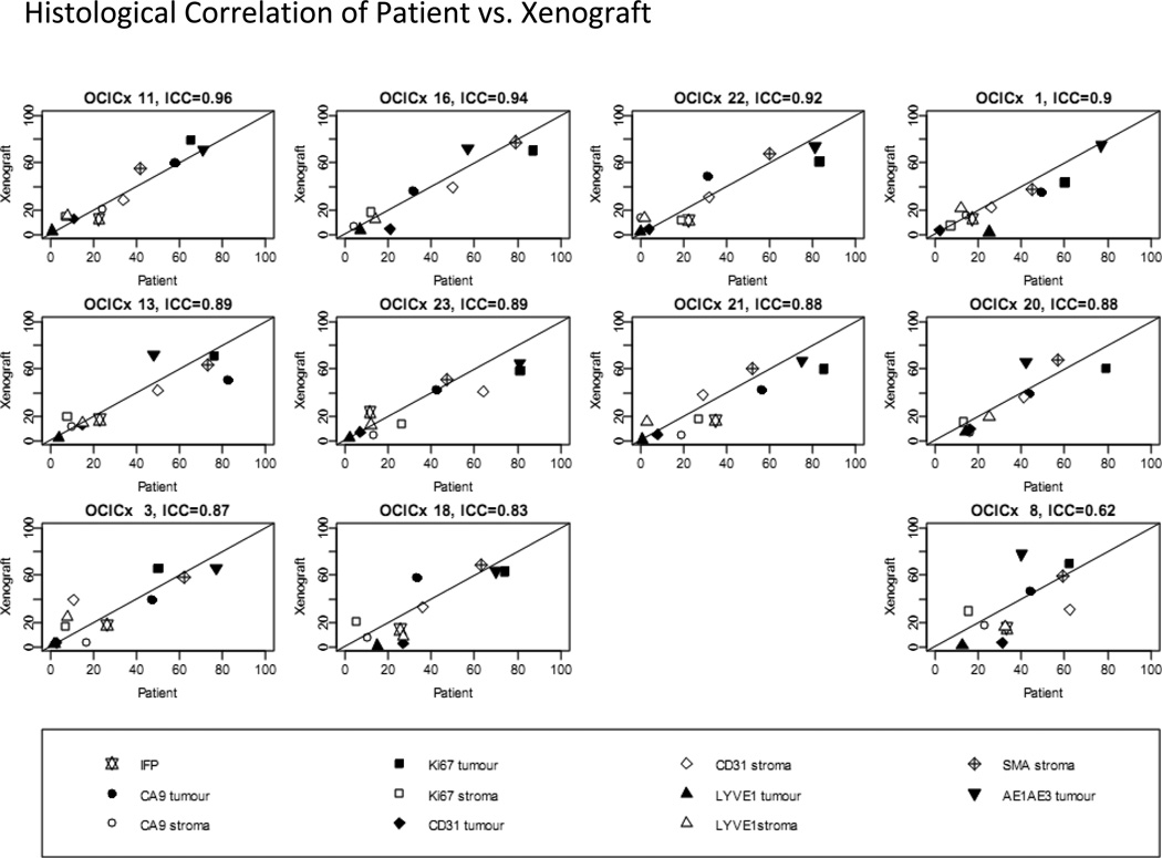
In the era of targeted therapies, patients with gynecologic malignancies have not yet been major beneficiaries of this new class of agents. This may reflect the fact that the main tumor types-ovarian, uterine, and cervical--are a highly heterogeneous group of cancers with variable response to standard chemotherapies and the lack of models in which to study the diversity of these cancers. Cancer-derived cell lines fail to adequately recapitulate molecular hallmarks of specific cancer subsets and complex microenvironments, which may be critical for sensitivity to targeted therapies. Patient-derived xenografts (PDX) generated from fresh human tumor without prior in vitro culture, combined with whole genome expression, gene copy number, and sequencing analyses, could dramatically aid the development of novel therapies for gynecologic malignancies. Gynecologic tumors can be engrafted in immunodeficient mice with a high rate of success and within a reasonable time frame. The resulting PDX accurately recapitulates the patient's tumor with respect to histologic, molecular, and in vivo treatment response characteristics. Orthotopic PDX develop complications relevant to the clinic, such as ascites and bowel obstruction, providing opportunities to understand the biology of these clinical problems. Thus, PDX have great promise for improved understanding of gynecologic malignancies, serve as better models for designing novel therapies and clinical trials, and could underpin individualized, directed therapy for patients from whom such models have been established.


Conflict of interest statement

Authors’ Disclosure of Potential Conflicts of Interests:

CLS receives in-kind collaborative laboratory support from Clovis Oncology.

PH is an unpaid consultant for Tesaro and Clovis Oncology and receives research funding from Tesaro, Clovis Oncology and Genentech.

Figures

Figure 1
Figure 1. Model Establishment Schema
The rationale for establishing PDX for understanding of gynecological malignancies is the assumption that each patient’s tumor is different from the others. This is represented by the varying colors. Aliquots of fresh tumors are immediately processed after surgical resection and injected into immunocompromised mice. Upon engraftment, tumors are resected and expanded in additional mice, once harvested; the tumors can be compared by a variety of molecular techniques to the source tumor specimens. By annotating specimens with clinical information from patients, correlations may be made including response to chemotherapy and survival time. Ultimately, tumors can be established to evaluating responses to treatment and potentially use these data to direct therapy in patients.
Figure 2
Figure 2. Drug Development Schema Using PDX Models
Using unselected PDX models (or sub-selected based on a pertinent characteristic, such as platinum-resistance or BRCA2 mutation), models can be screened for tumor regression in response to the experimental therapy. Using DNA microarray or other high-density whole-genome data, models would then be clustered based on response (+= responders, − = non-responders). The ‘response signature’ could then be developed and validated against the remaining pool of PDX models.
Figure 3
Figure 3. Individualized Therapy Directed by PDX Models
Using PDX models engrafted and expanded from patients, each model can be tested pre-emptively with investigational and/or standard therapies (depicted as treatments A, B and C). Such investigations could be initiated, for example, while patients are in remission from primary treatment. At the time of recurrence, the therapy determined to be most effective by the patients own ‘Avatar’ (denoted with ‘check’ sign), could then be delivered. This would require all PDX models to undergo a PDX therapy trial prior to knowing if patient would recur.
Figure 4
Figure 4. Surgical technique of orthotopic implantation onto the cervical site of SCID mice
(A). A 1-cm incision in the skin of the lower abdomen. (B) Incision in the peritoneum. (C) Accessing the cervix (D) The suture with the tumor fragment threaded onto it is passed from the inside of the uterus to the outside and the at the fragment sutured to the cervix (arrow). (E) The uterus is then placed back into the peritoneal cavity. The peritoneum is closed. (F) Post-implant, approximately following 3 weeks, the growth of the primary cervix tumor is shown (indicated by arrow).
Figure 5
Figure 5. Comparison of histology and imaging of patient and tumor compared to imaging and histology in mouse primary xenograft model
Figure 6
Figure 6. The relationship between passage 3 xenograft models and its matching biopsy
Plots for IFP and tumor and stroma measures for hypoxia, blood and lymphatic vessels, and proliferation and smooth muscle actin. Lines plotted are (0,1) line of perfect concordance.

References

    1. Adams RA. Heightened Immunity and Susceptibility toward Cheek Pouch Heterografts of a Mouse Leukemia in Syrian Hamsters. Cancer Res. 1963;23:1834–1840. - PubMed
    1. Carrel S, Sordat B, Merenda C. Establishment of a cell line (Co-115) from a human colon carcinoma transplanted into nude mice. Cancer Res. 1976;36:3978–3984. - PubMed
    1. Domcke S, Sinha R, Levine DA, Sander C, Schultz N. Evaluating cell lines as tumour models by comparison of genomic profiles. Nat Commun. 2013;4:2126. - PMC - PubMed
    1. Scott CL, Becker MA, Haluska P, Samimi G. Patient-Derived Xenograft Models to Improve Targeted Therapy in Epithelial Ovarian Cancer Treatment. Front Oncol. 2013;3:295. - PMC - PubMed
    1. Weroha SJ, Becker MA, Enderica-Gonzalez S, Harrington SC, Oberg AL, Maurer MJ, Perkins S, Al Hilli M, Butler K, McKinstry S, Fink SR, Jenkins RB, Hou X, Kalli KR, Goodman KM, Sarkaria JN, Karlan BY, Kumar A, Kaufmann SH, Hartmann LC, Haluska P. Tumorgrafts as in vivo surrogates for women with ovarian cancer. Clin Cancer Res. 2014 - PMC - PubMed

Publication types

LinkOut - more resources