

Pulsed focused ultrasound treatment of muscle mitigates paralysis-induced bone loss in the adjacent bone: a study in a mouse model
- PMID: 24857416
- PMCID: PMC4410740
- DOI: 10.1016/j.ultrasmedbio.2014.02.027
Pulsed focused ultrasound treatment of muscle mitigates paralysis-induced bone loss in the adjacent bone: a study in a mouse model
Abstract
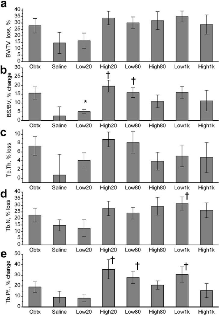
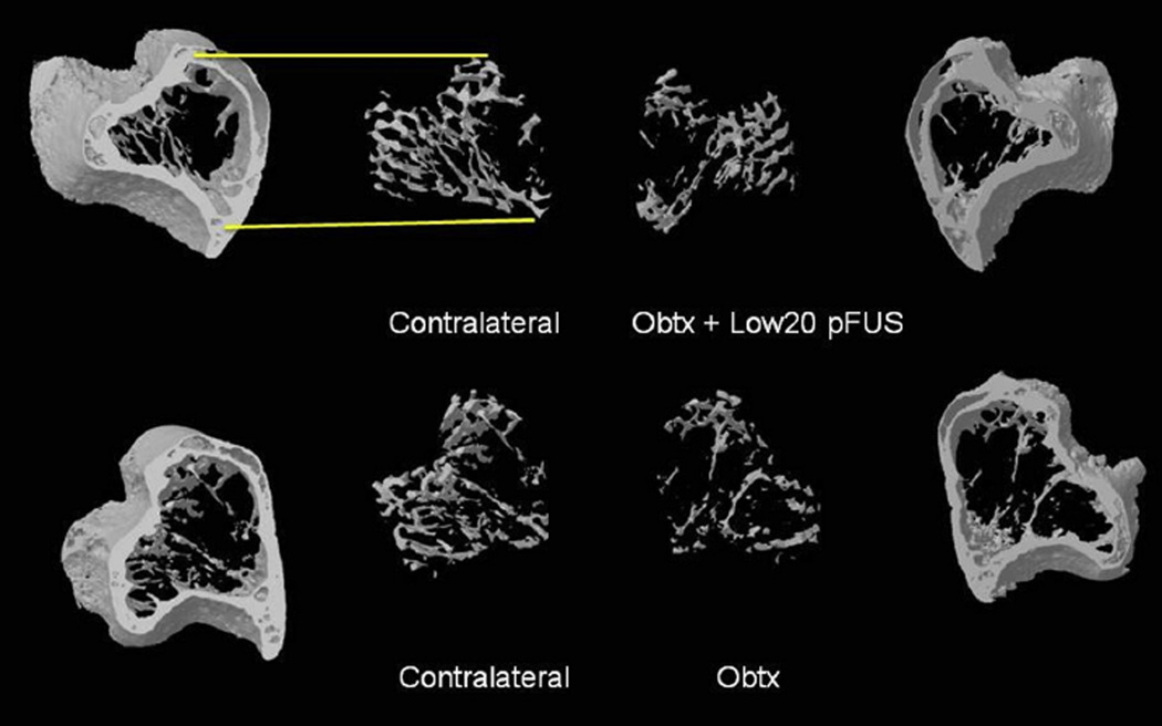
Bone loss can result from bed rest, space flight, spinal cord injury or age-related hormonal changes. Current bone loss mitigation techniques include pharmaceutical interventions, exercise, pulsed ultrasound targeted to bone and whole body vibration. In this study, we attempted to mitigate paralysis-induced bone loss by applying focused ultrasound to the midbelly of a paralyzed muscle. We employed a mouse model of disuse that uses onabotulinumtoxinA-induced paralysis, which causes rapid bone loss in 5 d. A focused 2 MHz transducer applied pulsed exposures with pulse repetition frequency mimicking that of motor neuron firing during walking (80 Hz), standing (20 Hz), or the standard pulsed ultrasound frequency used in fracture healing (1 kHz). Exposures were applied daily to calf muscle for 4 consecutive d. Trabecular bone changes were characterized using micro-computed tomography. Our results indicated that application of certain focused pulsed ultrasound parameters was able to mitigate some of the paralysis-induced bone loss.
Keywords: Micro-computed tomography; Mitigation of trabecular bone loss; Mouse model of disuse; Musculoskeletal; Paralysis; Pulsed focused ultrasound.
Copyright © 2014 World Federation for Ultrasound in Medicine & Biology. Published by Elsevier Inc. All rights reserved.
Figures
Similar articles
-
Metaphyseal and diaphyseal bone loss in the tibia following transient muscle paralysis are spatiotemporally distinct resorption events.Bone. 2013 Dec;57(2):413-22. doi: 10.1016/j.bone.2013.09.009. Epub 2013 Sep 21. Bone. 2013. PMID: 24063948 Free PMC article.
-
Dynamic acoustic radiation force retains bone structural and mechanical integrity in a functional disuse osteopenia model.Bone. 2015 Jun;75:8-17. doi: 10.1016/j.bone.2015.01.020. Epub 2015 Feb 7. Bone. 2015. PMID: 25661670 Free PMC article.
-
Acceleration of Bone Defect Healing and Regeneration by Low-Intensity Ultrasound Radiation Force in a Rat Tibial Model.Ultrasound Med Biol. 2018 Dec;44(12):2646-2654. doi: 10.1016/j.ultrasmedbio.2018.08.002. Epub 2018 Oct 1. Ultrasound Med Biol. 2018. PMID: 30286949
-
Effects of pulsed ultrasound on the mouse neonate: hind limb paralysis and lung hemorrhage.Ultrasound Med Biol. 1994;20(1):53-63. doi: 10.1016/0301-5629(94)90017-5. Ultrasound Med Biol. 1994. PMID: 8197627 Review.
-
The effects of spinal cord injury and exercise on bone mass: a literature review.NeuroRehabilitation. 2011;29(3):261-9. doi: 10.3233/NRE-2011-0702. NeuroRehabilitation. 2011. PMID: 22142760 Review.
Cited by
-
Mechanical damage thresholds for hematomas near gas-containing bodies in pulsed HIFU fields.Phys Med Biol. 2022 Oct 20;67(21):10.1088/1361-6560/ac96c7. doi: 10.1088/1361-6560/ac96c7. Phys Med Biol. 2022. PMID: 36179703 Free PMC article.
-
Botulinum Toxin A and Osteosarcopenia in Experimental Animals: A Scoping Review.Toxins (Basel). 2021 Mar 14;13(3):213. doi: 10.3390/toxins13030213. Toxins (Basel). 2021. PMID: 33799488 Free PMC article.
-
MRI-guided focused ultrasound surgery in musculoskeletal diseases: the hot topics.Br J Radiol. 2016;89(1057):20150358. doi: 10.1259/bjr.20150358. Epub 2015 Nov 26. Br J Radiol. 2016. PMID: 26607640 Free PMC article. Review.
References
-
- Al-Qraini MM, Canney MS, Oweis GF. Laser-induced fluorescence thermometry of heating in water from short bursts of high intensity focused ultrasound. Ultrasound Med Biol. 2013;39:647–659. - PubMed
-
- Andreev VG, Dmitriev VN, Pishchalnikov YA, Rudenko OV. Sapozhnikov OA, Sarvazyan AP Observation of shear waves excited by focused ultrasound in a rubber-like medium. Acoust Phys. 1997;43:123–128.
-
- Azuma Y, Ito M, Harada Y, Takagi H, Ohta T, Jingushi S. Low-intensity pulsed ultrasound accelerates rat femoral fracture healing by acting on the various cellular reactions in the fracture callus. J Bone Miner Res. 2001;16:671–680. - PubMed
-
- Belavy DL, Beller G, Armbrecht G, Perschel FH, Fitzner R, Bock O, Borst H, Degner C, Gast U, Felsenberg D. Evidence for an additional effect of whole-body vibration above resistive exercise alone in preventing bone loss during prolonged bed rest. Osteoporos Int. 2011;22:1581–1591. - PubMed
Publication types
MeSH terms
Grants and funding
LinkOut - more resources
Full Text Sources
Other Literature Sources
Medical
