

Comparison of biomechanical effects of small-incision lenticule extraction and laser in situ keratomileusis: finite-element analysis
- PMID: 24857440
- PMCID: PMC6030688
- DOI: 10.1016/j.jcrs.2013.08.065
Comparison of biomechanical effects of small-incision lenticule extraction and laser in situ keratomileusis: finite-element analysis
Abstract
Purpose: To theoretically compare the corneal stress distribution of laser in situ keratomileusis (LASIK) with the stress distribution of small-incision lenticule extraction.
Setting: Cleveland Clinic Cole Institute, Cleveland, and The Ohio State University, Columbus, Ohio, USA.
Design: Computational modeling study.
Methods: A finite-element anisotropic collagen fiber-dependent model of myopic surgery using patient-specific corneal geometry was constructed for LASIK, small-incision lenticule extraction, and a geometry analog model with unaltered material properties from preoperative but with postoperative geometry including thickness. Surgical parameters, magnitude of myopic correction, LASIK flap thickness, and lenticule depth in small-incision lenticule extraction were varied. Two sets of models, 1 with uniform and 1 with depth-dependent material properties, were constructed.
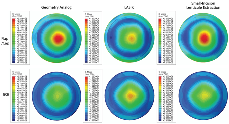
Results: Stress distribution between small-incision lenticule extraction simulations and the geometry analog model were similar. In contrast, LASIK consistently reduced stress in the flap and increased stress in the residual stromal bed (RSB) compared with the geometry analog model. An increase in flap thickness or lenticule depth resulted in a greater increase in RSB stress in the LASIK model than in the small-incision lenticule extraction model.
Conclusions: Small-incision lenticule extraction may present less biomechanical risk to the residual bed of susceptible corneas than comparable corrections involving LASIK flaps. Deeper corrections in the stroma may be possible in small-incision lenticule extraction without added risk for ectasia.
Financial disclosures: Proprietary or commercial disclosures are listed after the references.
Copyright © 2014 ASCRS and ESCRS. Published by Elsevier Inc. All rights reserved.
Figures


Similar articles
-
Corneal biomechanical effects: small-incision lenticule extraction versus femtosecond laser-assisted laser in situ keratomileusis.J Cataract Refract Surg. 2014 Jun;40(6):954-62. doi: 10.1016/j.jcrs.2013.07.056. Epub 2014 Apr 18. J Cataract Refract Surg. 2014. PMID: 24751146
-
Vector analysis of high (≥3 diopters) astigmatism correction using small-incision lenticule extraction and laser in situ keratomileusis.J Cataract Refract Surg. 2018 Jul;44(7):802-810. doi: 10.1016/j.jcrs.2018.04.038. Epub 2018 Jun 13. J Cataract Refract Surg. 2018. PMID: 29909252
-
Clinical evaluation of a new correction algorithm for dynamic Scheimpflug analyzer tonometry before and after laser in situ keratomileusis and small-incision lenticule extraction.J Cataract Refract Surg. 2018 May;44(5):581-588. doi: 10.1016/j.jcrs.2018.01.023. Epub 2018 Apr 22. J Cataract Refract Surg. 2018. PMID: 29685776
-
Comparison of corneal biomechanical changes after refractive surgery by noncontact tonometry: small-incision lenticule extraction versus flap-based refractive surgery - a systematic review.Acta Ophthalmol. 2019 Mar;97(2):127-136. doi: 10.1111/aos.13906. Epub 2018 Sep 10. Acta Ophthalmol. 2019. PMID: 30203530
-
Femtosecond laser refractive surgery: small-incision lenticule extraction vs. femtosecond laser-assisted LASIK.Curr Opin Ophthalmol. 2015 Jul;26(4):260-4. doi: 10.1097/ICU.0000000000000158. Curr Opin Ophthalmol. 2015. PMID: 26058022 Review.
Cited by
-
In Vivo Evaluation of the Effects of SMILE with Different Amounts of Stromal Ablation on Corneal Biomechanics by Optical Coherence Elastography.Diagnostics (Basel). 2022 Dec 22;13(1):30. doi: 10.3390/diagnostics13010030. Diagnostics (Basel). 2022. PMID: 36611322 Free PMC article.
-
Evaluation of corneal elastic modulus based on Corneal Visualization Scheimpflug Technology.Biomed Eng Online. 2019 Apr 4;18(1):42. doi: 10.1186/s12938-019-0662-1. Biomed Eng Online. 2019. PMID: 30947733 Free PMC article.
-
Comparison of corneal biomechanics after myopic small-incision lenticule extraction compared to LASIK: an ex vivo study.Clin Ophthalmol. 2018 Jan 25;12:237-245. doi: 10.2147/OPTH.S153509. eCollection 2018. Clin Ophthalmol. 2018. PMID: 29416315 Free PMC article.
-
Computational Biomechanical Analysis of Asymmetric Ectasia Risk in Unilateral Post-LASIK Ectasia.J Refract Surg. 2016 Dec 1;32(12):811-820. doi: 10.3928/1081597X-20160929-01. J Refract Surg. 2016. PMID: 27930791 Free PMC article.
-
Biomechanical Diagnostics of the Cornea.Int Ophthalmol Clin. 2017 Summer;57(3):75-86. doi: 10.1097/IIO.0000000000000172. Int Ophthalmol Clin. 2017. PMID: 28590282 Free PMC article. Review.
References
-
- Shah S Laiquzzaman M Yeung I Pan X Roberts C The use of the Ocular Response Analyzer to determine corneal hysteresis in eyes before and after excimer laser refractive surgery Cont Lens Anterior Eye 2009. 32 123–128 - PubMed
-
- Ambrósio R Jr Dawson DG Salomão M Guerra FP Caiado ALC Belin MW Corneal ectasia after LASIK despite low preoperative risk: tomographic and biomechanical findings in the unoperated, stable, fellow eye J Refract Surg 2010. 26 906–911 - PubMed
-
- Randleman JB Dawson DG Grossniklaus HE McCarey BE Edelhauser HF Depth-dependent cohesive tensile strength in human donor corneas: implications for refractive surgery J Refract Surg 2008. 24 S85–S89 - PubMed
-
- Abahussin M Hayes S Knox Cartwright NE Kamma-Lorger CS Khan Y Marshall J Meek KM Accessed October 14, 2013 3D collagen orientation study of the human cornea using x-ray diffraction and femtosecond laser technology Invest Ophthalmol Vis Sci 2009. 50 5159–5164 Available at: http://www.iovs.org/content/50/11/5159.full.pdf - PubMed
-
- Ahn H Kim J-K Kim CK Han GH Seo KY Kim EK Kim T-i Comparison of laser in situ keratomileusis flaps created by 3 femtosecond lasers and a microkeratome J Cataract Refract Surg 2011. 37 349–357 - PubMed
Publication types
MeSH terms
Grants and funding
LinkOut - more resources
Full Text Sources
Other Literature Sources
