. 2014 Jun 5;54(5):820-31.
doi: 10.1016/j.molcel.2014.04.026. Epub 2014 May 22.

O-GlcNAcylation regulates cancer metabolism and survival stress signaling via regulation of the HIF-1 pathway

Affiliations

O-GlcNAcylation regulates cancer metabolism and survival stress signaling via regulation of the HIF-1 pathway

Christina M Ferrer et al. Mol Cell. .

Abstract

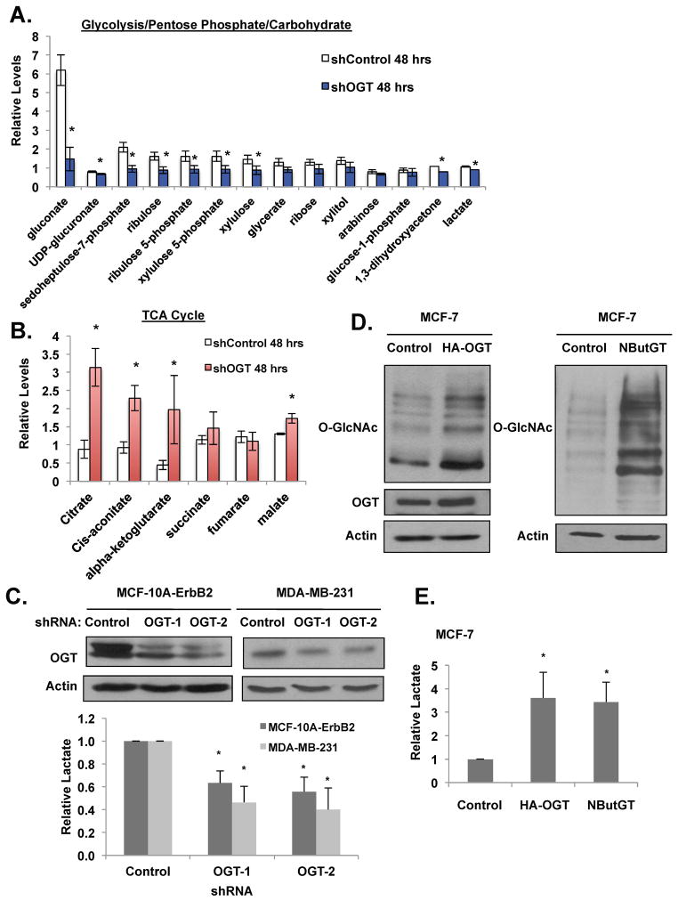
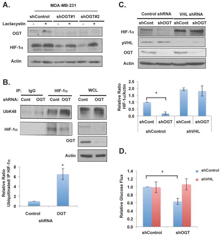
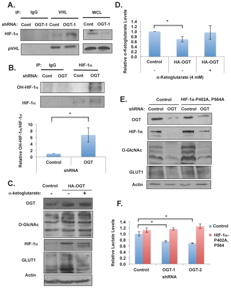
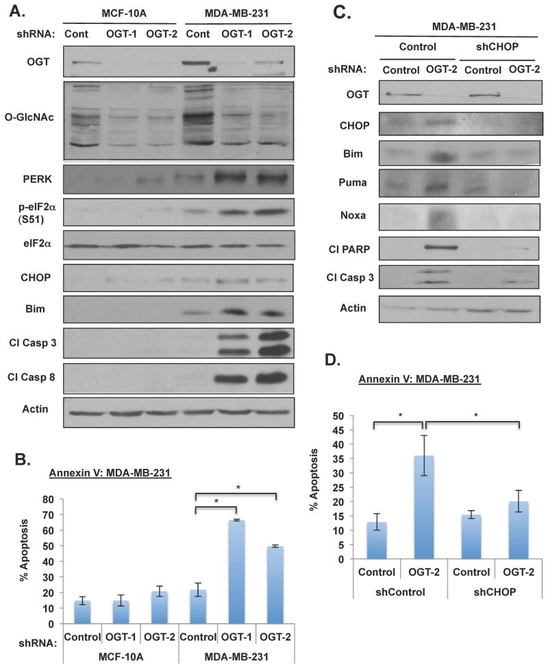
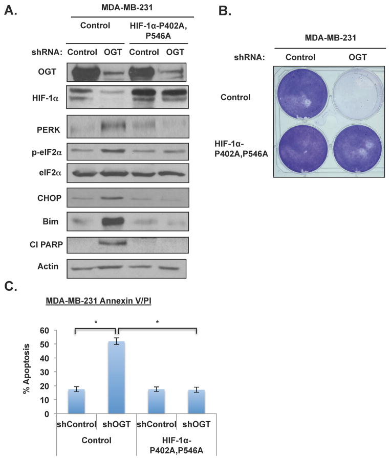
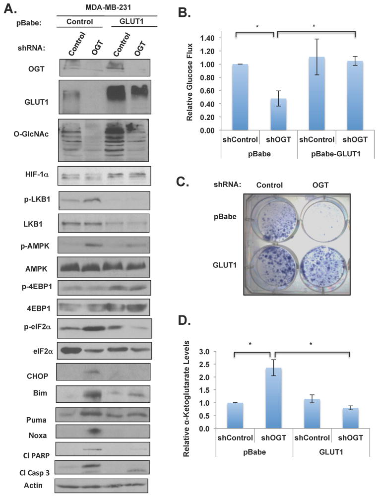
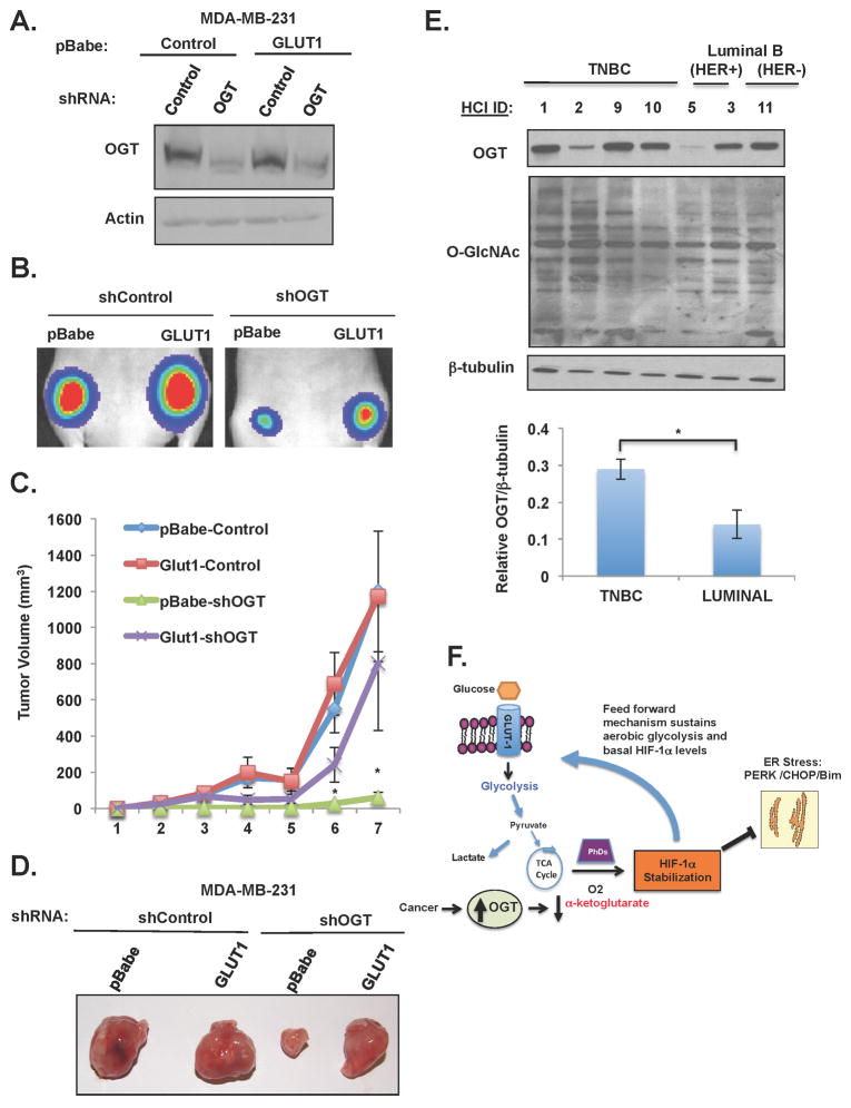
The hexosamine biosynthetic pathway elevates posttranslational addition of O-linked β-N-acetylglucosamine (O-GlcNAc) on intracellular proteins. Cancer cells elevate total O-GlcNAcylation by increasing O-GlcNAc transferase (OGT) and/or decreasing O-GlcNAcase (OGA) levels. Reducing O-GlcNAcylation inhibits oncogenesis. Here, we demonstrate that O-GlcNAcylation regulates glycolysis in cancer cells via hypoxia-inducible factor 1 (HIF-1α) and its transcriptional target GLUT1. Reducing O-GlcNAcylation increases α-ketoglutarate, HIF-1 hydroxylation, and interaction with von Hippel-Lindau protein (pVHL), resulting in HIF-1α degradation. Reducing O-GlcNAcylation in cancer cells results in activation of endoplasmic reticulum (ER) stress and cancer cell apoptosis mediated through C/EBP homologous protein (CHOP). HIF-1α and GLUT1 are critical for OGT-mediated regulation of metabolic stress, as overexpression of stable HIF-1 or GLUT1 rescues metabolic defects. Human breast cancers with high levels of HIF-1α contain elevated OGT, and lower OGA levels correlate independently with poor patient outcome. Thus, O-GlcNAcylation regulates cancer cell metabolic reprograming and survival stress signaling via regulation of HIF-1α.


Figures

Figure 1
Figure 1. OGT regulates glycolytic metabolism in breast cancer cells
(A) MDA-MB-231 cells expressing control or OGT-1 shRNA were collected and levels of glycolytic, pentose phosphate pathway, and carbohydrate metabolites and (B) TCA cycle intermediates were measured using LC-MS analysis. (C) Cell lysates from MDA-MB-231 and MCF-10A-ErbB2 expressing control, OGT-1 or OGT-2 shRNA were collected for immunoblot analysis with indicated antibodies (top). Lactate levels were measured and normalized to control shRNA treated cells (bottom). (D) Cell lysates from MCF-7 cells stably overexpressing control or HA-OGT (left) or MCF-7 cells were treated with DMSO control or 100 μM NButGT for 48 hours (right) were collected for immunoblot analysis with indicated antibodies. (E) Changes in lactate levels were measured from cells in (D) and normalized to control treated cells. (*p-value <0.05). See also Figure S1, S2.
Figure 2
Figure 2. O-GlcNAc Regulation of HIF-1α via Protein Degradation Requires VHL
(A) MDA-MB-231 cells expressing control or OGT shRNA were treated with DMSO control or 10mM lactacystin for 6 hours and lysates collected for immunoblot analysis. (B) Protein lysates from MDA-MB-231 cells expressing control or OGT shRNA were collected and subjected to immunoprecipitation with the indicated antibodies. Data are quantified and presented as an average from three or more independent experiments. (C) MDA-MB-231 cells expressing control or VHL shRNA and 48 hours later infected with control or OGT-2 shRNA. Protein lysates were collected for immunoblot analysis. Data are quantified and presented as an average from three or more independent experiments. (D) Glucose flux from cells described in (C) were measured and normalized to control shRNA treated cells. * p-value<0.05. See also Figure S3, S4.
Figure 3
Figure 3. O-GlcNAc Regulation of HIF-1α Hydroxylation is Required for OGT-mediated Regulation of Metabolism and Metabolic Signaling
(A) (B) Immunoprecipitation was performed with indicated antibodies from MDA-MB-231 cell lysates expressing control or OGT-1 shRNA. Data are quantified and presented as an average from three or more independent experiments. (C) MDA-MB-231 cells expressing control or HA-OGT plasmids and then treated with control or α-ketoglutarate (4 mM) for 16 hrs and lysates were collected for immunoblot analysis. (D) Cells in (C) were measured for changes in α-ketoglutarate levels. (E) Cell lysates were collected from MDA-MB-231 cells stably expressing control or HIF-1αP402A, P546A and either infected with control or OGT-1 shRNA and expression of proteins was analyzed by immunobloting. (F) Cells expressing control vector or HIF-1α (P402A-P546A) as in (E) were measured for changes in lactate. Levels were normalized to control shRNA treated cells. * p-value<0.05. See also Figure S5.
Figure 4
Figure 4. Targeting OGT Selectively Induces ER Stress-mediated Apoptosis in Cancer Cells
(A) Cell lysates were collected from MCF-10A and MDA-MB-231 cells after 7 days of lentiviral infection with control, OGT-1, or OGT-2 shRNA. Cells were lysed and expression of proteins were analyzed by immunobloting. (B) Cells expressing control, OGT-1 or OGT-2 shRNA were stained with Annexin V/PI and analyzed by flow cytometry. (C) Cell lysates were collected from cells expressing either control or CHOP shRNA after lentiviral infection with control or OGT-2 shRNA. Cells were lysed and proteins were analyzed by immunobloting. (D) Cells as in (C) were collected, stained with Annexin V/PI and analyzed by flow cytometry. * p-value<0.05. See also Figure S2, S5, S6.
Figure 5
Figure 5. Overexpression of Non-degradable HIF-1α Mutant Rescues ER Stress-mediated Apoptosis in Cancer Cells with Reduced OGT In Vitro.
(A) Cell lysates were collected from MDA-MB-231 cells overexpressing control or HIF-1αP402A, P546A and either infected with control or OGT-1 shRNA. Cells were lysed and proteins were analyzed by immunobloting. (B) MDA-MB-231 cells were treated with control or OGT shRNA after stable infection with control or HIF-1αP402A, P546A. Cell colonies were stained with crystal violet. (C) MDA-MB-231 cells as in (B) were analyzed for apoptosis after lentiviral infection with control or OGT shRNA. Cells were stained with Annexin V/PI and analyzed by flow cytometry. * p-value<0.05.
Figure 6
Figure 6. Overexpression of HIF-1α Transcriptional Target, GLUT1 is Sufficient to Rescue Stress Signaling, Glucose Uptake, α-ketoglutarate levels and ER Stress-induced Apoptosis in Breast Cancer Cells Caused by Targeting OGT
(A) Cell lysates were collected from cells expressing either control or GLUT1 and either infected with control or OGT shRNA. Cells were lysed and proteins were analyzed by immunobloting. (B) Glucose uptake levels in cells as in (A) were measured and normalized to control shRNA treated cells. (C) MDA-MB-231 cells were infected with control or OGT shRNA after stable expression of control or GLUT1. Cell colonies were stained with crystal violet. (D) α-ketoglutarate levels were measured from MDA-MB-231 cells stably overexpressing either control or GLUT1 and either infected with control or OGT shRNA. Levels were normalized to control shRNA treated cells. * p-value<0.05. See also Figure S6.
Figure 7
Figure 7. Overexpressing GLUT1 Partially Rescues Tumor Regression Induced by Targeting OGT In Vivo
(A) Cell lysates from indicated groups were collected before injected into mice and immunoblotted with the indicated antibodies. (B) Representative bioluminescent images from week 3 post-injection. (C) Mean tumor volume (mm3) of MDA-MB-231 cells with indicated treatment (n=13/group) shown at indicated week. (D) Representative tumors in mice 8 weeks after injection. (E) OGT/O-GlcNAc Levels in TNBC lysates. Cell lysates prepared from PDX tumor tissues representing various breast cancer subtypes and profiled for OGT and O-GlcNAc expression by immunoblotting; β-tubulin is included as a loading control. *p ≤0.05. (F) A schematic illustration of proposed model depicting OGT regulation of HIF-1α stabilization via regulation of α-ketoglutarate that promotes a feed forward mechanism increasing glycolytic flux and blocking ER stress-mediated apoptosis in cancer cells. See also Figure S7.

Similar articles

Cited by

References

    1. Brown RS, Wahl RL. Overexpression of Glut-1 glucose transporter in human breast cancer. An immunohistochemical study. Cancer. 1993;72:2979–2985. - PubMed
    1. Caldwell SA, Jackson SR, Shahriari KS, Lynch TP, Sethi G, Walker S, Vosseller K, Reginato MJ. Nutrient sensor O-GlcNAc transferase regulates breast cancer tumorigenesis through targeting of the oncogenic transcription factor FoxM1. Oncogene. 2010;29:2831–2842. - PubMed
    1. Dang CV, Semenza GL. Oncogenic alterations of metabolism. Trends Biochem Sci. 1999;24:68–72. - PubMed
    1. DeBerardinis RJ, Lum JJ, Hatzivassiliou G, Thompson CB. The biology of cancer: metabolic reprogramming fuels cell growth and proliferation. Cell Metab. 2008;7:11–20. - PubMed
    1. Dennis JW, Nabi IR, Demetriou M. Metabolism, cell surface organization, and disease. Cell. 2009;139:1229–1241. - PMC - PubMed

Publication types

MeSH terms

Substances