. 2014 Jun 12;7(5):1589-1600.
doi: 10.1016/j.celrep.2014.04.035. Epub 2014 May 22.

A role for dendritic mGluR5-mediated local translation of Arc/Arg3.1 in MEF2-dependent synapse elimination

Affiliations

A role for dendritic mGluR5-mediated local translation of Arc/Arg3.1 in MEF2-dependent synapse elimination

Julia R Wilkerson et al. Cell Rep. .

Abstract

Experience refines synaptic connectivity through neural activity-dependent regulation of transcription factors. Although activity-dependent regulation of transcription factors has been well described, it is unknown whether synaptic activity and local, dendritic regulation of the induced transcripts are necessary for mammalian synaptic plasticity in response to transcription factor activation. Neuronal depolarization activates the myocyte enhancer factor 2 (MEF2) family of transcription factors that suppresses excitatory synapse number. We report that activation of metabotropic glutamate receptor 5 (mGluR5) on the dendrites, but not cell soma, of hippocampal CA1 neurons is required for MEF2-induced functional and structural synapse elimination. We present evidence that mGluR5 is necessary for synapse elimination to stimulate dendritic translation of the MEF2 target gene Arc/Arg3.1. Activity-regulated cytoskeletal-associated protein (Arc) is required for MEF2-induced synapse elimination, where it plays an acute, cell-autonomous, and postsynaptic role. This work reveals a role for dendritic activity in local translation of specific transcripts in synapse refinement.


Figures

Figure 1
Figure 1. mGluR5 is necessary for MEF2-induced functional and structural synapse elimination and regulates CA1 synaptic transmissionin vivo.
(1A) Simultaneous voltage-clamp recordings from biolistically transfected (with MEF2-VP16; T) and neighboring, untransfected (U) CA1 pyramidal neurons in organotypic hippocampal slice cultures treated overnight (O/N) with vehicle or MPEP (10 μM)+LY367385 (100 μM). n=11-22 cell pairs. Right: Representative mEPSCs; scale bar=10pA/500ms. (1B) Representative images of secondary apical dendrites from live CA1 pyramidal neurons biolistically transfected with pA1-GFP with or without MEF2-VP16 from vehicle (control) and MPEP+LY367385-treated slice cultures. Scale bar=10μm. Group data of spine density (mean ±SEM) in each condition are plotted at right. n=13-15 cells/group. (1C) Synaptic function of CA1 neurons virally transfected with MEF2-VP16-ERtm and neighboring, untransfected neurons in slice cultures treated for 24 hrs with 4OHT, and then O/N with 4OHT and either vehicle or MPEP+ LY367385. n=9-10 cell pairs. (1D) Synaptic function of CA1 neurons virally transfected with MEF2-VP16-ERtm and neighboring, untransfected neurons treated O/N with 4OHT and either vehicle, MPEP, or LY367385. n=14-26 cell pairs. (1E) Synaptic function measurements from CA1 neurons biolistically transfected with MEF2-VP16-ERtm and neighboring, untransfected neurons from slice cultures of wildtype or mGluR5 KO littermates treated O/N with 4OHT. n=7-13 cell pairs. (1F) Synaptic function of CA1 neurons virally transfected with MEF2-VP16-ERtm and neighboring, untransfected neurons in slice cultures treated O/N with 4OHT and either vehicle or rapamycin (20 nM). n=8-20 cell pairs. (1G) Synaptic function measurements from AAV-Cre-GFP transfected and neighboring, untransfected CA1 pyramidal neurons in acute hippocampal brain slices from wildtype or mGluR5fl/fl mice. n=15-21 cell pairs. Group electrophysiological data are plotted as percent of the untransfected neuron in a cell pair (mean±SEM). Raw values are in Table S1. Averaged, representative evoked EPSCs (10-15 traces; scale bar=50pA/10ms) are shown as insets. *p<0.05, **p<0.01.
Figure 2
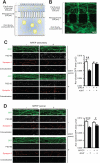
Figure 2. Dendritic mGluR5 activity is required for MEF2-induced synapse elimination.
(2A) Schematic of compartmentalized microfluidic device, which allows selective perfusion of cell somas or dendrites (Taylor et al., 2010). (2B) Representative image of hippocampal neuron culture grown in microfluidic device. Neurons were infected with lentivirus expressing GFP and MEF2-VP16-ERtm, then immunostained for GFP at 5-7 days post-transfection. Scale bar=10μm. (2C) Representative images of perfusion chamber dendrites from dissociated CA1 neurons virally transfected with lentivirus expressing GFP and MEF2-VP16-ERtm and stained for GFP, PSD-95 and Synapsin I following an overnight application of MPEP or vehicle to the perfusion channel (i.e., dendrites). The cell body compartment was simultaneously treated with vehicle or 4OHT to induce nuclear localization of MEF2-VP16-ERtm. n=7 microfluidic cultures. (2D) Same as Figure 2C, except that MPEP was applied to the cell body compartment. n=7 microfluidic cultures. All data are shown as mean±SEM. *p<0.05, **p<0.01; ***p<0.001.
Figure 3
Figure 3. Arc/Arg3.1 is necessary, but not sufficient for MEF2-induced functional and structural synapse elimination.
(3A) Synaptic function measurements from CA1 neurons virally transfected (T) with MEF2-VP16-ERtm and neighboring, untransfected (U) neurons in wildtype or Arc KO littermate slice cultures treated overnight with 4OHT. n=15 cell pairs/group. (3B) Representative images of live CA1 pyramidal neuron dendrites from slice cultures prepared from wildtype or Arc KO littermates. Cultures were biolistically transfected with pA1-GFP and MRE-mCherry with or without MEF2-VP16. n=15-24 cells/group. Scale bar=10μm. (3C) Synaptic function of CA1 neurons biolistically transfected for 24-48hr with pcDNA3-Arc and pA1-GFP compared to neighboring, untransfected neurons in wildtype slice cultures. n=26-30 cell pairs. (3D) Representative images of dendrites from living CA1 pyramidal neurons in slice cultures biolistically transfected for 24-48hr with pA1-GFP together with the pcDNA3 vector (control) or pcDNA3-Arc. n=32 cells/group. Scale bar=10μm. (3E) mEPSC measurements from Arc KO CA1 neurons biolistically transfected for 24-30hr with pcDNA3-Arc, MRE-mCherry, and pA1-GFP (transfection marker) with or without MEF2-VP16 and compared to neighboring, untransfected Arc KO neurons in slice cultures. n=19-23 cell pairs. (3F) Representative images of dendrites from live CA1 pyramidal neurons in Arc KO slice cultures that were biolistically transfected for 24-30hr with pcDNA3 vector (control), pcDNA3-Arc, or pcDNA3-Arc and MEF2-VP16, in addition to MRE-mCherry and pA1-GFP. n=22-29 cells/group. Scale bar=10μm. Electrophysiological group data are as in Figure 1; raw data are in Table S2. Representative mEPSC traces are shown to the right in Figures 3A & 3C; scale bar=10pA/500ms. All data are shown as mean±SEM. *p<0.05, **p<0.01, ****p<0.0001.
Figure 4
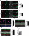
Figure 4. Dendritic mGluR5 activity is required for MEF2-driven increases in dendritic Arc/Arg3.1 protein, but not mRNA
(4A) Representative confocal images of dissociated wildtype hippocampal neurons virally transfected with MEF2-VP16-ERtm, treated with or without 4OHT and MPEP, then immunostained for Arc (red), GFP (green; transfection marker) and MAP2 (dendritic marker; gray). Magnified images of dendrites are shown to the right. Quantified group data of Arc/MAP2 fluorescence intensity in dendrites and cell soma are shown to the right. n=30 dendrites/somas per group. Scale bar=10μm. (4B) Representative images of dissociated wildtype hippocampal neurons prepared as in Figure 4A, but processed for MAP2 immunocytochemistry (blue) followed by fluorescence in situ hybridization for Arc mRNA (red). Dendrites from each channel and an overlay are shown below each image. Quantified group data of Arc FISH intensity normalized to dendritic length are shown to the right. Control: n=80 dendrites; 4OHT: n=90 dendrites; 4 cultures. Scale bar=10μm. (4C) Representative images of Arc and MAP2 immunocytochemistry from dendrites of wildtype neurons grown in microfluidic devices and virally transfected with MEF2-VP16-ERtm. MPEP was applied to the perfusion channel (i.e., dendrites) when 4OHT was applied to the cell body compartment. n=6 microfluidic cultures. (4D) Same as Figure 4C, except that MPEP was applied to the soma compartment. n=8 microfluidic cultures. (4E) RT-qPCR for Arc mRNA from the dendritic perfusion channel of wildtype cultures grown in microfluidic devices and virally transfected with MEF2-VP16-ERtm. The somal compartment was treated with vehicle (0.1% ethanol) or 4OHT; the dendritic compartment was treated with vehicle (ddH2O) or MPEP. n=11 cultures. For all cultures, the final drug concentration was 1μM for 4OHT and 10μM for MPEP; treatments lasted 6hr. All data are shown as mean±SEM. *p<0.05, **p<0.01, ***p<0.001.

Similar articles

Cited by

References

    1. BALLESTER-ROSADO CJ, ALBRIGHT MJ, WU CS, LIAO CC, ZHU J, XU J, LEE LJ, LU HC. mGluR5 in cortical excitatory neurons exerts both cell-autonomous and - nonautonomous influences on cortical somatosensory circuit formation. J Neurosci. 2010;30:16896–909. - PMC - PubMed
    1. BARBOSA AC, KIM MS, ERTUNC M, ADACHI M, NELSON ED, MCANALLY J, RICHARDSON JA, KAVALALI ET, MONTEGGIA LM, BASSEL-DUBY R, OLSON EN. MEF2C, a transcription factor that facilitates learning and memory by negative regulation of synapse numbers and function. Proc Natl Acad Sci U S A. 2008;105:9391–6. - PMC - PubMed
    1. BASSELL GJ, WARREN ST. Fragile X syndrome: loss of local mRNA regulation alters synaptic development and function. Neuron. 2008;60:201–14. - PMC - PubMed
    1. BASTRIKOVA N, GARDNER GA, REECE JM, JEROMIN A, DUDEK SM. Synapse elimination accompanies functional plasticity in hippocampal neurons. Proc Natl Acad Sci U S A. 2008;105:3123–7. - PMC - PubMed
    1. BEIQUE JC, NA Y, KUHL D, WORLEY PF, HUGANIR RL. Arc-dependent synapse-specific homeostatic plasticity. Proc Natl Acad Sci U S A. 2011;108:816–21. - PMC - PubMed

Publication types

MeSH terms

Substances

LinkOut - more resources