

Engineering vascularized bone grafts by integrating a biomimetic periosteum and β-TCP scaffold
- PMID: 24858072
- PMCID: PMC4075998
- DOI: 10.1021/am502056q
Engineering vascularized bone grafts by integrating a biomimetic periosteum and β-TCP scaffold
Abstract
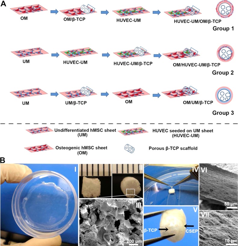
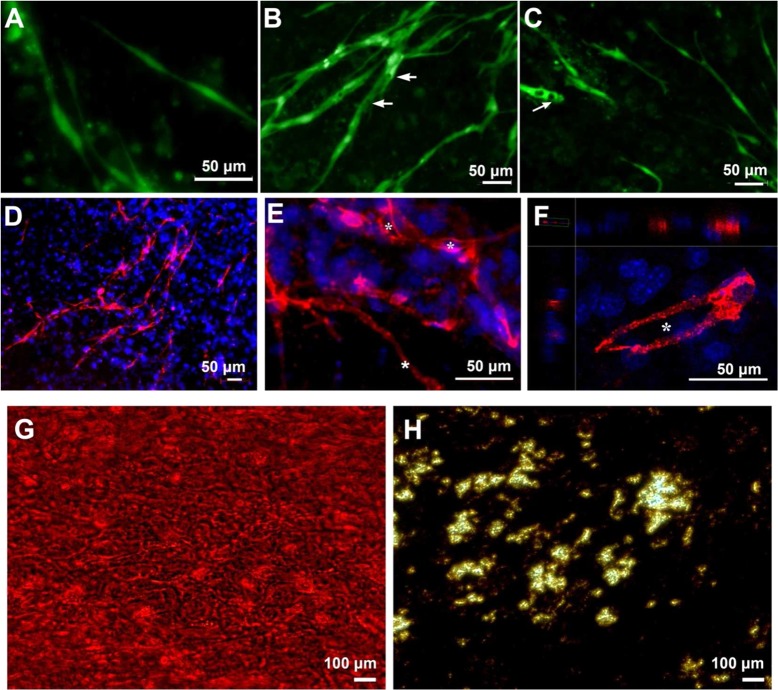
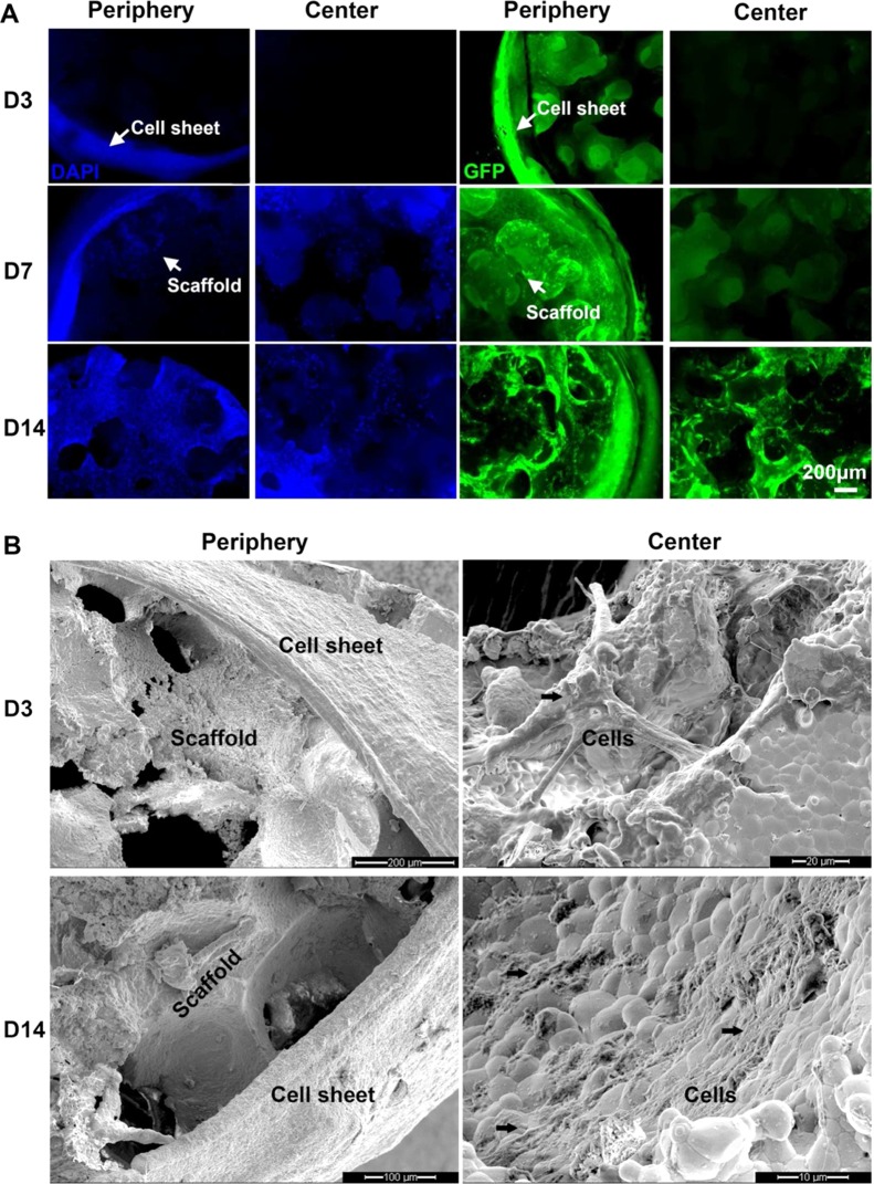
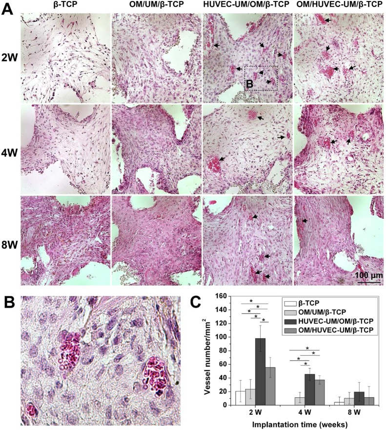
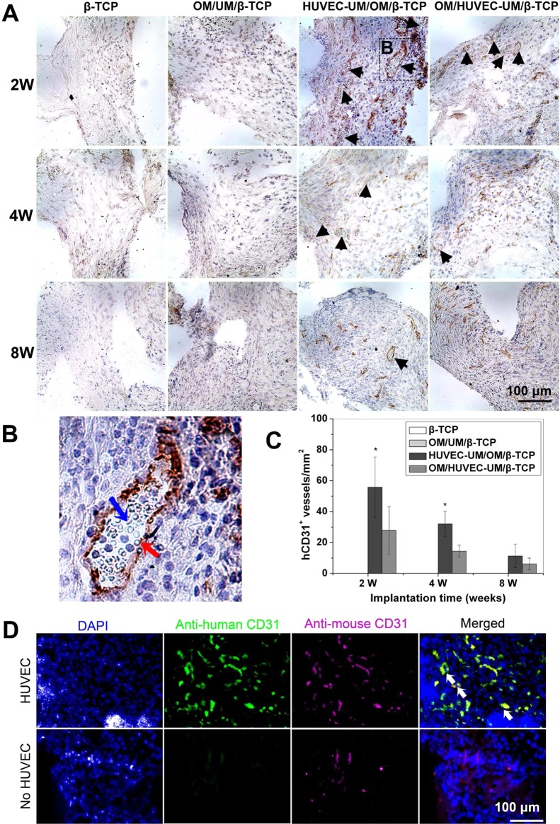
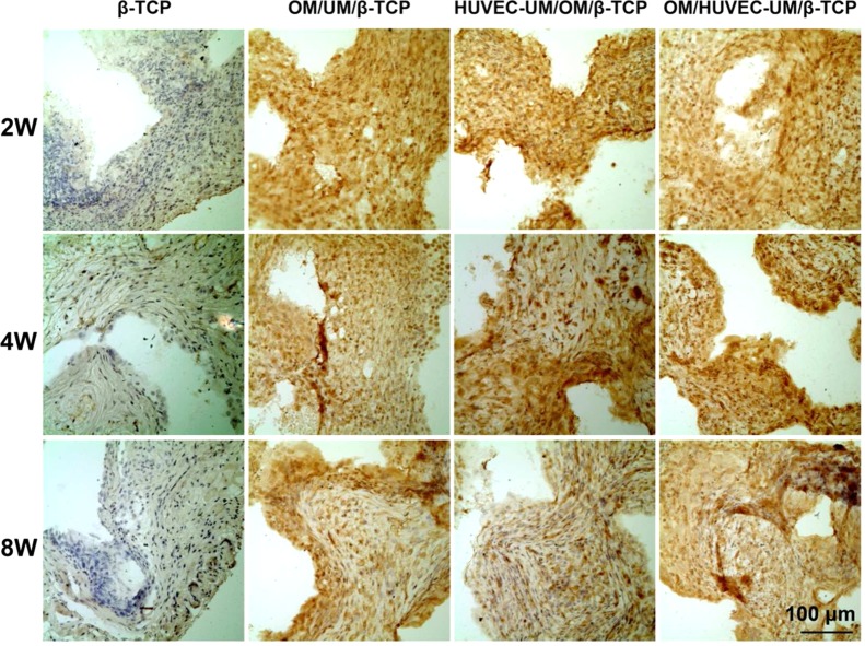
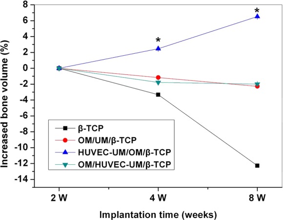
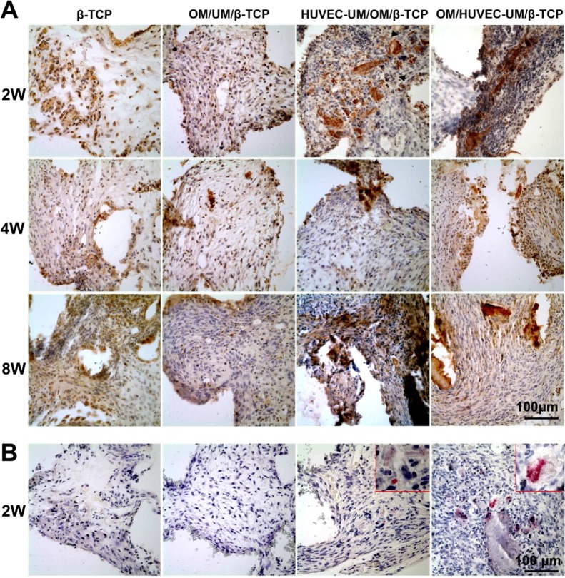
Treatment of large bone defects using synthetic scaffolds remain a challenge mainly due to insufficient vascularization. This study is to engineer a vascularized bone graft by integrating a vascularized biomimetic cell-sheet-engineered periosteum (CSEP) and a biodegradable macroporous beta-tricalcium phosphate (β-TCP) scaffold. We first cultured human mesenchymal stem cells (hMSCs) to form cell sheet and human umbilical vascular endothelial cells (HUVECs) were then seeded on the undifferentiated hMSCs sheet to form vascularized cell sheet for mimicking the fibrous layer of native periosteum. A mineralized hMSCs sheet was cultured to mimic the cambium layer of native periosteum. This mineralized hMSCs sheet was first wrapped onto a cylindrical β-TCP scaffold followed by wrapping the vascularized HUVEC/hMSC sheet, thus generating a biomimetic CSEP on the β-TCP scaffold. A nonperiosteum structural cell sheets-covered β-TCP and plain β-TCP were used as controls. In vitro studies indicate that the undifferentiated hMSCs sheet facilitated HUVECs to form rich capillary-like networks. In vivo studies indicate that the biomimetic CSEP enhanced angiogenesis and functional anastomosis between the in vitro preformed human capillary networks and the mouse host vasculature. MicroCT analysis and osteocalcin staining show that the biomimetic CSEP/β-TCP graft formed more bone matrix compared to the other groups. These results suggest that the CSEP that mimics the cellular components and spatial configuration of periosteum plays a critical role in vascularization and osteogenesis. Our studies suggest that a biomimetic periosteum-covered β-TCP graft is a promising approach for bone regeneration.
Figures
Similar articles
-
Engineering biomimetic periosteum with β-TCP scaffolds to promote bone formation in calvarial defects of rats.Stem Cell Res Ther. 2017 Jun 5;8(1):134. doi: 10.1186/s13287-017-0592-4. Stem Cell Res Ther. 2017. PMID: 28583167 Free PMC article.
-
Engineered periosteum-bone biomimetic bone graft enhances posterolateral spine fusion in a rabbit model.Spine J. 2019 Apr;19(4):762-771. doi: 10.1016/j.spinee.2018.09.013. Epub 2018 Sep 25. Spine J. 2019. PMID: 30266454
-
Fabrication, vascularization and osteogenic properties of a novel synthetic biomimetic induced membrane for the treatment of large bone defects.Bone. 2014 Jul;64:173-182. doi: 10.1016/j.bone.2014.04.011. Epub 2014 Apr 18. Bone. 2014. PMID: 24747351 Free PMC article.
-
Periosteum and development of the tissue-engineered periosteum for guided bone regeneration.J Orthop Translat. 2022 Feb 16;33:41-54. doi: 10.1016/j.jot.2022.01.002. eCollection 2022 Mar. J Orthop Translat. 2022. PMID: 35228996 Free PMC article. Review.
-
Current strategies in biomaterial-based periosteum scaffolds to promote bone regeneration: A review.J Biomater Appl. 2023 Feb;37(7):1259-1270. doi: 10.1177/08853282221135095. Epub 2022 Oct 17. J Biomater Appl. 2023. PMID: 36251764 Review.
Cited by
-
Biomimicking design of artificial periosteum for promoting bone healing.J Orthop Translat. 2022 Jul 11;36:18-32. doi: 10.1016/j.jot.2022.05.013. eCollection 2022 Sep. J Orthop Translat. 2022. PMID: 35891926 Free PMC article. Review.
-
Tissue Engineered Neurovascularization Strategies for Craniofacial Tissue Regeneration.ACS Appl Bio Mater. 2022 Jan 17;5(1):20-39. doi: 10.1021/acsabm.1c00979. Epub 2021 Nov 29. ACS Appl Bio Mater. 2022. PMID: 35014834 Free PMC article. Review.
-
[Vascular endothelial growth factor/polylactide-polyethyleneglycol-polylactic acid copolymer/basic fibroblast growth factor mixed microcapsules in promoting angiogenic differentiation of rat bone marrow mesenchymal stem cells in vitro].Zhongguo Xiu Fu Chong Jian Wai Ke Za Zhi. 2019 Feb 15;33(2):243-251. doi: 10.7507/1002-1892.201808099. Zhongguo Xiu Fu Chong Jian Wai Ke Za Zhi. 2019. PMID: 30739424 Free PMC article. Chinese.
-
Engineering biomimetic periosteum with β-TCP scaffolds to promote bone formation in calvarial defects of rats.Stem Cell Res Ther. 2017 Jun 5;8(1):134. doi: 10.1186/s13287-017-0592-4. Stem Cell Res Ther. 2017. PMID: 28583167 Free PMC article.
-
Periosteum Containing Implicit Stem Cells: A Progressive Source of Inspiration for Bone Tissue Regeneration.Int J Mol Sci. 2024 Feb 10;25(4):2162. doi: 10.3390/ijms25042162. Int J Mol Sci. 2024. PMID: 38396834 Free PMC article. Review.
References
-
- Schimming R.; Schmelzeisen R. Tissue-Engineered Bone for Maxillary Sinus Augmentation. J. Oral Maxillofac. Surg. 2004, 62, 724–729. - PubMed
-
- Lattermann C.; Romine S. E. Osteochondral Allografts: State of the Art. Clin. Sports Med. 2009, 28, 285–301. - PubMed
-
- Gitelis S.; Cole B. J. The Use of Allografts in Orthopaedic Surgery. Instr. Course Lect. 2002, 51, 507–520. - PubMed
-
- Li J.; Lin Z.; Zheng Q.; Guo X.; Lan S.; Liu S.; Yang S. Repair of Rabbit Radial Bone Defects Using True Bone Ceramics Combined with BMP-2-related Peptide and Type I Collagen. Mater. Sci. Eng., C 2010, 30, 1272–1279.
-
- Lozano D.; Trejo C. G.; Gomez-Barrena E.; Manzano M.; Doadrio J. C.; Salinas A. J.; Vallet-Regi M.; Garcia-Honduvilla N.; Esbrit P.; Bujan J. Osteostatin Loaded onto Mesoporous Ceramics Improves the Early Phase of Bone Regeneration in a Rabbit Osteopenia Model. Acta Biomater. 2012, 8, 2317–2323. - PubMed
Publication types
MeSH terms
Substances
Grants and funding
LinkOut - more resources
Full Text Sources
Other Literature Sources
