. 2014 May 23;9(5):e98208.
doi: 10.1371/journal.pone.0098208. eCollection 2014.

Integrated proteomics identified up-regulated focal adhesion-mediated proteins in human squamous cell carcinoma in an orthotopic murine model

Affiliations

Integrated proteomics identified up-regulated focal adhesion-mediated proteins in human squamous cell carcinoma in an orthotopic murine model

Daniela C Granato et al. PLoS One. .

Abstract

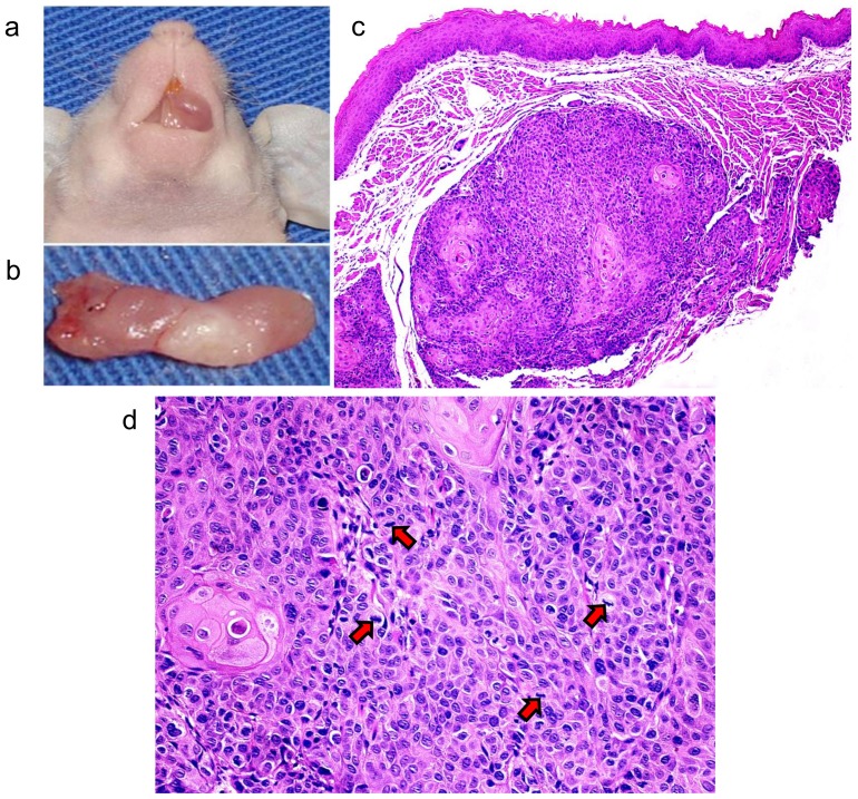
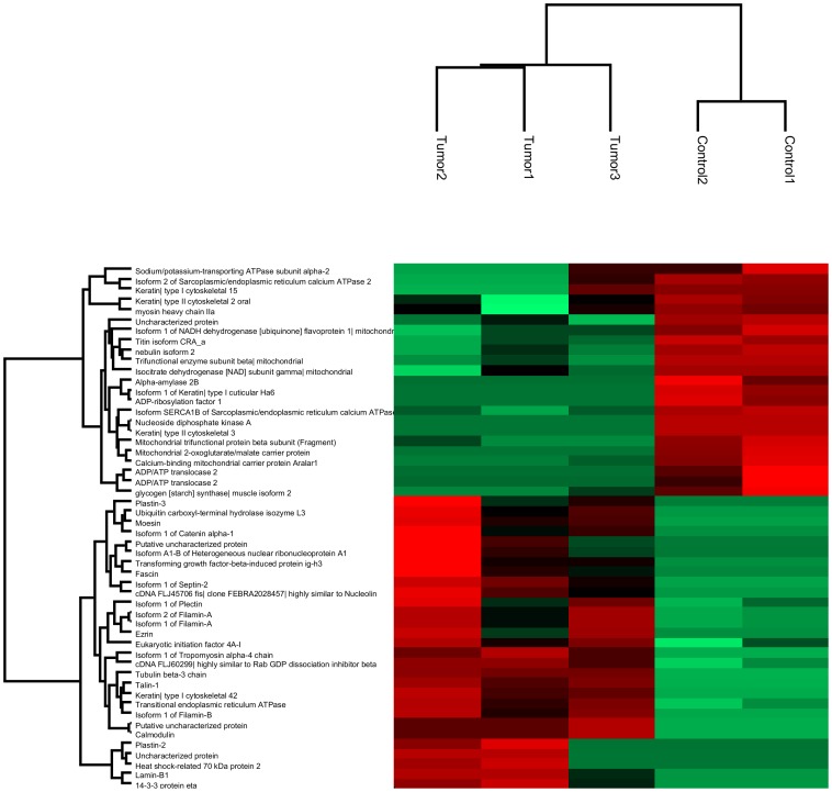
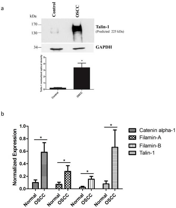
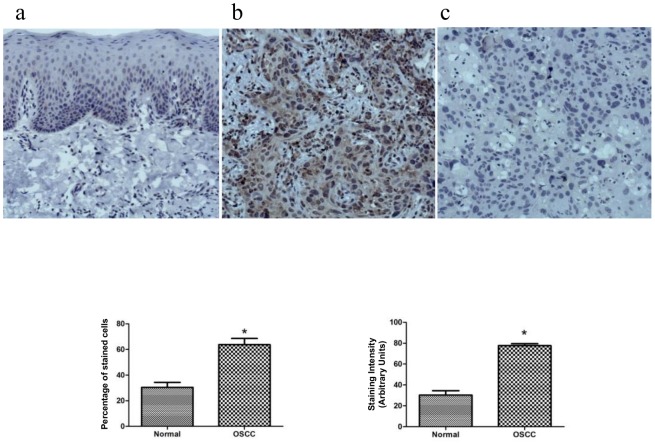
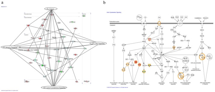
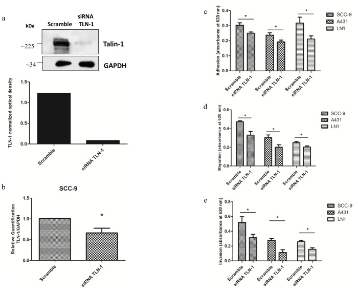
Understanding the molecular mechanisms of oral carcinogenesis will yield important advances in diagnostics, prognostics, effective treatment, and outcome of oral cancer. Hence, in this study we have investigated the proteomic and peptidomic profiles by combining an orthotopic murine model of oral squamous cell carcinoma (OSCC), mass spectrometry-based proteomics and biological network analysis. Our results indicated the up-regulation of proteins involved in actin cytoskeleton organization and cell-cell junction assembly events and their expression was validated in human OSCC tissues. In addition, the functional relevance of talin-1 in OSCC adhesion, migration and invasion was demonstrated. Taken together, this study identified specific processes deregulated in oral cancer and provided novel refined OSCC-targeting molecules.


Conflict of interest statement

Competing Interests: The authors have declared that no competing interests exist.

Figures

Figure 1
Figure 1. Development of OSCC in the tongue of an immunodeficient mouse.
(a) Shows the tumor developed in the oral cavity of BALBc/nude mouse by SCC-9 cells injection. (b) Shows the tumor dissected from the oral cavity of the BALBc/nude mouse. (c) Microscopical features of the OSCC included pleomorphic epithelial cells invading the muscle fibers of the tongue (50X, H&E). (d) Mitotic figures (arrows) were also observed (200X, H&E).
Figure 2
Figure 2. Bioinformatics analysis of differential expressed proteins in OSCC and control tissues identified by MS.
Clustering of significantly up- and down-regulated proteins (Fisher's exact test, p<0.05) in OSCC tissues compared to control tissues is shown as a Heat map, applying the Euclidian distance method and average linkage.
Figure 3
Figure 3. Talin-1 showed higher protein expression in OSCC tissues compared to control tissues in orthotopic murine model by immunoblotting (a).
The proteins (30 µg) were submitted to 1-D electrophoresis on 4–15% SDS-polyacrylamide gels, they were transferred onto nitrocellulose membrane and incubated with anti-talin-1 antibody. Anti-GAPDH antibody was used as loading control. The graph represents the normalized optical density of the average data of three immunoblotted samples (n = 3, * indicates p<0.05, Student's t-test). Talin-1, filamins A and B and catenin alpha-1 showed higher mRNA expression levels in human OSCC tumor tissues compared to control tissues by qRT-PCR (n = 12) (b). Relative mRNA expression levels were measured by the real-time quantitative PCR. The data were normalized with glyceraldehyde-3- phosphate dehydrogenase gene. Columns represent mean and SD (n = 12; Student's t-test, * indicates p<0.05).
Figure 4
Figure 4. Expression of talin-1 in the human normal oral mucosa and OSCC tissues by immunohistochemistry (n = 10).
Talin-1 demonstrated a weak and restrict cytoplasmic immunoreactivity in the basal and suprabasal layers of the normal oral tissue (a), whereas broad positivity with variable intensity was found in the neoplastic cells (b). As expected, some immune and inflammatory cells expressed talin-1. Panel c represents the negative control. The graphics represent the % of positive expression cells and expression intensity between the normal and OSCC tumor tissues (n = 10, Student's t-test, * indicates p<0.05).
Figure 5
Figure 5. Pathway analysis of differentially expressed proteins.
(a) The highest score network generated by IPA comprised 23 differentially expressed proteins (up-regulated proteins are displayed in red, whereas the down-regulated proteins are in green) plus additional interacting molecules that were not identified in this study (white). The network revealed 15 proteins in the context of cancer. Actin cytoskeleton signaling, integrin signaling and FAK signaling represent canonical pathways with 13, 10 and 6 identified proteins of the network, respectively. The proteins were grouped according to the canonical pathway or function. Fx: function and Cp: canonical pathway. (b) Representation of the top canonical pathway of actin cytoskeleton signaling.
Figure 6
Figure 6. Talin-1 knockdown decreased cell adhesion, migration and invasion of SCC-9, A431 and SCC-9 LN1 cells.
(a) Talin-1 showed lower protein expression in SCC-9/siRNA TLN-1 cells compared to SCC-9/control (scrambled) by immunoblotting. The total proteins (30 µg) were submitted to 1-D electrophoresis on 12% SDS-polyacrylamide gels, they were transferred onto nitrocellulose membrane and incubated with anti-talin-1 antibody. Anti-GAPDH antibody was used as loading control. The graph represents the normalized optical density. (b) Talin-1 mRNA expression levels in SCC-9/siRNA TLN-1 cells compared to SCC-9/control (scrambled) by qRT-PCR (n = 3, Student's t-test, p<0.05). The data were normalized with GAPDH gene. (c) SCC-9/control (scrambled) and SCC-9/siRNA TLN-1 cells, A431/control (scrambled) and A431/siRNA TLN-1 cells, SCC-9 LN1/control (scrambled) and SCC-9 LN1/siRNA TLN-1 cells were seeded in Matrigel coated 96-well plates. After 1 h, cells were stained and the cell adhesion was measured (n = 3, * indicates p<0.05, Student's t-test for each comparison). (d) SCC-9/control (scrambled) and SCC-9/siRNA TLN-1 cells, A431/control (scrambled) and A431/siRNA TLN-1 cells, SCC-9 LN1/control (scrambled) and SCC-9 LN1/siRNA TLN-1 cells were seeded in serum-free media in the upper chamber of transwell plates and were allowed to migrate towards the lower chamber containing 1% FBS supplemented media (n = 3, * indicates p<0.05, Student's t-test). (e) SCC-9/control (scrambled) and SCC-9/siRNA TLN-1 cells, A431/control (scrambled) and A431/siRNA TLN-1 cells, SCC-9 LN1/control (scrambled) and SCC-9 LN1/siRNA TLN-1 cells were seeded in serum-free media in the upper chamber of matrigel-coated transwell plates and were allowed to invade towards the lower chamber containing 10% FBS supplemented media (n = 2, * indicates p<0.05, Student's t-test).

References

    1. Siegel R, Naishadham D, Jemal A (2013) Cancer statistics, 2013. CA Cancer J Clin 63: 11–30. - PubMed
    1. Wong DT, Todd R, Tsuji T, Donoff RB (1996) Molecular biology of human oral cancer. Crit Rev Oral Biol Med 7: 319–328. - PubMed
    1. Warnakulasuriya S (2009) Global epidemiology of oral and oropharyngeal cancer. Oral Oncol 45: 309–316. - PubMed
    1. Choi S, Myers JN (2008) Molecular pathogenesis of oral squamous cell carcinoma: implications for therapy. J Dent Res 87: 14–32. - PubMed
    1. Panis C, Pizzatti L, Herrera AC, Cecchini R, Abdelhay E (2013) Putative circulating markers of the early and advanced stages of breast cancer identified by high-resolution label-free proteomics. Cancer Lett 330: 57–66. - PubMed

Publication types

MeSH terms

LinkOut - more resources