Review
. 2014 Jul;128(1):39-54.
doi: 10.1007/s00401-014-1288-9. Epub 2014 May 25.

A neuropathology-based approach to epilepsy surgery in brain tumors and proposal for a new terminology use for long-term epilepsy-associated brain tumors

Affiliations
Review

A neuropathology-based approach to epilepsy surgery in brain tumors and proposal for a new terminology use for long-term epilepsy-associated brain tumors

Ingmar Blumcke et al. Acta Neuropathol. 2014 Jul.

Abstract

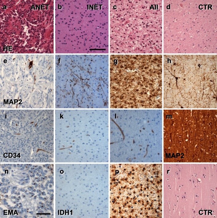
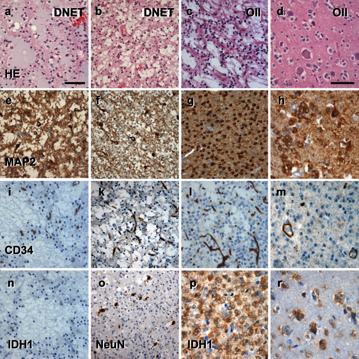
Every fourth patient submitted to epilepsy surgery suffers from a brain tumor. Microscopically, these neoplasms present with a wide-ranging spectrum of glial or glio-neuronal tumor subtypes. Gangliogliomas (GG) and dysembryoplastic neuroepithelial tumors (DNTs) are the most frequently recognized entities accounting for 65 % of 1,551 tumors collected at the European Epilepsy Brain Bank (n = 5,842 epilepsy surgery samples). These tumors often present with early seizure onset at a mean age of 16.5 years, with 77 % of neoplasms affecting the temporal lobe. Relapse and malignant progression are rare events in this particular group of brain tumors. Surgical resection should be regarded, therefore, also as important treatment strategy to prevent epilepsy progression as well as seizure- and medication-related comorbidities. The characteristic clinical presentation and broad histopathological spectrum of these highly epileptogenic brain tumors will herein be classified as "long-term epilepsy associated tumors-LEATs". LEATs differ from most other brain tumors by early onset of spontaneous seizures, and conceptually are regarded as developmental tumors to explain their pleomorphic microscopic appearance and frequent association with Focal Cortical Dysplasia Type IIIb. However, the broad neuropathologic spectrum and lack of reliable histopathological signatures make these tumors difficult to classify using the WHO system of brain tumors. As another consequence from poor agreement in published LEAT series, molecular diagnostic data remain ambiguous. Availability of surgical tissue specimens from patients which have been well characterized during their presurgical evaluation should open the possibility to systematically address the origin and epileptogenicity of LEATs, and will be further discussed herein. As a conclusion, the authors propose a novel A-B-C terminology of epileptogenic brain tumors ("epileptomas") which hopefully promote the discussion between neuropathologists, neurooncologists and epileptologists. It must be our future mission to achieve international consensus for the clinico-pathological classification of LEATs that would also involve World Health Organization (WHO) and the International League against Epilepsy (ILAE).


Figures

Fig. 1
Fig. 1
MRI characteristics of LEAT (CD34-positive BNET). ag 6-year-old boy with drug-resistant temporal lobe epilepsy. Coronal T2-weighted (a, b), sagittal T2-weighted (c), coronal FLAIR (d), and coronal T1-weighted contrast-enhanced images (e) show a cortical/subcortical tumor with a cortical cyst (a, c: arrow), a contrast-enhancing nodule (arrow in e), and a T2-/FLAIR hyperintense white matter portion (a, f, g: arrowhead). Histopathological diagnosis was CD34-positive ganglioglioma (BNET)
Fig. 2
Fig. 2
MRI characteristics of LEAT (DNET, ANET and INET). a–d 36-year-old man with temporal lobe seizures and histopathological diagnosis of DNET in the left amygdala and hippocampal head. The tumor revealed multiple tiny cysts, which can be resolved only by high-resolution T2-weighted (a, c: arrow), but not on FLAIR images (b, d). Axial T1-weighted (e), sagittal FLAIR (f), and axial T2-weighted images (g) showed a circumscript cortical and subcortical tumor at the base of the left parietal lobe dorsal to the cingulate gyrus. A ribbon-like cortical T1-hyperintensity (arrow in e) can be identified, and histopathology confirmed an ANET. h–k Isomorphic variant of astrocytoma (INET). The hippocampal and parahippocampal lesion has a space-occupying effect and a homogenous signal increase on T2 (h: arrow) and signal decrease on T1-weighted images (i: arrow), respectively. Signal changes suggested low cellularity with no contrast enhancement (k)
Fig. 3
Fig. 3
LEAT submission to the European Epilepsy Brain Bank (EEBB) in last 20 years. a During the last 20 years, submission frequencies have decreased for HS and increased for MCD (75 % FCD), whereas percentages of brain tumors associated with early epilepsy onset (LEAT) remained consistent over time. b Disease duration (time period from seizure onset until surgery; sz—seizures) has remained unchanged in HS and MCD during the last 20 years, whereas today’s patients with LEAT were operated 5 years earlier compared to mid 1990s (mean of 7.4 vs 12.9 years, respectively). EEBB submissions were grouped into four clusters of 5-year intervals between 1994 and 2013 to observe changes over time
Fig. 4
Fig. 4
The spectrum of histomorphological and immunohistochemical hallmarks in LEATs with a glio-neuronal phenotype (A–B–C terminology). a BNET, immunoreactive for CD34 class II epitope (mAB QBend10, hematoxylin counterstaining; first three columns). Three different patterns can be distinguished. Pattern 1: (TU)—the bulk tumor is densely stained for CD34 (magnification in f). Pattern 2: clusters of tumor cells are visible in adjacent cortical areas (arrow, magnification in g). Pattern 3: diffusely infiltrating CD34-positive cells can be found in distant remote areas from the bulk tumor (magnification in h). Note, that patterns 2 and 3 may be interpreted as FCD ILAE Type IIIb when not using CD34 immunohistochemistry. b, c Routine histology stainings (H&E) reveal a biphasic pattern in BNET and a predominant gangliocytic patter in GNET. d, e Immunohistochemistry for the embryonic MAP2 epitope (MAP2e) is helpful to identify the neuronal component. Compared to diffusely infiltrating gliomas, the glial component does not label for Map2e (see Figs. 5, 6). i no CD34 immunoreactivity is visible in GNETs. k–n Supplementary markers are helpful to distinguish LEAT entities. NeuN labels dysplastic neurons in BNETs (k). IDH1 staining is always recommended as it is detectable only in neoplastically transformed glial cells (diffuse astrocytomas AII and oligodendroglioma OII; see Figs. 5, 6), but not in BNET (l). Synaptophysin (SYN) may be helpful to visualize the neuronal component in BNET (m) and GNET (n). Scale bar in a 2 mm, d 50 µm applies also to b, c, e, in. Scale bar in g 200 µm, applies also to fh
Fig. 5
Fig. 5
The spectrum of histomorphological and immunohistochemical hallmarks in LEATs with an astrocytic phenotype (A–B–C terminology). a–c Routine histology stainings reveal an angiocentric pattern in ANET (first column), or a prevailing astrocytic differentiation in INET (second column; similar to diffuse astrocytoma (AII; third column) shown in c). d HE staining from normal white matter (control/CTR) obtained from epilepsy surgery specimens; forth column. eh Immunohistochemistry for the embryonic MAP2 epitope (MAP2e) is helpful to separate diffuse gliomas (AII, g) variants from any other LEAT entity (see also Figs. 4, 6). Only preexisting neurons and neuronal processes are visible in INET (f), comparable with heterotopic white matter neurons frequently seen in epilepsy surgery samples (h). Map2e-labeled cells in ANET (e) support the concept of a neuroepithelial tumor. i–l None of these variants shows CD34 immunoreactivity compared to BNET (see Fig. 4). m MAP2e staining in normal neocortex (layer 3; same specimen than d, h, and r). n–r Supplementary markers are helpful to distinguish LEAT entities. EMA dots can be frequently encountered in ANET variants. IDH1 staining is recommended to exclude neoplastically transformed glial cells in diffuse astrocytomas (p; see also Fig. 6), but not in INET (o). Scale bar in b 50 µm, applies to all images, with the exception of (n) 20 µm
Fig. 6
Fig. 6
The spectrum of histomorphological and immunohistochemical hallmarks in LEATs with a clear cell morphology (A–B–C terminology). a–c Routine histology stainings reveal the specific glio-neuronal element in DNET (a, b; first and second columns), which may be difficult to distinguish from some oligodendroglioma variants (OII; third and forth columns). d OII infiltrating diffusely into neocortex (also in h, m, r). e–h Immunohistochemistry for the embryonic MAP2 epitope (MAP2e) is variable in DNET variants with abundant labeling of glial and neuronal cells (e) or neurons only (f). Neoplastically transformed glial cells always label with MAP2e in OII (g, h). i–m CD34 immunoreactivity is always devoid in DNETs as well as clear cell glioma variants. Immunoreactivity of vascular endothelium can be used as positive control. n–r Supplementary markers are helpful to distinguish LEAT entities. IDH1 staining is always recommended as it is detectable only in neoplastically transformed glial cells (oligodendroglioma OII, pr), but not in DNET (n). NeuN may be helpful to visualize the neuronal component in DNETs (o). Scale bar in a 50 µm, applies to images b, c, e, f, g, i, k, l, n, o, p. Scale bar in d 20 µm, applies also to h, m and r

Similar articles

Cited by

References

    1. Alayev A, Holz MK. mTOR signaling for biological control and cancer. J Cell Physiol. 2013 - PMC - PubMed
    1. Arsene D, Ardeleanu C, Ogrezeanu I, Danaila L. Angiocentric glioma: presentation of two cases with dissimilar histology. Clin Neuropathol. 2008;27(6):391–395. doi: 10.5414/NPP27391. - DOI - PubMed
    1. Balss J, Meyer J, Mueller W, Korshunov A, Hartmann C, von Deimling A. Analysis of the IDH1 codon 132 mutation in brain tumors. Acta Neuropathol. 2008;116(6):597–602. doi: 10.1007/s00401-008-0455-2. - DOI - PubMed
    1. Becker AJ, Blumcke I, Urbach H, Hans V, Majores M. Molecular neuropathology of epilepsy-associated glioneuronal malformations. J Neuropathol Exp Neurol. 2006;65(2):99–108. doi: 10.1097/01.jnen.0000199570.19344.33. - DOI - PubMed
    1. Becker AJ, Lobach M, Klein H, Normann S, Nothen MM, von Deimling A, Mizuguchi M, Elger CE, Schramm J, Wiestler OD, Blumcke I. Mutational analysis of TSC1 and TSC2 genes in gangliogliomas. Neuropathol Appl Neurobiol. 2001;27(2):105–114. doi: 10.1046/j.0305-1846.2001.00302.x. - DOI - PubMed

Publication types