. 2014 Dec;71(24):4853-67.
doi: 10.1007/s00018-014-1647-7. Epub 2014 May 25.

TRPM6 kinase activity regulates TRPM7 trafficking and inhibits cellular growth under hypomagnesic conditions

Affiliations

TRPM6 kinase activity regulates TRPM7 trafficking and inhibits cellular growth under hypomagnesic conditions

Katherine Brandao et al. Cell Mol Life Sci. 2014 Dec.

Abstract

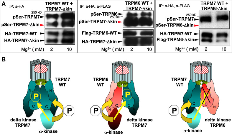
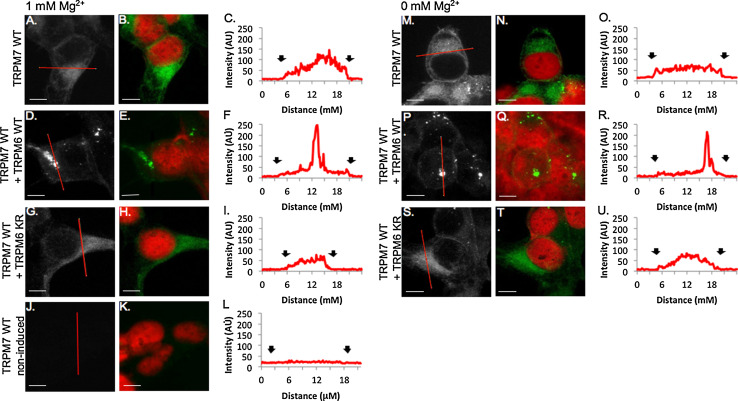
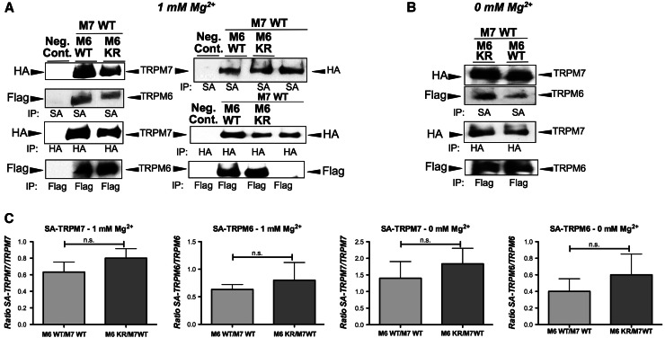
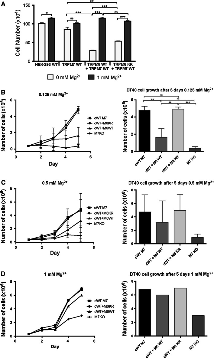
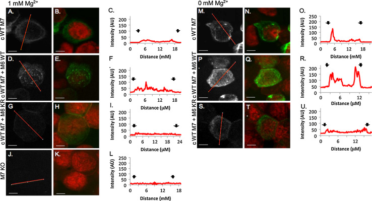
The channel kinases TRPM6 and TRPM7 are both members of the melastatin-related transient receptor potential (TRPM) subfamily of ion channels and the only known fusions of an ion channel pore with a kinase domain. TRPM6 and TRPM7 form functional, tetrameric channel complexes at the plasma membrane by heteromerization. TRPM6 was previously shown to cross-phosphorylate TRPM7 on threonine residues, but not vice versa. Genetic studies demonstrated that TRPM6 and TRPM7 fulfill non-redundant functions and that each channel contributes uniquely to the regulation of Mg(2+) homeostasis. Although there are indications that TRPM6 and TRPM7 can influence each other's cellular distribution and activity, little is known about the functional relationship between these two channel-kinases. In the present study, we examined how TRPM6 kinase activity influences TRPM7 serine phosphorylation, intracellular trafficking, and cell surface expression of TRPM7, as well as Mg(2+)-dependent cellular growth. We found TRPM7 serine phosphorylation via the TRPM6 kinase, but no TRPM6 serine phosphorylation via the TRPM7 kinase. Intracellular trafficking of TRPM7 was altered in HEK-293 epithelial kidney cells and DT40 B cells in the presence of TRPM6 with intact kinase activity, independently of the availability of extracellular Mg(2+), but TRPM6/7 surface labeling experiments indicate comparable levels of the TRPM6/7 channels at the plasma membrane. Furthermore, using a complementation approach in TRPM7-deficient DT40 B-cells, we demonstrated that wild-type TRPM6 inhibited cell growth under hypomagnesic cell culture conditions in cells co-expressing TRPM6 and TRPM7; however, co-expression of a TRPM6 kinase dead mutant had no effect-a similar phenotype was also observed in TRPM6/7 co-expressing HEK-293 cells. Our results provide first clues about how heteromer formation between TRPM6 and TRPM7 influences the biological activity of these ion channels. We show that TRPM6 regulates TRPM7 intracellular trafficking and TRPM7-dependent cell growth. All these effects are dependent upon the presence of an active TRPM6 kinase domain. Dysregulated Mg(2+)-homeostasis causes or exacerbates many pathologies. As TRPM6 and TRPM7 are expressed simultaneously in numerous cell types, understanding how their relationship impacts regulation of Mg(2+)-uptake is thus important knowledge.


Figures

Fig. 1
Fig. 1
The channel-kinase TRPM6 phosphorylates serine residues of associated TRPM7 but not vice versa. Human HA-tagged TRPM7 WT, human HA-tagged TRPM7 delta kinase (TRPM7-Δkin), human flag-tagged TRPM6 WT, and human flag-tagged TRPM6 delta kinase (TRPM6-Δkin) have been cloned, stably transfected and inducibly co-expressed in HEK-293 cells as indicated, see methods section and reference [11] for further details. In vitro phosphorylation reactions have been performed after HA and/or Flag immunoprecipiations under two different Mg2+ concentrations (2 and 10 mM) with 100 μM MgATP [11]. Phosphorylation of these proteins was subsequently analyzed after gel electrophoresis and immunoblotting with a general anti phospho serine antibody. Protein expression levels were verified after stripping of the membrane via reprobing with anti FLAG or anti HA antibodies. a Left panel TRPM7 kinase crossphosphorylates TRPM7 delta kinase channels in vitro upon TRPM7 WT and TRPM7 delta kinase co-expression. Middle panel TRPM6 kinase crossphosphorylates TRPM7 delta kinase channels in vitro upon TRPM6 WT and TRPM7 delta kinase co-expression. Right panel TRPM7 kinase does not crossphosphorylate TRPM6 delta kinase channels in vitro upon TRPM7 WT and TRPM6 delta kinase co-expression. These data are representative of three separate experiments. b Based on our results, model showing how TRPM6 and TRPM7 form heteromers and crossphosphorylate each other. TRPM6 and TRPM7 have to tetramerize in order to build a functional ion channel pore
Fig. 2
Fig. 2
TRPM7 cellular trafficking is regulated by TRPM6 kinase activity in HEK-293 cells. 100× confocal images of HA-tagged TRPM7 channels visualized with HA-FITC antibody (gray scale, green) and nuclei visualized with propidium iodide (red). Scale bars 5 μM. ai HEK cells overexpressing TRPM7 and TRPM6 channels grown with physiological Mg2+ levels (1 mM). Little immunoreactivity is detected in non-induced HEK mutant cells (jl). mu HEK-293 cells overexpressing TRPM7 and TRPM6 channels grown with 0 mM Mg2+ (c, f, i, l, o, r, u). Representative histograms of pixel intensity across cytoplasmic sections of cells (red line in a, d, g, j, m, p, s). Black arrows indicate membrane borders of the cell
Fig. 3
Fig. 3
TRPM6 kinase activity does not affect TRPM7 expression levels at the plasma membrane under normal or hypogmagnesic growth conditions in HEK-293 cells. Analysis of biotin cell surface labeled cells (co-)expressing TRPM6/7 WT or TRPM6 kinase dead mutant (M6 KR) channels. Cells were cultured under 1 or 0 mM Mg2+. The channels were subsequently immunoprecipitated with streptavidin magnetic beads (SA) and analyzed by immunoblotting with HA- or Flag-horseradish peroxidase. TRPM6 (flag tagged) and TRPM7 (HA tagged) protein expression levels were determined by HA- and Flag-immunoblotting accordingly. a At 1 mM Mg2+, left panel shows TRPM6 and TRPM7 at the plasma membrane, right panel shows additionally TRPM7 alone. b At 0 mM Mg2+, shows TRPM6/7 surface expression, w or w/o TRPM6’s active kinase. c Densitometric analysis of three independent experiments using ImageJ of TRPM6/7 surface expression levels as shown in a/b (ratio of SA-TRPM7 or SA-TRPM6 divided by corresponding total protein amount). (n.s. not significant, student t test)
Fig. 4
Fig. 4
TRPM6 kinase activity inhibits TRPM7 mediated cell growth in HEK-293 and DT40 cells growth under hypomagnesic conditions. a Growth curves of HEK-293 expressing human TRPM7 WT (M7 WT) alone, or co-expressing hTRPM7 WT with either human TRPM6 WT (M6/7 WT), or the TRPM6 K1804R kinase dead mutant (M6KR + M7 WT). Cells were cultured in normal HEK-293 media, prior to Mg2+ deprivation protein overexpression was induced for 6 h with doxycycline (1 μg/ml). Cells were spun down and equal number of cells transferred for 24 h into fresh, complete, serum-free growth media with 0 or 1 mM Mg2+, as indicated. Note: TRPM7 overexpressing HEK-293 cells die after 36–48 h doxycycline induced TRPM7 overexpression. bd growth curves of TRPM7 deficient DT40 cells complemented with human TRPM7 WT (cWT M7) or cKR M7 (kinase dead) alone, or with hTRPM7 WT co-expressed with either human TRPM6 WT (cWT M7 + M6 WT), or the TRPM6 K1804R kinase dead mutant (cWT M7 + M6 KR). Cells were cultured in complete, chemically defined, Mg2+-free media without serum requirement. Cells were grown under various Mg2+ concentrations for several days as indicated. Prior to Mg2+ deprivation protein overexpression has been induced for 24–48 h with doxycycline (final concentration: 1 μg/ml) in complete growth media with additional 10 mM MgCl2 (Note: TRPM7−/− DT40 cells would die without Mg2+ supplementation of the growth media). Cells were spun down and equal number of cells (2 × 105) transferred into fresh, complete growth media with different Mg2+ concentrations, as indicated. For statistical analysis, a two-tailed t test was performed. Data shown are the mean ± SEM; *p < 0.05, **p < 0.01, ***p < 0.001. Note: Growth data at physiological (1 mM Mg2+) and hyerpmagnesic conditions have been published in 2003/2005 and did not show any differences
Fig. 5
Fig. 5
TRPM7 membrane trafficking pattern is altered by Mg2+ concentration and the TRPM6 kinase in DT40 B cells. 100× confocal images of HA-tagged TRPM7 channels visualized with HA-FITC antibody (gray scale, green) and nuclei visualized with propidium iodide (red). Scale bars 5 μM. TRPM7−/− DT40 B cells complemented with human TRPM7 WT and co-expressing TRPM6 WT or TRPM6 KR grown in 1 mM Mg2+ (al) or 0 mM Mg2+ (mu). Histograms correspond to one-pixel wide lines (red line in gray scale images) drawn across the center of the cell. ac cWT M7 cells in 1 mM Mg2+ TRPM7 immunoreactivity is diffuse throughout the cytoplasm with a small peak at the edge of the cell (black arrows, c). df. TRPM7 immunoreactivity in 1 mM Mg2+ is also low and diffuse in cWT M7 + M6WT cells, except for the presence of TRPM7 clusters near the nuclear membrane. cWT M7 + M6 KR cells (jl) express low levels of TRPM7 at 1 mM Mg2+. In cells grown in 0 mM Mg2+, TRPM7 immunoreactivity increases near the plasma and nuclear membranes in cWT M7 cells (mu). cWT M7 + M6 KR cells have increased expression levels and clustered distribution of TRPM7 (pr). There is a small increase in TRPM7 fluorescence near the periphery of cWT M7 + M6 KR cells (su). TRPM7 immunoreactivity is controlled for in TRPM7−/− DT40 B cells that have no detectable fluorescence (jl)

Similar articles

Cited by

References

    1. Voets T, et al. TRPM6 forms the Mg2+influx channel involved in intestinal and renal Mg2+ absorption. J Biol Chem. 2004;279(1):19–25. doi: 10.1074/jbc.M311201200. - DOI - PubMed
    1. Jiang J, Li M, Yue L. Potentiation of TRPM7 inward currents by protons. J Gen Physiol. 2005;126(2):137–150. doi: 10.1085/jgp.200409185. - DOI - PMC - PubMed
    1. Li M, Jiang J, Yue L. Functional characterization of homo- and heteromeric channel kinases TRPM6 and TRPM7. J Gen Physiol. 2006;127(5):525–537. doi: 10.1085/jgp.200609502. - DOI - PMC - PubMed
    1. Schlingmann KP, et al. Hypomagnesemia with secondary hypocalcemia is caused by mutations in TRPM6, a new member of the TRPM gene family. Nat Genet. 2002;31(2):166–170. doi: 10.1038/ng889. - DOI - PubMed
    1. Walder RY, et al. Mutation of TRPM6 causes familial hypomagnesemia with secondary hypocalcemia. Nat Genet. 2002;31(2):171–174. doi: 10.1038/ng901. - DOI - PubMed

Publication types

MeSH terms

LinkOut - more resources