

Nasopharyngeal tonsils (adenoids) contain extrathymic corticothymocytes
- PMID: 24858437
- PMCID: PMC4032319
- DOI: 10.1371/journal.pone.0098222
Nasopharyngeal tonsils (adenoids) contain extrathymic corticothymocytes
Abstract
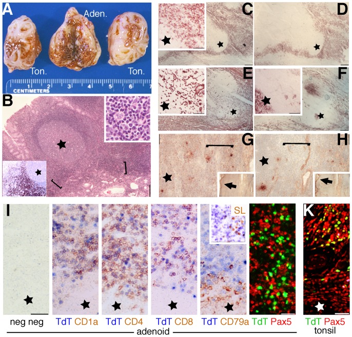
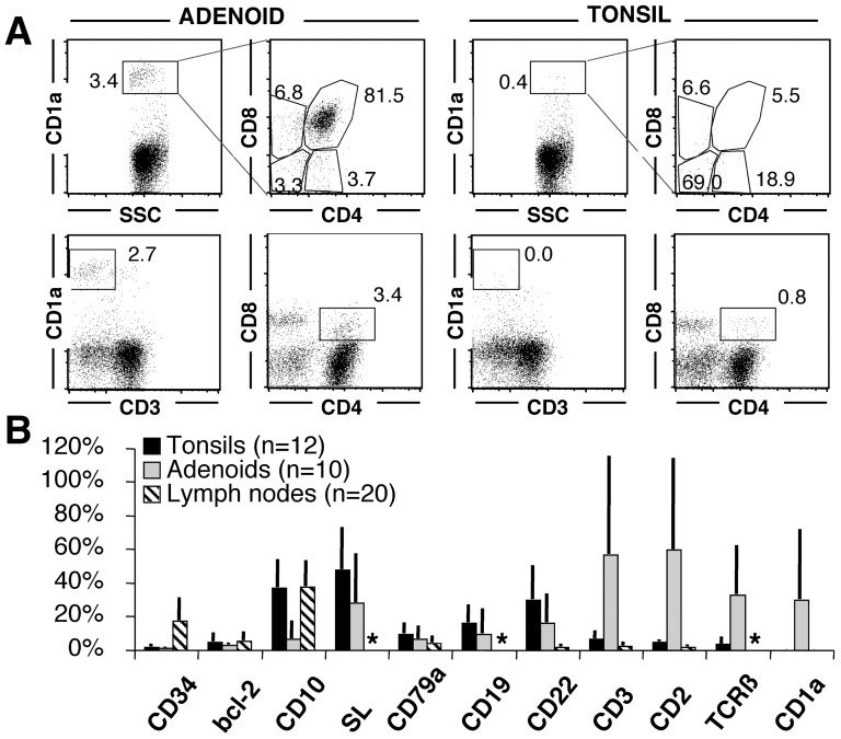
Adenoidal tissue (also known as nasopharyngeal tonsils) of 58% of humans in the pediatric age group contains immature T-lymphoid cells with the phenotype of thymocytes (TdT+, CD1abc+, cytoplasmic CD3+, coexpressing CD4 and CD8, lacking an Intraepithelial Lymphocyte-associated phenotype). The notable difference in comparison to palatine tonsils is the clustering in groups and sheets, comprising hundreds or thousands of cells (1.7%±0.2 of total T cells). The thymic epithelium is morphologically and phenotypically absent. Adenoids share with tonsils and lymph nodes the presence of immature B cell precursors (TdT+, CD1a-, Pax5+, Surrogate light chain±), however in these latter the presence of TdT+, CD1a+, Pax5- precursors is absent or limited to individual cells. Human adenoids are distinct among the Waldeyer's ring lymphoid tissue because of the known embryogenic derivation from the third pharyngeal pouch, from which the thymus develops; in addition, they may display phenotypic incomplete features of a vestigial thymus.
Conflict of interest statement
Figures
Similar articles
-
Human nasopharyngeal-associated lymphoreticular tissues. Functional analysis of subepithelial and intraepithelial B and T cells from adenoids and tonsils.Am J Pathol. 2000 Dec;157(6):2023-35. doi: 10.1016/S0002-9440(10)64841-9. Am J Pathol. 2000. PMID: 11106575 Free PMC article.
-
Lymphocyte subsets in human adenoids and tonsils. Rosette formation, alpha-naphthyl acetate esterase cytochemistry, monoclonal antibodies and peanut lectin reactivity.Immunol Lett. 1984;8(1):1-9. doi: 10.1016/0165-2478(84)90096-8. Immunol Lett. 1984. PMID: 6746014
-
Immunohistochemical characterization of intraepithelial and subepithelial mononuclear cells of the upper airways.Am J Pathol. 1993 Nov;143(5):1416-22. Am J Pathol. 1993. PMID: 8238257 Free PMC article.
-
Mapping Human Immunity and the Education of Waldeyer's Ring.Annu Rev Genomics Hum Genet. 2024 Aug;25(1):161-182. doi: 10.1146/annurev-genom-120522-012938. Epub 2024 Aug 6. Annu Rev Genomics Hum Genet. 2024. PMID: 38594932 Review.
-
The problems of tonsils and adenoids.J Singapore Paediatr Soc. 1989;31(3-4):97-102. J Singapore Paediatr Soc. 1989. PMID: 2700589 Review.
Cited by
-
Histogenesis of Merkel Cell Carcinoma: A Comprehensive Review.Front Oncol. 2019 Jun 10;9:451. doi: 10.3389/fonc.2019.00451. eCollection 2019. Front Oncol. 2019. PMID: 31245285 Free PMC article. Review.
-
Neonatal infection with species C adenoviruses confirmed in viable cord blood lymphocytes.PLoS One. 2015 Mar 12;10(3):e0119256. doi: 10.1371/journal.pone.0119256. eCollection 2015. PLoS One. 2015. PMID: 25764068 Free PMC article.
-
Differential changes in the adenoids and tonsils in Japanese children and teenagers: a cross-sectional study.Sci Rep. 2017 Aug 29;7(1):9734. doi: 10.1038/s41598-017-09893-9. Sci Rep. 2017. PMID: 28851993 Free PMC article.
References
-
- Gill J, Malin M, Sutherland J, Gray D, Hollander G, et al. (2003) Thymic generation and regeneration. Immunol Rev 195: 28–50. - PubMed
-
- Suzuki K, Oida T, Hamada H, Hitotsumatsu O, Watanabe M, et al. (2000) Gut cryptopatches: direct evidence of extrathymic anatomical sites for intestinal T lymphopoiesis. Immunity 13: 691–702. - PubMed
-
- Lundqvist C, Baranov V, Hammarstrom S, Athlin L, Hammarstrom M-L (1995) Intra-epithelial lymphocytes. Evidence for regional specialization and extrathymic T cell maturation in the human gut epithelium. Int Immunol 7: 1473–1487. - PubMed
Publication types
MeSH terms
Substances
LinkOut - more resources
Full Text Sources
Other Literature Sources
Medical
Research Materials
