Timing of cisplatin administration for chemoradiotherapy in transgenic mice bearing lens tumors
- PMID: 24858487
- PMCID: PMC4067430
- DOI: 10.3892/or.2014.3202
Timing of cisplatin administration for chemoradiotherapy in transgenic mice bearing lens tumors
Abstract
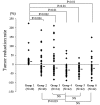
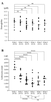
Cisplatin-based concurrent chemoradiotherapy (CCRT) has become a standard treatment for cancer of the uterine cervix. However, there have been no investigations into the optimum timing for administration of anticancer drugs using animal models. The aim of the present study was to determine the appropriate timing for administration of the anticancer drug cisplatin in relation to delivery of radiation by assessing the antitumor activity and adverse effects of 3 different regimens in αT3 transgenic mice bearing lens epithelial tumors. CCRT showed the strongest antitumor activity. There was a significant difference between CCRT and administration of cisplatin before radiotherapy (neoadjuvant therapy) with regard to the apoptotic effect detected by TUNEL staining, but there was no significant difference between CCRT and administration of cisplatin after radiotherapy (adjuvant therapy). Assessment of adverse effects showed that there were no significant differences in the mortality rate, weight loss, anemia and leukopenia among the 3 regimens. In conclusion, these findings obtained in an animal model suggest that cisplatin should probably not be administered before irradiation, since the antitumor effect is significantly weaker than that of CCRT or administration after irradiation, while adverse effects are similar.
Figures


Similar articles
-
Concurrent weekly cisplatin plus external beam radiotherapy and high-dose rate brachytherapy for advanced cervical cancer: a control cohort comparison with radiation alone on treatment outcome and complications.Int J Radiat Oncol Biol Phys. 2006 Dec 1;66(5):1370-7. doi: 10.1016/j.ijrobp.2006.07.004. Epub 2006 Sep 18. Int J Radiat Oncol Biol Phys. 2006. PMID: 16979836
-
The Dynamic Alternation of Local and Systemic Tumor Immune Microenvironment During Concurrent Chemoradiotherapy of Cervical Cancer: A Prospective Clinical Trial.Int J Radiat Oncol Biol Phys. 2021 Aug 1;110(5):1432-1441. doi: 10.1016/j.ijrobp.2021.03.003. Epub 2021 Mar 10. Int J Radiat Oncol Biol Phys. 2021. PMID: 33713744 Clinical Trial.
-
Randomized phase III trial of radiotherapy or chemoradiotherapy with topotecan and cisplatin in intermediate-risk cervical cancer patients after radical hysterectomy.BMC Cancer. 2015 May 4;15:353. doi: 10.1186/s12885-015-1355-1. BMC Cancer. 2015. PMID: 25935645 Free PMC article. Clinical Trial.
-
Why Concurrent CDDP and Radiotherapy Has Synergistic Antitumor Effects: A Review of In Vitro Experimental and Clinical-Based Studies.Int J Mol Sci. 2021 Mar 19;22(6):3140. doi: 10.3390/ijms22063140. Int J Mol Sci. 2021. PMID: 33808722 Free PMC article. Review.
-
[Chemoradiotherapy in cancers of the uterine cervix].Cancer Radiother. 1998 Dec;2(6):718-22. doi: 10.1016/s1278-3218(99)80014-6. Cancer Radiother. 1998. PMID: 9922779 Review. French.
Cited by
-
Divergent effects of AKI to CKD models on inflammation and fibrosis.Am J Physiol Renal Physiol. 2018 Oct 1;315(4):F1107-F1118. doi: 10.1152/ajprenal.00179.2018. Epub 2018 Jun 13. Am J Physiol Renal Physiol. 2018. PMID: 29897282 Free PMC article.
-
p53 as a unique target of action of cisplatin in acute leukaemia cells.J Cell Mol Med. 2022 Sep;26(17):4727-4739. doi: 10.1111/jcmm.17502. Epub 2022 Aug 9. J Cell Mol Med. 2022. PMID: 35946055 Free PMC article.
References
-
- Nees M, Geoghegan JM, Munson P, et al. Human papillomavirus type 16 E6 and E7 proteins inhibit differentiation-dependent expression of transforming growth factor-β2 in cervical keratinocytes. Cancer Res. 2000;60:4289–4298. - PubMed
-
- Parkin DM, Pisani P, Ferlay J. Estimates of the worldwide incidence of eighteen major cancers in 1985. Int J Cancer. 1993;54:594–606. - PubMed
-
- Runowicz CD, Wadler S, Rodriguez-Rodriguez L, et al. Concomitant cisplatin and radiotherapy in locally advanced cervical carcinoma. Gynecol Oncol. 1989;34:395–401. - PubMed
-
- Alberts DS, Garcia D, Mason-Liddil N. Cisplatin in advanced cancer of the cervix: an update. Semin Oncol. 1991;18:11–24. - PubMed
Publication types
MeSH terms
Substances
LinkOut - more resources
Full Text Sources
Other Literature Sources
Medical

