

Regulatory T cells control diabetes without compromising acute anti-viral defense
- PMID: 24858581
- PMCID: PMC4889438
- DOI: 10.1016/j.clim.2014.05.006
Regulatory T cells control diabetes without compromising acute anti-viral defense
Abstract
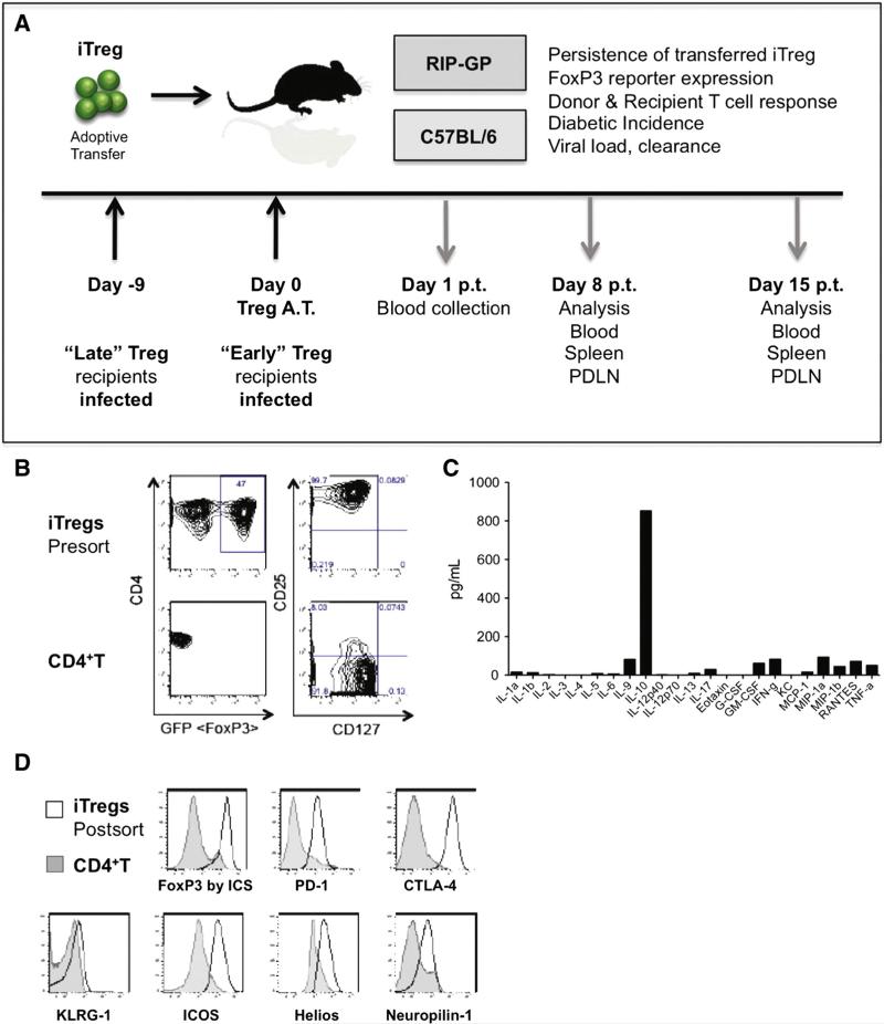
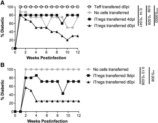
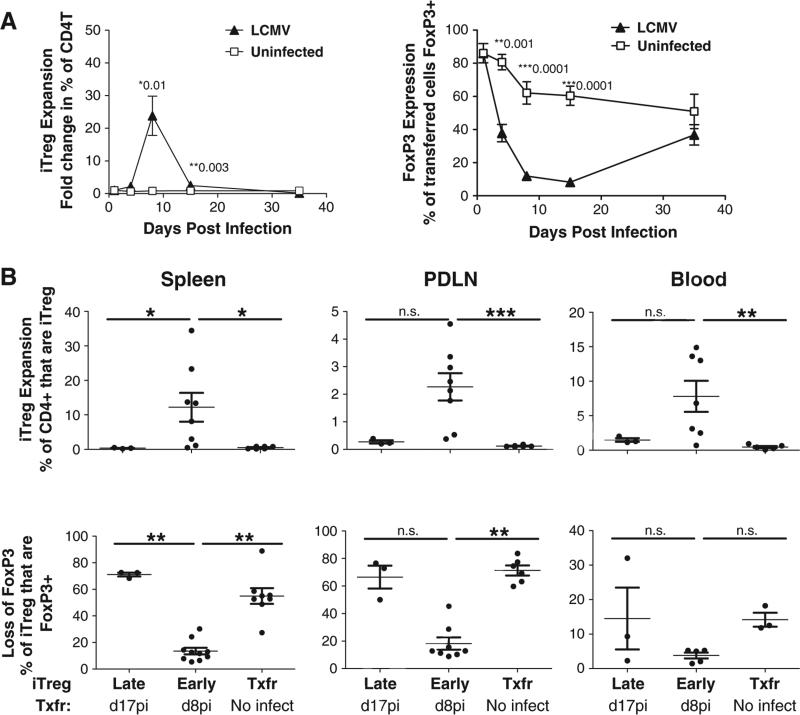
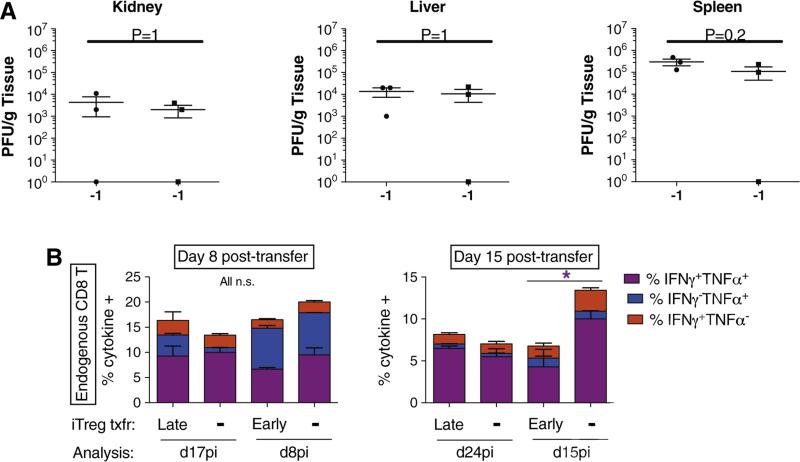
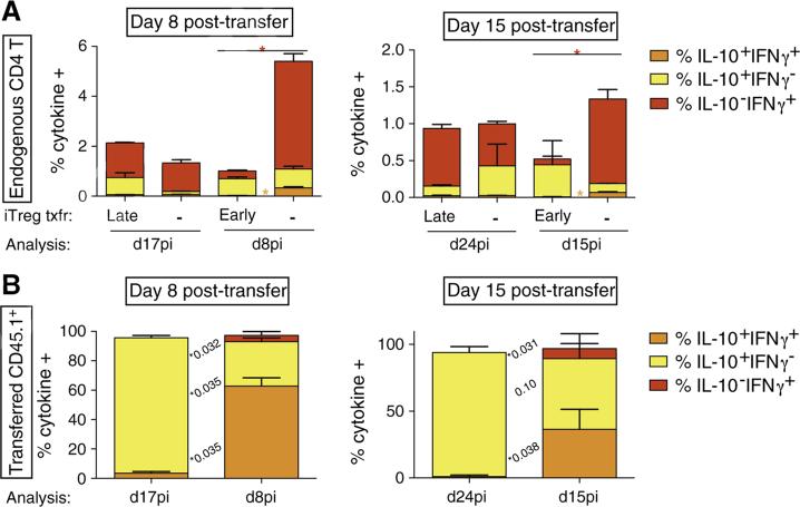
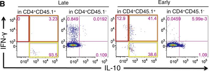
While previous reports have demonstrated the efficacy of regulatory T cell therapy in the prevention of diabetes, systemic immunocompromise and Treg instability remain key safety concerns. Here we examined the influence of induced Treg (iTreg) cell therapy on anti-viral host defense and autoimmune T cell responses during acute viral infection in a murine model of autoimmune diabetes. Protective transfers of iTregs maintained IL-10 expression, expanded in vivo and controlled diabetes, despite losing FoxP3 expression. Adoptive transfer of iTregs affected neither the primary anti-viral CD8 T cell response nor viral clearance, although a significant and sustained suppression of CD4 T cell responses was observed. Following acute viral clearance, iTregs transferred early suppressed both CD4 and CD8 T cell responses, which resulted in the reversion of diabetes. These observations indicate that iTregs suppress local autoimmune processes while preserving the immunocompetent host's ability to combat acute viral infection.
Keywords: Diabetes;; Regulatory T cells;; Safety;; Stability;; Therapy; Viral infection;.
Copyright © 2014 Elsevier Inc. All rights reserved.
Figures
Similar articles
-
Acute and chronic B cell depletion disrupts CD4+ and CD8+ T cell homeostasis and expansion during acute viral infection in mice.J Immunol. 2014 Jul 15;193(2):746-56. doi: 10.4049/jimmunol.1302848. Epub 2014 Jun 13. J Immunol. 2014. PMID: 24928986 Free PMC article.
-
Production of IL-10 by CD4(+) regulatory T cells during the resolution of infection promotes the maturation of memory CD8(+) T cells.Nat Immunol. 2015 Aug;16(8):871-9. doi: 10.1038/ni.3224. Epub 2015 Jul 6. Nat Immunol. 2015. PMID: 26147684 Free PMC article.
-
Destruction of lymphoid organ architecture and hepatitis caused by CD4+ T cells.PLoS One. 2011;6(9):e24772. doi: 10.1371/journal.pone.0024772. Epub 2011 Sep 23. PLoS One. 2011. PMID: 21966366 Free PMC article.
-
IL-2 contributes to maintaining a balance between CD4+Foxp3+ regulatory T cells and effector CD4+ T cells required for immune control of blood-stage malaria infection.J Immunol. 2011 Apr 15;186(8):4862-71. doi: 10.4049/jimmunol.1003777. Epub 2011 Mar 9. J Immunol. 2011. PMID: 21389253
-
In vivo treatment with a MHC class I-restricted blocking peptide can prevent virus-induced autoimmune diabetes.J Immunol. 1998 Nov 1;161(9):5087-96. J Immunol. 1998. PMID: 9794447
Cited by
-
Viral Infections and Autoimmune Disease: Roles of LCMV in Delineating Mechanisms of Immune Tolerance.Viruses. 2019 Sep 21;11(10):885. doi: 10.3390/v11100885. Viruses. 2019. PMID: 31546586 Free PMC article. Review.
-
Glucocorticoid receptor in T cells mediates protection from autoimmunity in pregnancy.Proc Natl Acad Sci U S A. 2017 Jan 10;114(2):E181-E190. doi: 10.1073/pnas.1617115114. Epub 2017 Jan 3. Proc Natl Acad Sci U S A. 2017. PMID: 28049829 Free PMC article.
-
Stem cell-derived tissue-associated regulatory T cells ameliorate the development of autoimmunity.Sci Rep. 2016 Feb 5;6:20588. doi: 10.1038/srep20588. Sci Rep. 2016. PMID: 26846186 Free PMC article.
-
Expansion of Human Tregs from Cryopreserved Umbilical Cord Blood for GMP-Compliant Autologous Adoptive Cell Transfer Therapy.Mol Ther Methods Clin Dev. 2016 Dec 24;4:178-191. doi: 10.1016/j.omtm.2016.12.003. eCollection 2017 Mar 17. Mol Ther Methods Clin Dev. 2016. PMID: 28345003 Free PMC article.
-
Commensal bacteria education history calibrates the naivety and activation threshold of adaptive antiviral immune system.Front Immunol. 2025 Feb 7;16:1519023. doi: 10.3389/fimmu.2025.1519023. eCollection 2025. Front Immunol. 2025. PMID: 39991160 Free PMC article.
References
-
- Shevach EM. From vanilla to 28 flavors: multiple varieties of T regulatory cells. Immunity. 2006;25:195–201. - PubMed
-
- Sakaguchi S. Naturally arising CD4+ regulatory t cells for immunologic self-tolerance and negative control of immune responses. Annu. Rev. Immunol. 2004;22:531–562. - PubMed
-
- Shevach EM. Mechanisms of foxp3+ T regulatory cell-mediated suppression. Immunity. 2009;30:636–645. - PubMed
Publication types
MeSH terms
Substances
Grants and funding
LinkOut - more resources
Full Text Sources
Other Literature Sources
Medical
Research Materials