Review
. 2014 Sep;36(5):581-94.
doi: 10.1007/s00281-014-0433-9. Epub 2014 May 24.

Autoimmune and autoinflammatory mechanisms in uveitis

Affiliations
Review

Autoimmune and autoinflammatory mechanisms in uveitis

Richard W Lee et al. Semin Immunopathol. 2014 Sep.

Abstract

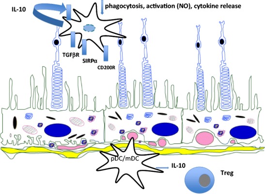
The eye, as currently viewed, is neither immunologically ignorant nor sequestered from the systemic environment. The eye utilises distinct immunoregulatory mechanisms to preserve tissue and cellular function in the face of immune-mediated insult; clinically, inflammation following such an insult is termed uveitis. The intra-ocular inflammation in uveitis may be clinically obvious as a result of infection (e.g. toxoplasma, herpes), but in the main infection, if any, remains covert. We now recognise that healthy tissues including the retina have regulatory mechanisms imparted by control of myeloid cells through receptors (e.g. CD200R) and soluble inhibitory factors (e.g. alpha-MSH), regulation of the blood retinal barrier, and active immune surveillance. Once homoeostasis has been disrupted and inflammation ensues, the mechanisms to regulate inflammation, including T cell apoptosis, generation of Treg cells, and myeloid cell suppression in situ, are less successful. Why inflammation becomes persistent remains unknown, but extrapolating from animal models, possibilities include differential trafficking of T cells from the retina, residency of CD8(+) T cells, and alterations of myeloid cell phenotype and function. Translating lessons learned from animal models to humans has been helped by system biology approaches and informatics, which suggest that diseased animals and people share similar changes in T cell phenotypes and monocyte function to date. Together the data infer a possible cryptic infectious drive in uveitis that unlocks and drives persistent autoimmune responses, or promotes further innate immune responses. Thus there may be many mechanisms in common with those observed in autoinflammatory disorders.


Figures

Fig. 1
Fig. 1
Diagnostic imaging depicting manifestations of uveitis. a Vitreous haze seen in the right eye of a 39-year-old African American female with sarcoidosis associated panuveitis (i, left panel) clears following treatment (ii, right panel). Please note that the borders of optic nerve and details of retinal vasculature are not clearly visible due to vitreous haze. b Peripheral fundus photographs of an African American male with neurosarcoidosis and panuveitis show significant perivascular exudates and chorioretinal granulomas. c Fluorescein angiogram of the same patient in b shows no staining in the very early phase but diffuse involvement of the entire retinal vasculature with staining of the exudates in early-mid phase (upper right and lower left panels) and leakage in late phase (lower right panel) is evident. d Spectral domain optical coherence tomography (SD OCT) of a 28-year-old Hispanic male with noninfectious uveitis and cystoid macular edema shows intra retinal cysts as well as subretinal fluid (arrows). Please note the detailed visibility of different retinal layers and the disruption of outer segment (ellipsoid) layer in the area of subretinal fluid (arrow). e Fundus photograph of the left eye of a 58-year-old Caucasian female with Birdshot chorioretinopathy shows multiple, deep, yellowish choroidal lesions scattered in the posterior pole, particularly nasal to the optic nerve
Fig. 2
Fig. 2
Regulation and setting of the threshold of myeloid cell responses within the retina and choroid. Microglia and choroidal myeloid cells (dendritic cells and macrophages) sense the environment and regulate inflammatory responses. The healthy tissue sets the threshold for response through inhibitory receptors (e.g. CD200R, SIRPα) or via the TGF-β rich environment. The regulation via neuronal cognate interaction is augmented by the regulatory functions of RPE, via mediators such as PD-1 and PD-L1 interactions, TGF-β secretion, and inhibitory peptides. The response to activation of myeloid cells is dominated by IL-10 release; whilst other pro-inflammatory cytokines are also produced, the default response is downregulation [–75]

References

    1. Jabs DA, Nussenblatt RB, Rosenbaum JT. Standardization of uveitis nomenclature for reporting clinical data. Results of the First International Workshop. Am J Ophthalmol. 2005;140:509–516. doi: 10.1016/j.ajo.2005.03.057. - DOI - PMC - PubMed
    1. The World Health Report, WHO (2010) Global data on visual impairments. http://www.who.int/blindness/GLOBALDATAFINALforweb.pdf. Accessed 21 Jan 2014
    1. Nussenblatt RB. The natural history of uveitis. Int Ophthalmol. 1990;14:303–308. doi: 10.1007/BF00163549. - DOI - PubMed
    1. Bodaghi B, Cassoux N, Wechsler B, Hannouche D, Fardeau C, et al. Chronic severe uveitis: etiology and visual outcome in 927 patients from a single center. Medicine (Baltimore) 2001;80:263–270. doi: 10.1097/00005792-200107000-00005. - DOI - PubMed
    1. Rothova A, Buitenhuis HJ, Meenken C, Brinkman CJ, Linssen A, et al. Uveitis and systemic disease. Br J Ophthalmol. 1992;76:137–141. doi: 10.1136/bjo.76.3.137. - DOI - PMC - PubMed

Publication types