. 2014 Jul 15;307(2):H252-8.
doi: 10.1152/ajpheart.00149.2014. Epub 2014 May 23.

HDAC6 contributes to pathological responses of heart and skeletal muscle to chronic angiotensin-II signaling

Affiliations

HDAC6 contributes to pathological responses of heart and skeletal muscle to chronic angiotensin-II signaling

Kimberly M Demos-Davies et al. Am J Physiol Heart Circ Physiol. .

Abstract

Little is known about the function of the cytoplasmic histone deacetylase HDAC6 in striated muscle. Here, we addressed the role of HDAC6 in cardiac and skeletal muscle remodeling induced by the peptide hormone angiotensin II (ANG II), which plays a central role in blood pressure control, heart failure, and associated skeletal muscle wasting. Comparable with wild-type (WT) mice, HDAC6 null mice developed cardiac hypertrophy and fibrosis in response to ANG II. However, whereas WT mice developed systolic dysfunction upon treatment with ANG II, cardiac function was maintained in HDAC6 null mice treated with ANG II for up to 8 wk. The cardioprotective effect of HDAC6 deletion was mimicked in WT mice treated with the small molecule HDAC6 inhibitor tubastatin A. HDAC6 null mice also exhibited improved left ventricular function in the setting of pressure overload mediated by transverse aortic constriction. HDAC6 inhibition appeared to preserve systolic function, in part, by enhancing cooperativity of myofibrillar force generation. Finally, we show that HDAC6 null mice are resistant to skeletal muscle wasting mediated by chronic ANG-II signaling. These findings define novel roles for HDAC6 in striated muscle and suggest potential for HDAC6-selective inhibitors for the treatment of cardiac dysfunction and muscle wasting in patients with heart failure.

Keywords: cardiac dysfunction; deacetylase; muscle atrophy.


Figures

Fig. 1.
Fig. 1.
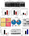
Histone deacetylase 6 (HDAC6) deletion improves cardiac function in mice treated chronically with angiotensin II (ANG II). Wild-type (WT) and HDAC6 knockout (KO) mice were treated with ANG II for 8 wk via osmotic minipumps. Control animals were given sham pumps lacking ANG II. M-mode echocardiographic images (A) were used to quantify ejection fraction (EF; B). C: left ventricular (LV) weight-to-tibia length (TL) ratios were determined at necropsy. D and E: fibrosis in LV sections was quantified using picrosirius red dye. F: flow cytometry was used to quantify the presence of the indicated cell populations in ventricles from ANG II-treated mice. Mac, macrophages; Neut, neutrophils; NK, NK cells; Non-Gran, nongranulocytes. G: homogenates of LVs were assessed by immunoblotting with the indicated antibodies. H: autophagic activity in LVs was measured by immunoblotting to detect conversion of LC3-I to LC3-II. LC3-II levels were quantified by densitometry. I: representative LC3 immunoblot. J: WT mice were treated with ANG II for 2 wk and were injected daily with the HDAC6 inhibitor tubastatin A (Tub A) or vehicle control. Tub A rescued ANG II-mediated impairment of LV EF. For B–D, H, and J, values represent means ± SE. *P < 0.05 vs. sham controls.
Fig. 2.
Fig. 2.
HDAC6 deletion improves cardiac function in mice subjected to LV pressure overload. Mice underwent sham surgery or transverse aortic constriction (TAC) and were analyzed after 4 wk. A–D: M-mode echocardiographic images were used to quantify EF, interventricular septum (IVS) thickness and LV posterior wall (LVPW) thickness (d, diastole). E: LV weight-to-TL ratios were determined at necropsy. F: fibrosis in LV sections was quantified using picrosirius red dye. Values represent means ± SE. *P < 0.05 vs. sham controls.
Fig. 3.
Fig. 3.
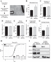
HDAC6 (HD6) deletion augments myofibril inotropy. A: representative image of a small bundle of LV myofibrils mounted on a force recording apparatus. B–G: mechanical characteristics of LV myofibrils from WT and HDAC6 null mice treated with ANG II for 8 wk. H: to determine myofibril calcium sensitivity (pCa50) and the Hill coefficient, the ratio of force generated at submaximal (P) and maximal calcium (P0) activation was plotted; multiple calcium concentrations were used (pCa: 9, 5.8, 5.7, 5.6, and 4.5; and pCa = −log [Ca2+]). I: immunoblotting was performed to compare the amount of HDAC6 and HDAC3 in total LV homogenates vs. purified myofibrils. GAPDH (cytoplasmic marker) and troponin I (sarcomeric protein) served as controls to confirm myofibril enrichment. *P < 0.05 vs. WT + ANG II.
Fig. 4.
Fig. 4.
HDAC6 deletion blocks ANG II-mediated skeletal muscle wasting. A: representative image of WT and HDAC6 knockout mice after 8 wk of treatment with ANG II. B: change in animal body weight over the course of the 8-wk study. C: total mouse body weight at the beginning and end of the study. D–F: dual-energy X-ray absorptiometry (DEXA) scan analysis of mice after 8 wk of treatment with ANG II or sham osmotic minipumps. G: gastrocnemius-to-TL ratios were determined at the time of necropsy. H and I: quantitative PCR analysis of atrogin-1 and muscle RING-finger protein-1 (MURF1) ubiquitin ligase mRNA expression in gastrocnemius homogenates from WT and HDAC6 knockout mice treated with ANG II for 2 or 8 wk. J: autophagic activity in gastrocnemius muscle was measured by immunoblotting to detect conversion of LC3-I to LC3-II. LC3-II levels were quantified by densitometry. K: representative LC3 immunoblot. Values for all graphs represent means ± SE. *P < 0.05 vs. WT + ANG II (H–J).

References

    1. Alamdari N, Smith IJ, Aversa Z, Hasselgren PO. Sepsis and glucocorticoids upregulate p300 and downregulate HDAC6 expression and activity in skeletal muscle. Am J Physiol Regul Integr Comp Physiol 299: R509–R520, 2010 - PMC - PubMed
    1. Butler KV, Kalin J, Brochier C, Vistoli G, Langley B, Kozikowski AP. Rational design and simple chemistry yield a superior, neuroprotective HDAC6 inhibitor, tubastatin A. J Am Chem Soc 132: 10842–10846, 2010 - PMC - PubMed
    1. Cabello-Verrugio C, Cordova G, Salas JD. Angiotensin II: role in skeletal muscle atrophy. Curr Protein Pept Sci 13: 560–569, 2012 - PubMed
    1. Cohen TJ, Waddell DS, Barrientos T, Lu Z, Feng G, Cox GA, Bodine SC, Yao TP. The histone deacetylase HDAC4 connects neural activity to muscle transcriptional reprogramming. J Biol Chem 282: 33752–33759, 2007 - PubMed
    1. Di Bari M, van de Poll-Franse LV, Onder G, Kritchevsky SB, Newman A, Harris TB, Williamson JD, Marchionni N, Pahor M. Antihypertensive medications and differences in muscle mass in older persons: the Health, Aging and Body Composition Study. J Am Geriatr Soc 52: 961–966, 2004 - PubMed

Publication types

MeSH terms