

Deletion of ameloblastin exon 6 is associated with amelogenesis imperfecta
- PMID: 24858907
- PMCID: PMC4168819
- DOI: 10.1093/hmg/ddu247
Deletion of ameloblastin exon 6 is associated with amelogenesis imperfecta
Abstract
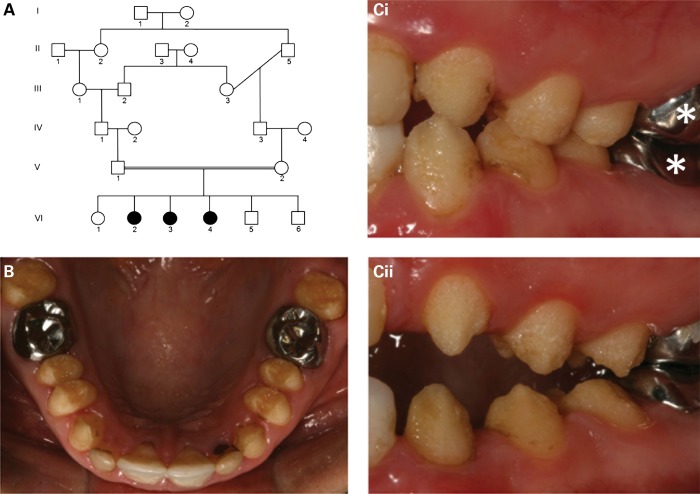
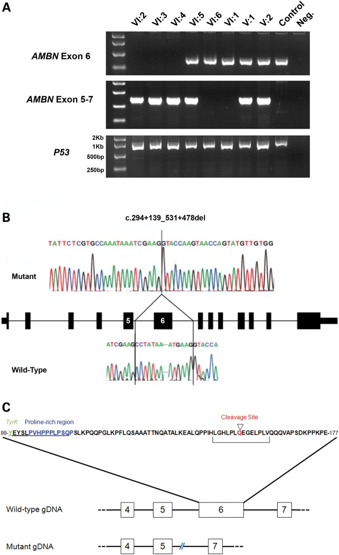
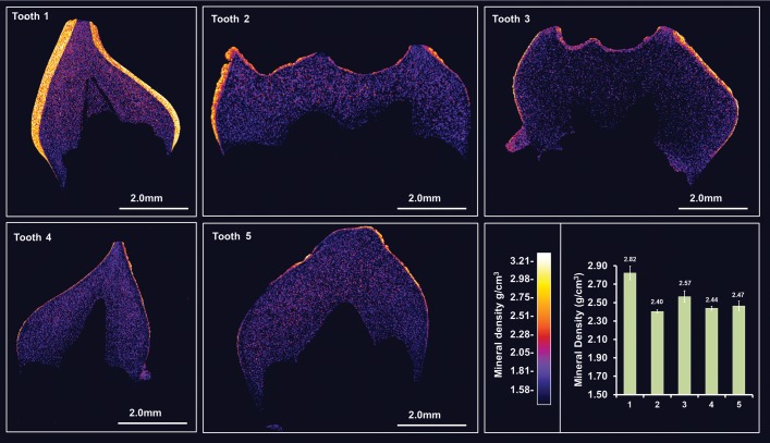
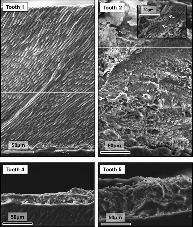
Amelogenesis imperfecta (AI) describes a heterogeneous group of inherited dental enamel defects reflecting failure of normal amelogenesis. Ameloblastin (AMBN) is the second most abundant enamel matrix protein expressed during amelogenesis. The pivotal role of AMBN in amelogenesis has been confirmed experimentally using mouse models. However, no AMBN mutations have been associated with human AI. Using autozygosity mapping and exome sequencing, we identified genomic deletion of AMBN exon 6 in a second cousin consanguineous family with three of the six children having hypoplastic AI. The genomic deletion corresponds to an in-frame deletion of 79 amino acids, shortening the protein from 447 to 368 residues. Exfoliated primary teeth (unmatched to genotype) were available from family members. The most severely affected had thin, aprismatic enamel (similar to that reported in mice homozygous for Ambn lacking exons 5 and 6). Other teeth exhibited thicker but largely aprismatic enamel. One tooth had apparently normal enamel. It has been suggested that AMBN may function in bone development. No clinically obvious bone or other co-segregating health problems were identified in the family investigated. This study confirms for the first time that AMBN mutations cause non-syndromic human AI and that mouse models with disrupted Ambn function are valid.
© The Author 2014. Published by Oxford University Press.
Figures
Similar articles
-
Novel Ameloblastin Variants, Contrasting Amelogenesis Imperfecta Phenotypes.J Dent Res. 2024 Jan;103(1):22-30. doi: 10.1177/00220345231203694. Epub 2023 Dec 6. J Dent Res. 2024. PMID: 38058155 Free PMC article.
-
Whole exome sequencing identifies an AMBN missense mutation causing severe autosomal-dominant amelogenesis imperfecta and dentin disorders.Int J Oral Sci. 2018 Sep 3;10(3):26. doi: 10.1038/s41368-018-0027-9. Int J Oral Sci. 2018. PMID: 30174330 Free PMC article.
-
Enamelin/ameloblastin gene polymorphisms in autosomal amelogenesis imperfecta among Syrian families.J Investig Clin Dent. 2011 Feb;2(1):16-22. doi: 10.1111/j.2041-1626.2010.00038.x. Epub 2010 Nov 8. J Investig Clin Dent. 2011. PMID: 25427323
-
Ameloblastin and its multifunctionality in amelogenesis: A review.Matrix Biol. 2024 Aug;131:62-76. doi: 10.1016/j.matbio.2024.05.007. Epub 2024 May 28. Matrix Biol. 2024. PMID: 38815936 Free PMC article. Review.
-
Enamelin and autosomal-dominant amelogenesis imperfecta.Crit Rev Oral Biol Med. 2003;14(6):387-98. doi: 10.1177/154411130301400602. Crit Rev Oral Biol Med. 2003. PMID: 14656895 Review.
Cited by
-
A Novel De Novo SP6 Mutation Causes Severe Hypoplastic Amelogenesis Imperfecta.Genes (Basel). 2021 Feb 26;12(3):346. doi: 10.3390/genes12030346. Genes (Basel). 2021. PMID: 33652941 Free PMC article.
-
Mutations in the pH-Sensing G-protein-Coupled Receptor GPR68 Cause Amelogenesis Imperfecta.Am J Hum Genet. 2016 Oct 6;99(4):984-990. doi: 10.1016/j.ajhg.2016.08.020. Epub 2016 Sep 29. Am J Hum Genet. 2016. PMID: 27693231 Free PMC article.
-
Novel Ameloblastin Variants, Contrasting Amelogenesis Imperfecta Phenotypes.J Dent Res. 2024 Jan;103(1):22-30. doi: 10.1177/00220345231203694. Epub 2023 Dec 6. J Dent Res. 2024. PMID: 38058155 Free PMC article.
-
An Intron c.103-3T>C Variant of the AMELX Gene Causes Combined Hypomineralized and Hypoplastic Type of Amelogenesis Imperfecta: Case Series and Review of the Literature.Genes (Basel). 2022 Jul 18;13(7):1272. doi: 10.3390/genes13071272. Genes (Basel). 2022. PMID: 35886055 Free PMC article.
-
Ameloblastin Binds to Phospholipid Bilayers via a Helix-Forming Motif within the Sequence Encoded by Exon 5.ACS Omega. 2019 Feb 28;4(2):4405-4416. doi: 10.1021/acsomega.8b03582. ACS Omega. 2019. PMID: 30873509 Free PMC article.
References
-
- Smith C.E. Cellular and chemical events during enamel maturation. Crit. Rev. Oral Biol. Med. 1998;9:128–161. - PubMed
-
- Simmer J.P., Fincham A.G. Molecular mechanisms of dental enamel formation. Crit. Rev. Oral Biol. Med. 1995;6:84–108. - PubMed
-
- Smith C.E., Warshawsky H. Quantitative analysis of cell turnover in the enamel organ of the rat incisor. Evidence for ameloblast death immediately after enamel matrix secretion. Anat. Rec. 1977;187:63–98. - PubMed
-
- Backman B., Holm A.K. Amelogenesis imperfecta: prevalence and incidence in a northern Swedish county. Community Dent. Oral Epidemiol. 1986;14:43–47. - PubMed
-
- Witkop C.J., Sauk J.J., Jr . In: Oral Facial Genetics. Stewart R., Prescott G., editors. St Louis: CV Mosby Company; 1976. pp. 151–226.
Publication types
MeSH terms
Substances
Grants and funding
LinkOut - more resources
Full Text Sources
Other Literature Sources
Molecular Biology Databases
