. 2014 Sep 15;23(18):4985-94.
doi: 10.1093/hmg/ddu213. Epub 2014 May 8.

X inactivation plays a major role in the gender bias in somatic expansion in a mouse model of the fragile X-related disorders: implications for the mechanism of repeat expansion

Affiliations

X inactivation plays a major role in the gender bias in somatic expansion in a mouse model of the fragile X-related disorders: implications for the mechanism of repeat expansion

Rachel Adihe Lokanga et al. Hum Mol Genet. .

Abstract

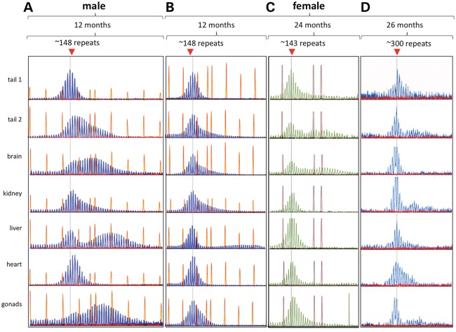
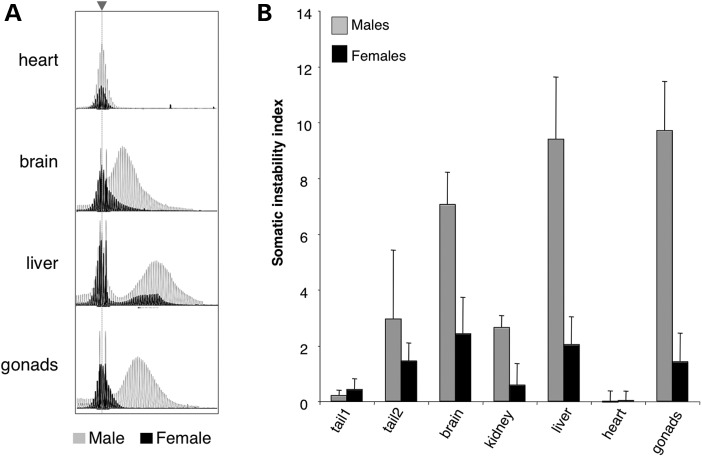
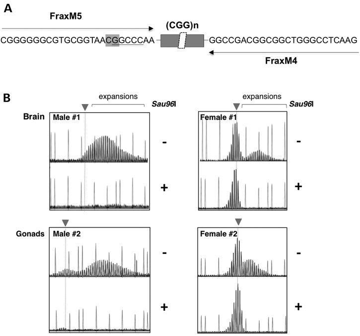
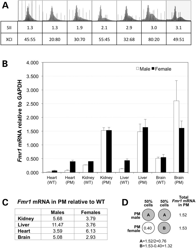
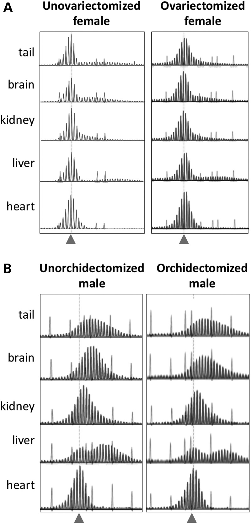
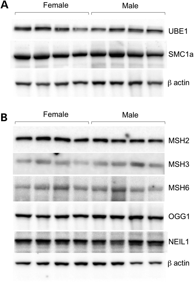
The Fragile X-related disorders are X-linked disorders resulting from the inheritance of FMR1 alleles with >54 CGG/CCG repeats in their 5' UTR. The repeats expand both somatically and on intergenerational transmission and increased repeat numbers are associated with increased risk of disease and increased risk of further expansion. The mechanism responsible for expansion is unknown. Here, we show in a knockin mouse model of these disorders that somatic expansion is much less common in females than in males. We show that this is due in large part to the fact that expansions occur only when the repeat is on the active X chromosome. However, even when this is taken into account, expansions in females are still less common than expected. This additional gender effect is not due to a protective effect of estrogen, a deleterious effect of testosterone or to differences in the expression of the Fmr1 gene or a variety of X-linked and autosomal DNA repair genes. However, our data do suggest that a higher level of expression of genes that protect against oxidative damage in females may contribute to their lower levels of expansion. Whatever the basis, our data suggest that the risk for somatic expansion may be lower in women than it is in men. This could help explain the reduced penetrance of some aspects of disease pathology in women. The fact that expansion only occurs when the Fmr1 allele is on the active X chromosome has important implications for the mechanism of repeat expansion.


Figures

Figure 1.
Figure 1.
Somatic instability is more extensive in male FX PM mice than in females. Comparison of the GeneMapper profiles for different organs of a 12-month-old male mouse (A) with the profiles for different organs from an age-matched female with a similar repeat number (B), a 24-month-old female with a similar repeat number (C) and a 26-month-old female with ∼300 repeats (D). The dotted gray line indicates the position of the original inherited allele. Either 500 ROX™ or the 1200 LIZ® molecular weight markers were used. The choice of marker did not affect the GeneMapper profile. Tail 1 refers to the tail DNA at weaning, while tail 2 is the tail DNA sample taken at the time of euthanasia.
Figure 2.
Figure 2.
Comparison of somatic instability and SII for male and female mice 12 months of age. (A) Overlay of representative GeneMapper profiles from the heart, brain, liver and gonads of a 12-month-old male (gray) and female (black) mouse with ∼146 repeats. The dotted gray line and arrow head indicates the position of the original inherited allele. (B) The SII in different organs of 12-month-old males (n = 8) and females (n = 8) with ∼145 repeats. Tail 1 refers to the tail DNA at weaning, while tail 2 is the tail DNA sample taken at 12 months of age.
Figure 3.
Figure 3.
Relationship between expansion and presence of the PM on the active X chromosome. (A) Schematic showing the region of the FX PM mouse Fmr1 gene amplified with FraxM4 and FraxM5, illustrating the principle behind using Sau96I-predigestion to examine expansion on the inactive X chromosome. FraxM4 and FraxM5 amplify the PM allele but not the WT allele since the 3′ ends of each primer correspond to the bases added to the PM allele when the mouse line was first generated. The sequence corresponding to the primer in the case of FraxM5 and the primer binding site for FraxM4 is shown. The position of a CpG residue that overlaps with a Sau96I cleavage site in the FraxM5 sequence is indicated in the boxed region. Since Sau96I cleavage is blocked by overlapping methylation, the FraxM5 region will be cut by Sau96I only when the PM allele is on the active X chromosome. As a result when the DNA is digested with Sau96I prior to PCR, the FraxM5 primer will only be able to amplify the PM on the inactive X chromosome. Note that a Sau96I cleavage site is also present in the region corresponding to the FraxM4 sequence. However, this site does not overlap a CpG residue and thus is cleaved whether the allele is methylated or not but since the cleavage site leaves 18 bases adjacent to the repeat for the primer to bind, this primer amplifies the repeat in Sau96I treated material whether the allele is methylated or not. Since males only possess an active X chromosome, Sau96I digestion eliminates all traces of the PM allele. In females, only the PM allele on the inactive X chromosome remains to be amplified after digestion by Sau96I. (B) GeneMapper profiles of two males and two females without (−) and with (+) pretreatment with Sau96I along with the 1200 LIZ® molecular weight markers. The dotted gray line and arrowhead indicates the position of the original inherited allele.
Figure 4.
Figure 4.
SII, XCI ratios and Fmr1 transcription. (A) The GeneMapper profiles, SII and XCI for the brains of seven age-matched females with similar repeat sizes run with either the 500 ROX™ or 1200 LIZ® molecular weight markers. The XCI ratio is shown as the fraction of the PM allele on the active X versus the fraction of the PM on the inactive X. (B) Comparison of Fmr1 mRNA expression in various organs of male and female mice with and without the PM allele. The Fmr1 mRNA levels were determined by quantitative real-time PCR and are expressed relative to GAPDH. The standard deviation for each group is shown. (C) The Fmr1 mRNA levels in PM mice relative to the levels of Fmr1 mRNA in WT mice. The Fmr1 levels in different organs of male and female mice were normalized to GAPDH and then expressed as the ratio of the levels in PM to the levels in WT mice. (D) Illustration of how the amount of mRNA produced from the PM allele was deduced in female kidney by using the level of mRNA seen in WT mice. The open circle represents the WT allele and the grey circles the PM alleles.
Figure 5.
Figure 5.
Effect of removal of female and male gonads on the extent of somatic expansion. The GeneMapper profiles for 12-month-old male and female mice with repeat numbers of ∼145 who were either untreated or had their gonads removed at 7 weeks of age. Samples were run with either a 500 ROX™ (unovariectomized female) or 1200 LIZ® molecular weight marker. The dotted gray line and arrowhead indicate the position of the original inherited allele.
Figure 6.
Figure 6.
Western blots showing the levels of various DNA repair proteins in male and female FX PM mice. Protein isolated from the livers of 12-month-old male and female FX PM mice was subjected to electrophoresis, transferred to membrane and challenged with antibodies to the indicated proteins as described in Materials and Methods. (A) Levels of X-linked DNA repair genes. (B) Levels of DNA repair genes implicated in the repeat expansion diseases.

Similar articles

Cited by

References

    1. Chonchaiya W., Schneider A., Hagerman R.J. Fragile X: a family of disorders. Adv. Pediatr. 2009;56:165–186. - PMC - PubMed
    1. Hagerman P. Fragile X-associated tremor/ataxia syndrome (FXTAS): pathology and mechanisms. Acta Neuropathol. 2013;126:1–19. - PMC - PubMed
    1. Sherman S.L. Premature ovarian failure in the fragile X syndrome. Am. J. Med. Genet. 2000;97:189–194. - PubMed
    1. Murray A., Webb J., Grimley S., Conway G., Jacobs P. Studies of FRAXA and FRAXE in women with premature ovarian failure. J. Med. Genet. 1998;35:637–640. - PMC - PubMed
    1. Fry M., Usdin K. Human Nucleotide Expansion Disorders. Springer, Heidelberg; 2006.

Publication types

Substances