. 2014 May 23;9(5):e98344.
doi: 10.1371/journal.pone.0098344. eCollection 2014.

Role of lipid rafts and GM1 in the segregation and processing of prion protein

Affiliations

Role of lipid rafts and GM1 in the segregation and processing of prion protein

Laura Botto et al. PLoS One. .

Abstract

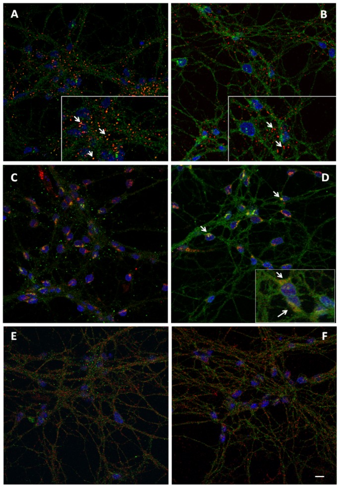
The prion protein (PrPC) is highly expressed within the nervous system. Similar to other GPI-anchored proteins, PrPC is found in lipid rafts, membrane domains enriched in cholesterol and sphingolipids. PrPC raft association, together with raft lipid composition, appears essential for the conversion of PrPC into the scrapie isoform PrPSc, and the development of prion disease. Controversial findings were reported on the nature of PrPC-containing rafts, as well as on the distribution of PrPC between rafts and non-raft membranes. We investigated PrPC/ganglioside relationships and their influence on PrPC localization in a neuronal cellular model, cerebellar granule cells. Our findings argue that in these cells at least two PrPC conformations coexist: in lipid rafts PrPC is present in the native folding (α-helical), stabilized by chemico-physical condition, while it is mainly present in other membrane compartments in a PrPSc-like conformation. We verified, by means of antibody reactivity and circular dichroism spectroscopy, that changes in lipid raft-ganglioside content alters PrPC conformation and interaction with lipid bilayers, without modifying PrPC distribution or cleavage. Our data provide new insights into the cellular mechanism of prion conversion and suggest that GM1-prion protein interaction at the cell surface could play a significant role in the mechanism predisposing to pathology.


Conflict of interest statement

Competing Interests: The authors have declared that no competing interests exist.

Figures

Figure 1
Figure 1. Characterization of PrPC distribution in CGCs.
Panel A- Schematic diagram of the proteolysis of PrPC and the epitope recognized by the antibodies used in this study. The native full-length PrPC is shown with its C-terminal GPI-anchor, the two N-linked glycosylation sites, the three helical regions (HA, HB and HC), the octapeptide repeat region (black), and the “toxic” 106–126 domain (white). The epitopes for antibody SAF32 (residues 79–92, in the unstructured octapetidic stretch) and 6H4 (residues 114–152, localized in HA) are indicated. The two cleavage sites generating N1/C1 (α-cleavage) and N2/C2 (β-cleavage) are shown by arrows. C1 is recognized only by 6H4. Panel B Characterization of PrPC localization in gradient fractions prepared from control CGCs. Cells were incubated with 1% Triton X-100-containing buffer for 30 min on ice. The suspension was subjected to discontinuous sucrose density gradient centrifugation. One-milliliter fractions were withdrawn from the gradient, submitted to 15% SDS-PAGE, transferred to nitrocellulose membranes, and immunoblotted with 6H4 or SAF32 antibodies against PrPC (20 µg proteins/lane), followed by ECL detection. Representative blots from three independent experiments are shown. g: glycosylated PrPC; u: unglycosylated PrPC. Panel C and D: immunofluorescence images showing PrPC distribution visualized by 6H4Ab (C) and SAF32Ab (D) in 8 DIV CGCs. Note the clusterized pattern visualized by 6H4Ab with respect to the diffuse staining of SAF32Ab. DAPI staining (blu) evidences nuclei. Scale bar: 10 µm.
Figure 2
Figure 2. Immunofluorescence analysis of PrPC distribution in CGCs.
Panel A: CGCs were immunolabelled with PrPC 6H4Ab (green) and the Alexa Fluor 594 cholerae toxin B (red) to visualize lipid rafts. Panel B: CGCs were doubly immunolabelled with PrPC SAF32 (green) and cholerae toxin B subunit (CTB). Insets show the different colocalization of the Abs with lipid rafts, indicating that 6H4Ab preferentially recognizes PrPC resident in lipid rafts, while SAF32Ab show PrPC that is widespread throughout the membrane. Arrows mark the position of CTB. Panel C and D: double immunofluorescence of PrPC antibodies with Giantin (red) denoting a major presence of SAF32Ab-positive PrPC in the Golgi apparatus with respect to 6H4Ab. Arrows mark the colocalization. Panels E and F: double immunofluorescence of PrPC antibodies (green) showing a lack of colocalization with Na+-K+/ATPase, a non-lipid raft plasmamembrane marker (red). Scale bar: 10 µm.; insets: 20 µm.
Figure 3
Figure 3. Cellular gangliosides treatment.
CGCs were incubated with different gangliosides (GM3, GM1 or GD1a) and correspondent radiolabelled gangliosides ([3H]GM3, [3H]GM1 or [3H]GD1a), at a final concentration of 2×10⁻⁶ M at 37°C for 4 h. At the end of incubation, the ganglioside solution was removed and cells were washed 3 times with Locke's solution and maintained at 37°C for 20 min in 3 mL of FBS-BME. The lipids extract, from cell homogenates, were analysed to determine the ganglioside incorporation (panel A) and metabolism by HPTLC following radioactivity imaging (panel B). Lane 1: granule cell ganglioside pattern; lane 2: granule cells ganglioside extracted after incubation with GM3/[3H]GM3 2×10⁻⁶ M at 37°C for 4 h; lane 3: granule cells ganglioside extracted after incubation with GM1/[3H]GM1 2×10⁻⁶ M at 37°C for 4 h; lane 4: granule cells ganglioside extracted after incubation with GD1a/[3H]GD1a 2×10⁻⁶ M at 37°C for 4 h; lane 5: [3H]GM3 standard; lane 6. [3H]GM1 and [3H]GD1a standards. * Gangliosides endogenous content as reported by Palestini et al., 1991.
Figure 4
Figure 4. Distribution of gangliosides radioactivity and proteins in the different fractions of the sucrose gradient.
CGCs, after incubation with different gangliosides (GM3, GM1 or GD1a) and correspondent radiolabelled gangliosides ([3H]GM3, [3H]GM1 or [3H]GD1a), at a final concentration of 2×10⁻⁶ M at 37°C for 4 h, were treated with 1% Triton X-100-containing buffer for 30 min on ice. The cellular lysate was submitted to discontinuous sucrose density gradient centrifugation. One-milliliter fractions were withdrawn from the gradient and submitted to [3H]GM3, [3H]GM1 or [3H]GD1a radioactivity determination (panel A) and evaluation of proteins distribution (panel B). Data are means ± SD from at least three independent experiments performed in triplicate.
Figure 5
Figure 5. Characterization of PrPC in gradient fractions obtained from treated CGCs.
Panel A Cells, after incubation with different gangliosides (GM3, GM1 or GD1a) and correspondent radiolabelled gangliosides ([3H]GM3, [3H]GM1 or [3H]GD1a), at a final concentration of 2×10⁻⁶ M at 37°C for 4 h (Standard treatment, St), were treated with 1% Triton X-100-containing buffer for 30 min on ice. The cellular lysate was submitted to discontinuous sucrose density gradient centrifugation. One-milliliter fractions were withdrawn from the gradient, submitted to 15% SDS-PAGE (20 µg protein/lane), transferred to nitrocellulose membranes and immunoblotted with 6H4 or SAF32 antibodies against PrPC followed by ECL detection. Representative blots from three independent experiments are shown. g: glycosylated PrPC; u: unglycosylated PrPC. GM3 = GM3-treated CGCs; GM1 = GM1-treated CGCs; GD1a = GD1a-treated CGCs. Panel B-E: immunofluorescence analysis of PrPC in CGCs with 6H4Ab (B and C) and SAF32Ab (D and E) in the presence (C and E) or in the absence (B and D) of GM1. Note that after ganglioside treatment, PrPC recognized by 6H4Ab appears generally less clusterized being more widespread and less concentrated around cell bodies and proximal dendrites (C, green), while PrPC distribution detected by SAF32Ab does not differ from that of control cells. Insets show double staining of PrPC and CTB. Arrows mark the position of CTB. Scale bar: 10 µm; insets: 20 µm.
Figure 6
Figure 6. Effect of gangliosides treatment on the localization of PKC, ADAM10 and Thy1 in gradient fractions from gangliosides treated-CGCs.
Cells, after incubation with different gangliosides (GM3, GM1 or GD1a) and correspondent radiolabelled gangliosides ([3H]GM3, [3H]GM1 or [3H]GD1a), at a final concentration of 2×10⁻⁶ M at 37°C for 4 h, were treated with 1% Triton X-100-containing buffer for 30 min on ice. The cellular lysate was subjected to discontinuous sucrose density gradient centrifugation. One-milliliter fractions were analyzed by immunoblotting with anti-PKC (panel A), anti-ADAM10 (panel B) and anti-Thy1 (panel C) antibodies. Immunoblot bands were analyzed and quantified by Kodak Image Station 2000R interfaced with a Kodak Molecular Imaging Software. The enrichment of the proteins in DRM was calculated as previously reported . The data reported for each protein are the mean of 3 immunoblots ± S.D. obtained from 3 independent sucrose gradients. Ctrl =  CGCs control; GM3 = CGCs treated with GM3; GM1 =  CGCs treated with GM1; GD1a =  CGCs treated with GD1a. Ctrl vs GM1 *p<0.01 (one way ANOVA).
Figure 7
Figure 7. Influence of GM1 cells treatment in PrPC processing.
CGCs, after incubation with GM1/[3H]GM1 at a final concentration of 2×10⁻⁶ M at 37°C for 4 h, were treated with 1% Triton X-100-containing buffer for 30 min on ice. The cellular lysate was submitted to discontinuous sucrose density gradient centrifugation. 50 µg of proteins from gradient fractions were subjected to protein deglycosylation by PNGase F treatment and immunoblotted with 6H4Ab (panel A), SAF32Ab (panel B) and 8G8Ab (panel C). Bands were analyzed and quantified by Kodak Image Station 2000R interfaced with a Kodak Molecular Imaging Software. Representative blots from three independent experiments are shown. 5TQ =  fraction 5 not subjected to PNGase F treatment; f.l.  = full length-PrPC; u  =  unglycosylated PrPC; g =  glycosylated PrPC.
Figure 8
Figure 8. Temperature and GM1 dose dependence of PrPC distribution in GM1-treated CGCs.
Cells after incubation with GM1/[3H]GM1 2×10⁻⁶ M at 4°C for 4 h or 1×10⁻⁶ M at 37°C for 4 h, were treated with 1% Triton X-100-containing buffer for 30 min on ice. A small amount of cells homogenates were analyzed to determine the gangliosides incorporation (panel A) and the residual was submitted to discontinuous sucrose density gradient centrifugation. One-milliliter fractions were withdrawn from the gradient and submitted to proteins and [3H]GM1 radioactivity determination (panel B). 20 µg of proteins from different fractions were submitted to 15% SDS-PAGE, transferred to nitrocellulose membranes and immunoblotted with 6H4Ab or SAF32Ab followed by ECL detection (panel C and D). Immunoblot bands were analyzed and quantified by Kodak Image Station 2000R interfaced with a Kodak Molecular Imaging Software. Representative blots from three independent experiments are shown. Ctrl  = control CGCs; GM1  =  GM1-treated CGCs.
Figure 9
Figure 9. GM1-containing liposomes alter PrPC structure.
Panel A- Circular dichroism spectra of recPrP (23–231) alone (black line) or mixed with POPC liposomes (blue line) or GM1-containing POPC liposomes (red line). Panel B- Results obtained replacing POPC with DPPC liposomes.

Similar articles

Cited by

References

    1. Oesch B, Westaway D, Wälchli M, McKinley MP, Kent SBH, et al. (1985) A cellular gene encodes scrapie PrP 27–30 protein. Cell 40: 735–746 doi:10.1016/0092-8674(85)90333-2 - DOI - PubMed
    1. Biasini E, Turnbaugh JA, Unterberger U, Harris DA (2012) Prion protein at the crossroads of physiology and disease. Trends Neurosci 36: 92–103 doi: 10.1016/j.tins.2011.10.002 - DOI - PMC - PubMed
    1. Simons K, Ikonen E (1997) Functional rafts in cell membranes. Nature 387: 569–572 doi:10.1038/42408 - DOI - PubMed
    1. Allen JA, Halverson-Tamboli RA, Rasenick MM (2007) Lipid raft microdomains and neurotransmitter signalling. Nature, Neuroscience 8: 128–140 doi:10.1038/nrn2059 - DOI - PubMed
    1. Prusiner SB (1998) Prions. Proc Natl Acad Sci USA. 95: 13363–13383 doi: 10.1073/pnas.95.23.13363 - DOI - PMC - PubMed

Publication types

LinkOut - more resources