. 2014 May 23;9(5):e97383.
doi: 10.1371/journal.pone.0097383. eCollection 2014.

Prolactin induces apoptosis of lactotropes in female rodents

Affiliations

Prolactin induces apoptosis of lactotropes in female rodents

Jimena Ferraris et al. PLoS One. .

Abstract

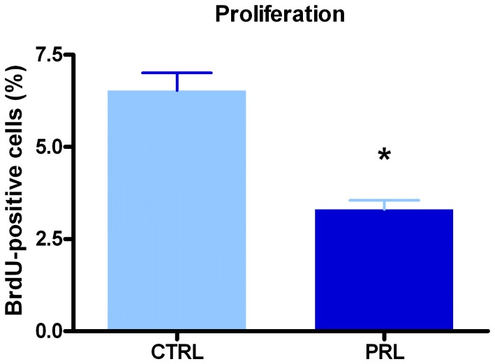
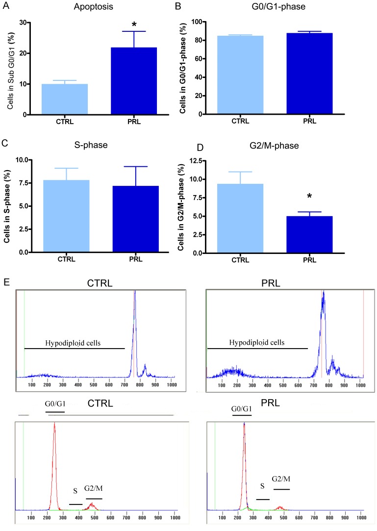
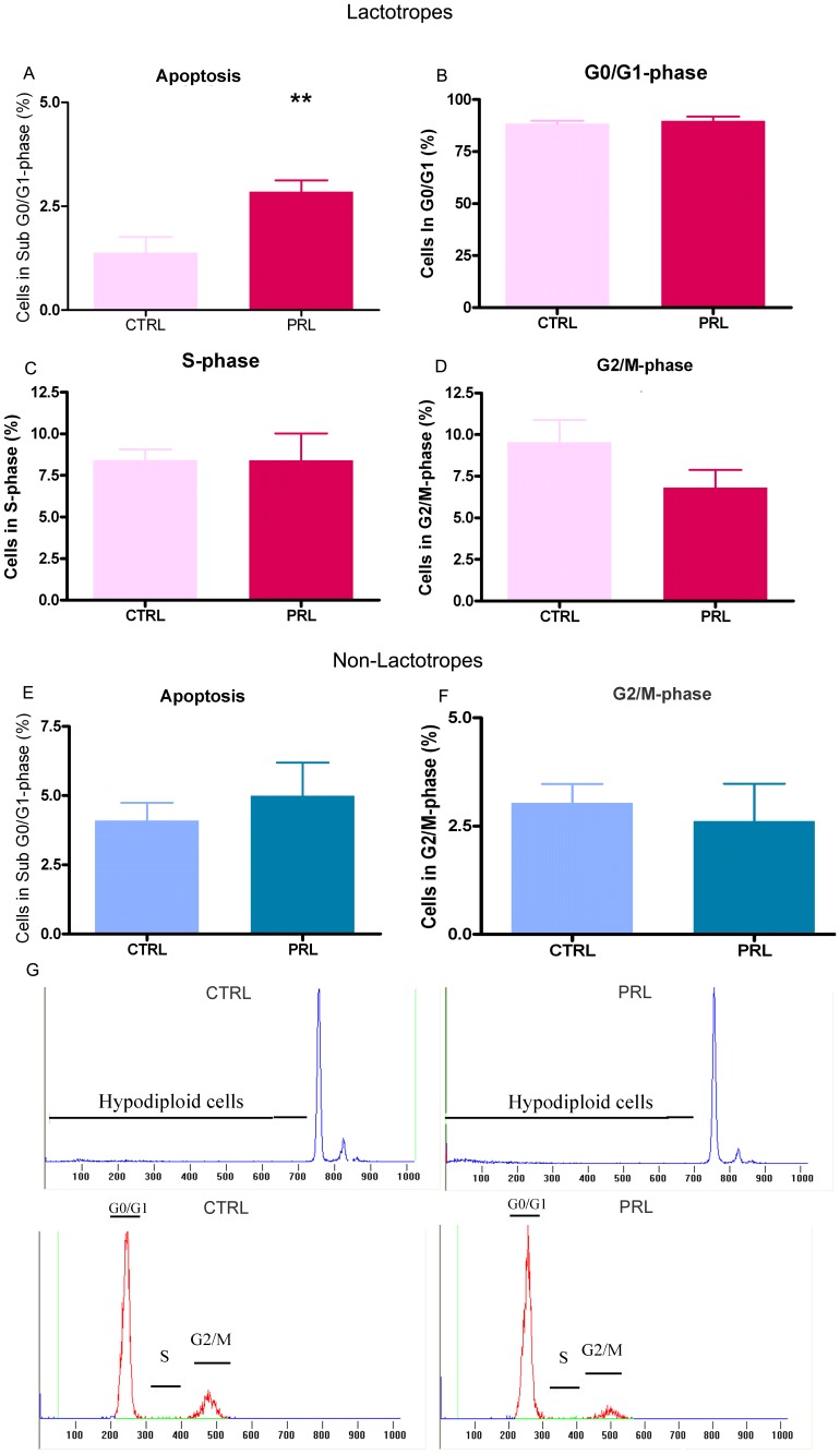
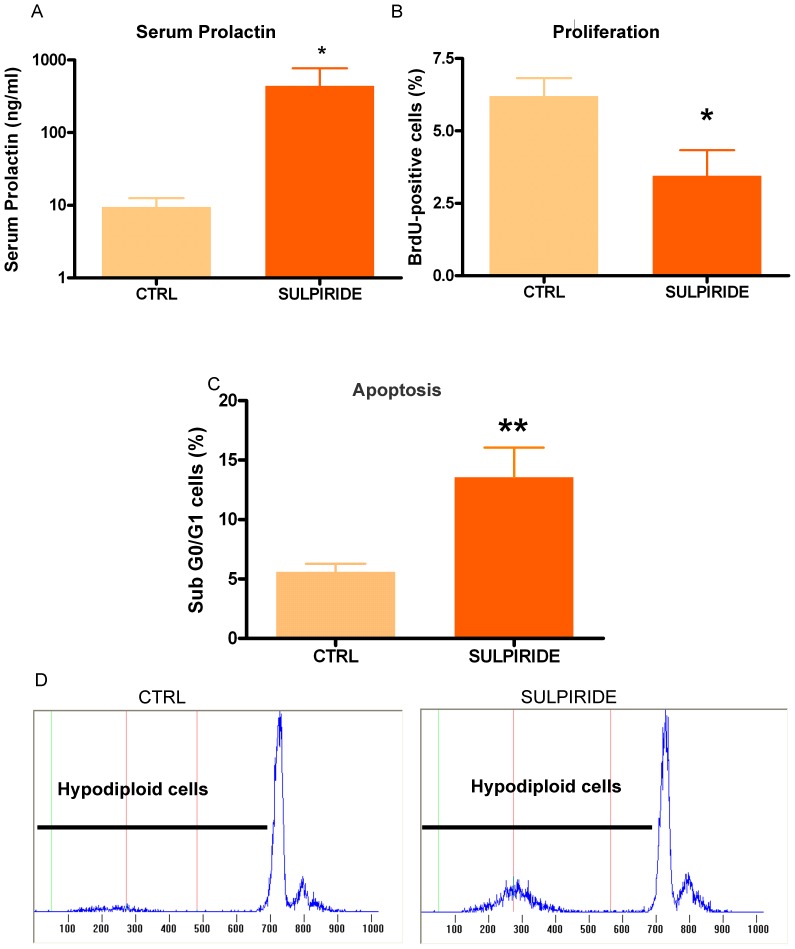
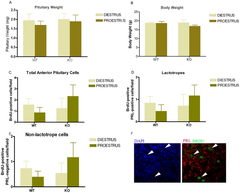
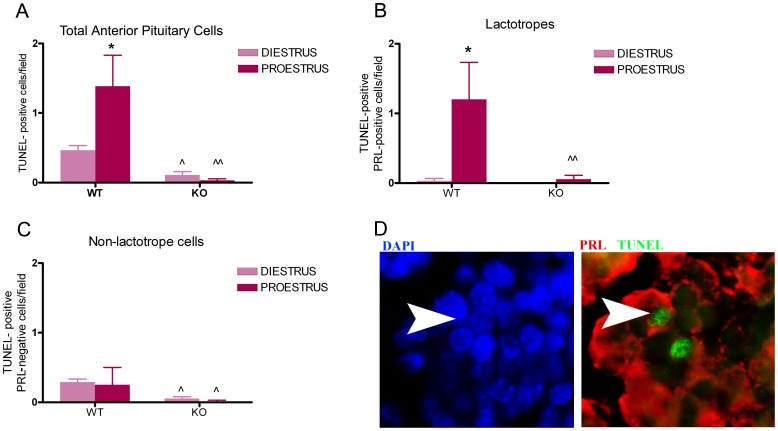
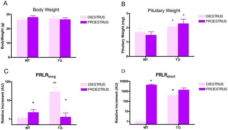
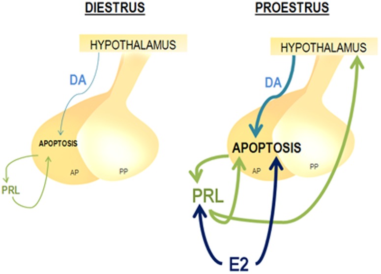
Anterior pituitary cell turnover occurring during female sexual cycle is a poorly understood process that involves complex regulation of cell proliferation and apoptosis by multiple hormones. In rats, the prolactin (PRL) surge that occurs at proestrus coincides with the highest apoptotic rate. Since anterior pituitary cells express the prolactin receptor (PRLR), we aimed to address the actual role of PRL in the regulation of pituitary cell turnover in cycling females. We showed that acute hyperprolactinemia induced in ovariectomized rats using PRL injection or dopamine antagonist treatment rapidly increased apoptosis and decreased proliferation specifically of PRL producing cells (lactotropes), suggesting a direct regulation of these cell responses by PRL. To demonstrate that apoptosis naturally occurring at proestrus was regulated by transient elevation of endogenous PRL levels, we used PRLR-deficient female mice (PRLRKO) in which PRL signaling is totally abolished. According to our hypothesis, no increase in lactotrope apoptotic rate was observed at proestrus, which likely contributes to pituitary tumorigenesis observed in these animals. To decipher the molecular mechanisms underlying PRL effects, we explored the isoform-specific pattern of PRLR expression in cycling wild type females. This analysis revealed dramatic changes of long versus short PRLR ratio during the estrous cycle, which is particularly relevant since these isoforms exhibit distinct signaling properties. This pattern was markedly altered in a model of chronic PRLR signaling blockade involving transgenic mice expressing a pure PRLR antagonist (TGΔ1-9-G129R-hPRL), providing evidence that PRL regulates the expression of its own receptor in an isoform-specific manner. Taken together, these results demonstrate that i) the PRL surge occurring during proestrus is a major proapoptotic signal for lactotropes, and ii) partial or total deficiencies in PRLR signaling in the anterior pituitary may result in pituitary hyperplasia and eventual prolactinoma development, as observed in TGΔ1-9-G129R-hPRL and PRLRKO mice, respectively.


Conflict of interest statement

Competing Interests: The authors have declared that no competing interests exist.

Figures

Figure 1
Figure 1. PRL decreases anterior pituitary cell proliferation in vivo.
Ovariectomized rats (n = 4 rats/group) were injected with oPRL (1 mg/kg, 6 h) or saline and BrdU (50 mg/kg, 6 h). Proliferation rate was determined by the detection of BrdU incorporation and FACS analysis. Each column represents the mean ± SEM of the percentage of BrdU-positive cells. *p<0.05 vs. respective control (CTRL) animals injected with vehicle, Student's t test.
Figure 2
Figure 2. PRL increases anterior pituitary cells apoptosis in vivo and decreases the percentage of total anterior pituitary cells in G2/M-phase.
Ovariectomized rats (n = 12 rats/group) were injected with oPRL (1 mg/kg, 6 h) or saline. A: Apoptosis was determined as the percentage of hypodiploid cells, using PI and FACS. Each column represents the mean ± SEM of the percentage of sub G0–G1 cells. *p<0.05 vs. respective control animals injected with vehicle, Student's t test. B–D: Cell cycle was analyzed by FACS using PI. Each column represents the mean ± SEM of the percentage of cells in G0/G1-phase (B), cells in S-phase (C) and cells in G2/M-phase (D). * p<0.05 vs. control animals injected with vehicle, Student's t test. E: Representative histograms showing hypodiploidy or cells in each cell cycle stage in CTRL and oPRL-treated animals.
Figure 3
Figure 3. PRL increases lactotrope apoptosis in vivo.
Lactotropes were identified by PRL immunostaining and analyzed by FACS. Apoptosis and cell cycle analysis were performed in the PRL-positive subpopulation. A: Apoptosis was determined by FACS using PI. Each column represents the mean ± SEM of the percentage of sub cells in G0–G1. *p<0.05 vs. CTRL animals injected with vehicle, Student's t test. B–D: Cell cycle was analyzed by FACS using PI. Each column represents the mean ± SEM of the percentage of cells in G0/G1-phase (B), cells in S-phase (C) and cells in G2/M-phase (D) p = 0.05 for G2/M in oPRL respect to CTRL, Student's t test. E–F: oPRL treatment did not change the apoptosis rate (E) or the percentage of cells in G2/M in the PRL-negative subpopulation. G: Representative histograms showing hypodiploidy or cells in each stage of the cell cycle in CTRL and PRL-treated animals.
Figure 4
Figure 4. Sulpiride decreases anterior pituitary cell proliferation in vivo.
Ovariectomized rats (n = 5–7 rats/group) were injected with sulpiride (5 mg/kg, 6 h) or saline, and with BrdU (50 mg/kg, 6 h). A: Sulpiride treatment induced hiperprolactinemia in OVX rats. Each column represents serum PRL levels ± SEM in CTRL or sulpiride-treated animals. *p<0.05 vs. CTRL animals injected with vehicle, Student t test. B: Proliferation rate determined by the detection of BrdU incorporation and FACS. Each column represents the mean ± SEM of the percentage of BrdU-positive cells. *p<0.05 vs. CTRL animals injected with vehicle, Student t test. C: Apoptosis was determined by FACS, using PI. Each column represents the mean ± SEM of the percentage of sub-G0-G1 cells. **p<0.01 vs. CTRL animals injected with vehicle, Student's t test. D: Representative histograms showing hypodiploidy in CTRL and sulpiride-treated animals.
Figure 5
Figure 5. Anterior pituitary cell proliferation in WT and PRLR KO mice at proestrus or diestrus.
Wild type and PRLRKO mice (6–10 animals per group) were injected with BrdU (50 mg/kg, 24 h) and sacrificed at proestrus or diestrus. Proliferation was determined by BrdU incorporation in tissue sections. A, B: Body weight (A) and pituitary weight (B) of WT and PRLRKO mice euthanized at diestrus or proestrus, expressed as mean ± SEM, two-way ANOVA. C: Each column represents the media ± SEM of proliferating total anterior pituitary cells (BrdU-positive cells/field). Two-way ANOVA. D: Each column represents the media ± SEM of proliferating lactotropes (BrdU-positive PRL positive cells/field), Two-way ANOVA. E: Each column represents the media ± SEM of proliferating non-lactotrope cells (BrdU-positive PRL-negative cells/field). Two-way ANOVA. F: Representative microphotographs of anterior pituitaries from KO mice euthanized at proestrus. Arrow heads indicate BrdU-positive lactotropes. Arrows indicate BrdU-positive cells.
Figure 6
Figure 6. Apoptosis in lactotropes but not in non-lactotrope cells varies during the estrous cycle and decreases in PRLRKO mice.
Wild type and PRLRKO mice (6–10 animals per group) were euthanized at proestrus or diestrus. Apoptosis was determined by TUNEL assay in tissue sections. A: Each column represents the media ± SEM of TUNEL-positive cells/field. *p<0.05 vs. diestrus WT, ∧p<0.01 vs. diestrus WT, ∧∧p<0.01 vs. proestrus WT. Two-way ANOVA, followed by Tukey's test. B: Each column represents the media ± SEM of apoptotic lactotropes (TUNEL-positive PRL-positive cells/field). *p<0.05 vs. diestrus, ∧∧p<0.01 vs. proestrus WT. Two-way ANOVA, followed by Tukey's test. C: Each column represents the media ± SEM of apoptotic non-lactotrope cells (TUNEL-positive PRL-negative cells/field). ∧p<0.05 vs. diestrus WT or proestrus WT. Two-way ANOVA, followed by Tukey's test. D: Representative microphotographs of anterior pituitaries from WT mice euthanized at proestrus. Arrow heads indicate a TUNEL-positive lactotrope.
Figure 7
Figure 7. PRLR antagonism increases the expression of the long and short isoforms of PRLR.
A, B: Body weight and pituitary weight of WT and TGΔ1–9-G129R-hPRL mice, euthanized at diestrus or proestrus ∧p<0.05 vs. respective WT at diestrus or proestrus. Two-way ANOVA followed by Tukey's test. C, D: Expression of PRLR isoforms in the anterior pituitary from WT and TGΔ1–9-G129R-hPRL mice, euthanized at diestrus or proestrus. Real Time PCR was performed using specific primers for long and short (S1, S2, S3) PRLR isoforms. The S1 short isoform was not detected. Each column represents the relative increment ± SEM with respect to the expression of PRLRlong (C) or PRLRshort (D) in control animals (WT mice at diestrus) (n≥5 animals/group). *p<0.05 vs. respective diestrus, ∧p<0.0.5 and ∧∧p<0.01 vs. respective WT at diestrus. Two-way ANOVA, followed by Tukey's test.
Figure 8
Figure 8. Proposed model of anterior pituitary apoptosis modulation during the estrous cycle.
During proestrus the surge of circulating estradiol (E2) (1) increases PRL secretion (2) which, together with E2 itself induces apoptosis of anterior pituitary cells (AP) (3, 3′). PRL stimulates hypothalamic (4) dopamine (DA) release (5) which also contributes to the apoptosis observed in the gland at this stage of the estrous cycle (6). PP: Posterior Pituitary.

References

    1. Bole-Feysot C, Goffin V, Edery M, Binart N, Kelly PA (1998) Prolactin (PRL) and its receptor: actions, signal transduction pathways and phenotypes observed in PRL receptor knockout mice. Endocr Rev 19: 225–268. - PubMed
    1. Ben-Jonathan N, LaPensee CR, LaPensee EW (2008) What can we learn from rodents about prolactin in humans? Endocr Rev 29: 1–41. - PMC - PubMed
    1. Fernandez I, Touraine P, Goffin V (2010) Prolactin and human tumourogenesis. J Neuroendocrinol 22: 771–777. - PubMed
    1. Bernichtein S, Touraine P, Goffin V (2010) New concepts in prolactin biology. J Endocrinol 206: 1–11. - PubMed
    1. Goffin V, Hoang DT, Bogorad RL, Nevalainen MT (2011) Prolactin regulation of the prostate gland: a female player in a male game. Nat Rev Urol 8: 597–607. - PMC - PubMed

Publication types

LinkOut - more resources