Review
. 2014 Dec;1839(12):1416-32.
doi: 10.1016/j.bbagrm.2014.05.009. Epub 2014 May 21.

Targeting histone lysine demethylases - progress, challenges, and the future

Affiliations
Review

Targeting histone lysine demethylases - progress, challenges, and the future

Cyrille C Thinnes et al. Biochim Biophys Acta. 2014 Dec.

Abstract

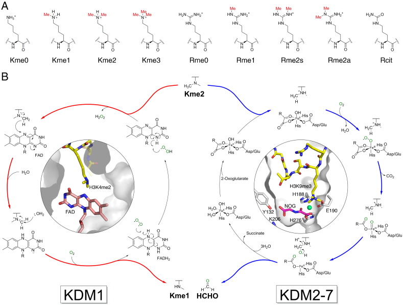
N-Methylation of lysine and arginine residues has emerged as a major mechanism of transcriptional regulation in eukaryotes. In humans, N(ε)-methyllysine residue demethylation is catalysed by two distinct subfamilies of demethylases (KDMs), the flavin-dependent KDM1 subfamily and the 2-oxoglutarate- (2OG) dependent JmjC subfamily, which both employ oxidative mechanisms. Modulation of histone methylation status is proposed to be important in epigenetic regulation and has substantial medicinal potential for the treatment of diseases including cancer and genetic disorders. This article provides an introduction to the enzymology of the KDMs and the therapeutic possibilities and challenges associated with targeting them, followed by a review of reported KDM inhibitors and their mechanisms of action from kinetic and structural perspectives.

Keywords: Demethylase; Epigenetics; Histone; Inhibition; Lysine; Methylation.


Figures

Fig. 1
Fig. 1
Histone lysyl demethylation is catalysed by histone demethylases (KDMs). (A) Methylation states of lysine and arginine residues in histones. Lysine residues may be mono-, di- or trimethylated on their Nε-amino groups (Kme1, Kme2 and Kme3 respectively), whereas arginine residues may be either mono- or dimethylated on their Nω-guanidino nitrogens (Rme1 and Rme2 respectively). Dimethylated arginine exists in either symmetric (Rme2s) or asymmetric (Rme2a) forms, depending upon the positions of methylation. Arginine may also be deaminated to form citrulline (Rcit), as catalysed by peptidylarginine deiminase activity. (B) Mechanisms of lysyl demethylation catalysed by the KDM1 and JmjC (KDM2-7) subfamilies. The KDM1 subfamily only accepts mono- and dimethylated lysines as substrates.
Fig. 2
Fig. 2
Mechanism-based inhibitors of the KDM1 subfamily. (A) Structures of representative mechanism-based KDM1 inhibitors. The MAO inhibitors phenelzine 1, tranylcypromine 2 and pargyline 3 were among the first reported inhibitors of KDM1, which led to the development of KDM1-selective analogues. (B) Views from X-ray crystal structures of tranylcypromine (2) and peptidic inhibitor (14) cross-linked to FAD in the active site of KDM1A (PDB IDs: 2Z3Y and 2UXN respectively). Views from both structures are overlaid. (C) Views from crystal structures of tranylcypromine analogues (FAD adducts) in the active site of KDM1A (PDB IDs: 2XAJ, 2XAG, 2XAH, 2XAS and 2XAQ2XAS2XAQ) . The analogues protrude into the substrate binding pocket. (D) Proposed structures of adducts formed by reaction of tranylcypromine with FAD in the active site of KDM1s. There is evidence for three of the structures from crystallographic analyses (see section E). (E) Structures of FAD adducts of mechanism-based inhibitors bound in the active sites of KDM1s (views of structures are from PDB IDs: 2Z3Y, 2XAJ, 2XAG, 2XAH, 2XAS, 2XAQ, 2Z5U, 3ABT, 3ABU, 4GUU and 2UXN[155]).
Fig. 3
Fig. 3
Structures of representative non-tranylcypromine-based KDM1 subfamily inhibitors. Bisguanidines, bisbiguanides, polyamines and (thio)urea-containing compounds (for examples see 2124) have all been shown to inhibit KDM1 activity in vitro, with some showing effects in cells. A detailed mechanistic/structural understanding of KDM1 inhibition by these compounds is presently not available. Recently, a series of other inhibitors of the KDM1 subfamily have been identified, including Namolin (26), aminothiazoles (2729), benzohydrazides (30) and a tricyclic pyridine (31).
Fig. 4
Fig. 4
Structures of representative iron-chelating inhibitors of the JmjC KDMs. (A) Structures of tricarboxylic acid (TCA) cycle intermediates and 2OG mimetics. 2OG is a co-substrate of the 2OG oxygenases. Some 2OG oxygenases are inhibited by succinate (33, a co-product of catalysis), fumarate (34), and 2-hydroxyglutarate (35 and 36). Levels of these small molecules can be substantially increased in some tumour cells. N-Oxalylglycine (NOG, 32) is a close isostere of 2OG. C-α derivatisation of NOG can confer selectivity for different 2OG oxygenase subfamilies. (B) Structures of hydroxamic acid-containing inhibitors and Daminozide (45). Hydroxamic acids are established metal chelators and with appropriate functionalisation can be potent and selective 2OG oxygenase inhibitors. Examples include the JmjC KDM inhibitor Methylstat (44), and SAHA (40), which is a clinically used inhibitor of histone deacetylases that also inhibits JmjC KDMs in vitro. Daminozide (45), a small achiral hydrazide, inhibits the KDM2/7 subfamily of JmjC KDMs. (C) Examples of pyridine-based KDM inhibitors. Pyridine-2,4-dicarboxylic acid (2,4-PDCA, 47) is a broad-spectrum 2OG oxygenase inhibitor that chelates active site-bound iron via its pyridyl nitrogen and 2-carboxylate. KDM4-selective derivatives of 2,4-PDCA have been prepared via substitution at the 3-position (e.g. 48). Recently, pyridine-containing fragments without a 2-carboxylate group (e.g. 49) have also been reported to inhibit JmjC KDMs via monodentate iron chelation. Other pyridine-containing inhibitors include bipyridyl compounds (e.g. 51 and 52), which chelate iron via both pyridyl nitrogens, and the pyridylhydrazone 50, which was recently identified from a high-throughput screen. (D) Structures of 8-hydroxyquinoline derivatives. 8-Hydroxyquinolines chelate iron in a bidentate manner via their pyridyl nitrogen and phenolic oxygen atoms. 5-Carboxy-8-hydroxyquinoline (IOX1, 54), which was identified from a high-throughput screen against KDM4E, is a broad-spectrum 2OG oxygenase inhibitor that exhibits moderate selectivity for some JmjC KDM subfamilies (KDM2/7, KDM3, KDM4, KDM6). Substitution at the 2-, 4-, 5- and 7- positions of the 8-hydroxyquinoline ring has resulted in improved selectivity for KDMs (e.g. 56-59). Crystallographic studies with KDM4A, KDM6B and the HIF asparaginyl hydroxylase FIH reveal that IOX1 can induce translocation of the active-site iron (see Fig. 5, Fig. 7). Such metal movement is not observed for 4-carboxy-8-hydroxyquinoline (4C8HQ, 55) binding to KDM4A (Fig. 5).
Fig. 5
Fig. 5
Crystal structures of inhibitors bound to KDM4A. KDM4 inhibitor structures reported to date are for bidentate iron chelators. One chelating group from the inhibitor coordinates opposite to the iron-binding glutamate residue (Glu190 in KDM4A); however, the second chelating group may coordinate either opposite the first iron-binding histidine (His188 in KDM4A), as observed for 2OG chelation (and for NOG, see top left), or opposite the second histidine (His276 in KDM4A), as observed for 2,4-PDCA (see middle right). The preference of the two binding modes is likely determined by steric factors. Binding of IOX1 in the active site of KDM4A induces translocation of the active site metal (red arrow, bottom right). In all structures, the active site iron is substituted for nickel. PDB IDs: 2OQ6, 4AI9, 2WWJ, 2YBK, 2YBS, 2VD7, 3PDQ, 4BIS and 3NJY.
Fig. 6
Fig. 6
Structures of the KDM6 subfamily inhibitor GSK-J1 and derivatives. GSK-J1 (61) is an iron chelator, which induces metal movement within the active site of KDM6B (for a view of a crystal structure of GSK-J1 bound in the active site of KDM6B, see Fig. 11). The ethyl ester prodrug (GSK-J4, 62) is active in cells, whereas the prodrug form of the inactive analogue GSK-J2 (63) shows no cellular effects (GSK-J5, 64).
Fig. 7
Fig. 7
Structures of 2-4(4-methylphenyl)-1,2-benzisothiazol-3(2H)-one (PBIT) and ebselen. PBIT (65) and ebselen (66) inhibit both the KDM4 and KDM5 subfamilies of KDMs, probably via removal of the enzyme-bound zinc.
Fig. 8
Fig. 8
Structures of histone substrate-competitive JmjC KDM inhibitors. Compounds 67-69 comprise both iron-chelating groups and histone fragments to enable potency and selectivity for JmjC KDMs. The lysine methyltransferase inhibitor BIX-01294 (70) also inhibits JmjC KDMs; development of BIX-01294 analogues has resulted in JmjC KDM-specific inhibitors (71 and 72). Crystallographic studies of 71 with KDM7A suggest that the compound may compete with the histone substrate for binding (Fig. 11).
Fig. 9
Fig. 9
Structures of flavonoid, catechol, tripartin, and pyrido[1,2-a]indole inhibitors of JmjC KDMs. Numerous flavonoid and catechol compounds have been identified as JmjC KDM inhibitors from high-throughput screening, including myricetin (73) and dopamine (76). Precise mode(s) of inhibition, however, are unclear. Tripartin (79) is an indanone-based natural product that was shown to inhibit KDM4. Pyrido[1,2-a]indoles have been recently reported to inhibit KDM4C via unknown mechanism(s).
Fig. 10
Fig. 10
View from a crystal structure of KDM4A with bound peptide substrate analogue (sequence ARK(me3)SCGGK, yellow) and N-oxalyl-D-cysteine (DNOC, pink). The peptide and DNOC form a disulfide linkage within the active site (dashed line, circled). Replacing the disulfide bond with a sulfide led to the development of a stable analogue exhibiting potent and selective inhibition for the KDM4 subfamily (68, Fig. 8). PDB ID: 3U4S.
Fig. 11
Fig. 11
Views from crystal structures of inhibitors bound to the KDM6 and KDM2/7 subfamilies. As observed in inhibitor structures with KDM4A, bidentate iron chelators may adopt one of two coordination modes around the iron, with one iron-binding group positioned opposite to the iron-binding carboxylate residue (Glu1148 in KDM6A, Glu1389 in KDM6B, Asp249 in PHF8 and Asp284 in KDM7A) and the other opposite to one of the iron-binding histidines. Both IOX1 (54, Fig. 4) and GSK-J1 (61, Fig. 7) induce metal translocation in KDM6B (note: the crystal structure of GSK-J1 binding was solved using mouse KDM6B). E67 (71, an analogue of the lysine methyltransferase inhibitor BIX-01294, 70, Fig. 8) binds away from the iron binding site in KDM7A, suggesting no metal chelation. PDB IDs: KDM6A + NOG, 3AVS (metal = nickel) ; KDM6B + IOX1, 2XXZ (metal = nickel) ; mKDM6B + GSK-J1, 4ASK (metal = cobalt) ; PHF8 + NOG, 3KV4 (metal = iron) ; PHF8 + Daminozide, 4DO0 (metal = zinc) ; KDM7A + E67, 3U78 (metal = nickel) .
Fig. 12
Fig. 12
Analysis of heterocyclic iron chelators binding to JmjC KDMs. Overlays of reported structures of heterocyclic inhibitors bound to JmjC KDMs reveal preferential positioning of 2 aromatic groups within the active sites. In structures of KDM4A with bound 2,4-PDCA (section A), IOX1 (section B), 4C8HQ (section C) and the bipyridine compound 52 (section D), an aromatic group is positioned between the metal site and the 2OG C-5 carboxylate binding site defined by residues Tyr132 and Lys206 (ring position A, section E). In KDM4A, the position of this aromatic group is likely stabilised by π-π stacking interactions with Phe185. For bicyclic chelators (IOX1, 4C8HQ and 52), the other aromatic groups are positioned in one of two discrete orientations (ring positions B or C, sections E and F). Despite IOX1 inducing translocation of the active site metal, the position of quinolyl bicyclic system is identical to that adopted by 4C8HQ, suggesting metal movement may be preferred to altered ring-positioning, at least in KDM4A.
Fig. 13
Fig. 13
Structures of 'pan-KDM' inhibitors. Compounds 80 and 81 (as racemates) combine KDM1 inhibitor tranylcypromine 2 with the JmjC KDM inhibitors 4-carboxy-4′-carboxymethoxy-2,2′-bipyridine 51 and IOX1 54 respectively. These dual inhibitors induce cell growth inhibition and apoptosis in prostate and colon cancer cell lines, but not in noncancer mesenchymal progenitor cells.

References

    1. Berdasco M., Esteller M. Aberrant epigenetic landscape in cancer: how cellular identity goes awry. Dev. Cell. 2010;19:698–711. - PubMed
    1. Suganuma T., Workman J.L. Signals and combinatorial functions of histone modifications. Annu. Rev. Biochem. 2011;80:473–499. - PubMed
    1. Zentner G.E., Henikoff S. Regulation of nucleosome dynamics by histone modifications. Nat. Struct. Mol. Biol. 2013;20:259–266. - PubMed
    1. Greer E.L., Shi Y. Histone methylation: a dynamic mark in health, disease and inheritance. Nat. Rev. Genet. 2012;13:343–357. - PMC - PubMed
    1. Portela A., Esteller M. Epigenetic modifications and human disease. Nat. Biotechnol. 2010;28:1057–1068. - PubMed

Publication types

MeSH terms