. 2014 Aug;13(8):812-6.
doi: 10.1038/nmat3979. Epub 2014 May 25.

Internal dynamics of a supramolecular nanofibre

Affiliations

Internal dynamics of a supramolecular nanofibre

Julia H Ortony et al. Nat Mater. 2014 Aug.

Abstract

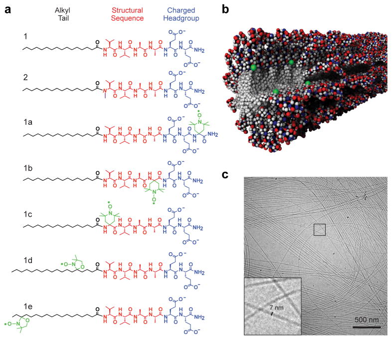
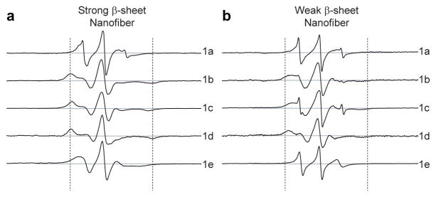
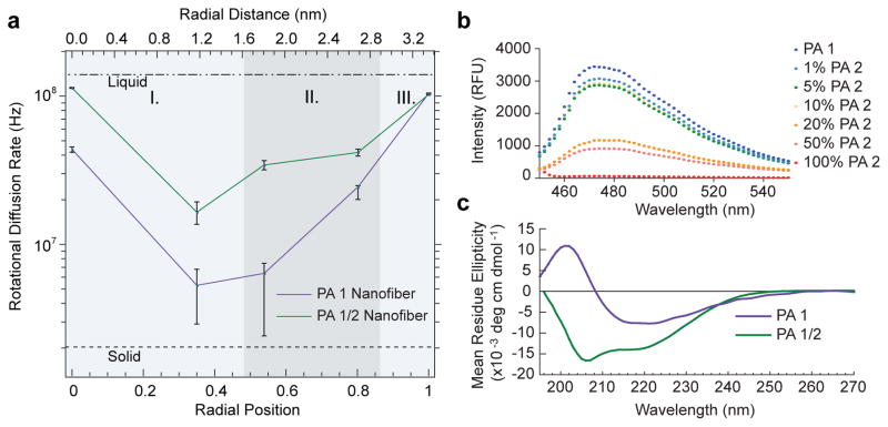
A large variety of functional self-assembled supramolecular nanostructures have been reported over recent decades. The experimental approach to these systems initially focused on the design of molecules with specific interactions that lead to discrete geometric structures, and more recently on the kinetics and mechanistic pathways of self-assembly. However, there remains a major gap in our understanding of the internal conformational dynamics of these systems and of the links between their dynamics and function. Molecular dynamics simulations have yielded information on the molecular fluctuations of supramolecular assemblies, yet experimentally it has been difficult to obtain analogous data with subnanometre spatial resolution. Using site-directed spin labelling and electron paramagnetic resonance spectroscopy, we measured the conformational dynamics of a self-assembled nanofibre in water through its 6.7 nm cross-section. Our measurements provide unique insight for the design of supramolecular functional materials.


Figures

Figure 1
Figure 1
Peptide amphiphiles self-assemble into high-aspect-ratio nanofibers. a) Chemical structures of the PA molecules investigated where the aliphatic region is shown in black, structural sequence shown in red, and charged residues in blue. PA 1 contains six residues VVAAEE, where the valines are expected to contribute to intermolecular hydrogen bonding. PA 2 contains an N-methylated valine, VN(me)VAAEE, to reduce intermolecular hydrogen bonding propensity. PAs 1a1e are labeled with radical electron spin labels (radicals shown in green); b) Molecular graphics rendering of a coassembled supramolecular nanofiber with PA molecules containing spin labels at the innermost residue corresponding to PA 1/1c; c) Cryogenic transmission electron micrograph of PA 1 in water reveals high-aspect-ratio nanofibers with diameters of approx. 7 nm; the area outlined with a square is shown magnified in the inset.
Figure 2
Figure 2
Electron paramagnetic resonance spectra at specific sites through the cross-section of peptide amphiphile nanofibers. a) EPR spectra of strong β-sheet nanofiber composed of 99.6% PA 1 and 0.4% of each spin labeled PA (1a1e). Spectra of nanofibers produced by co-assembly of PA 1 with PA 1a (top) through PA 1e (bottom) correspond to the cross-section from the nanofiber surface to the core, respectively; b) weak β-sheet nanofiber composed of 49.8% PA 1, 49.8% PA 2, and 0.4% of each PA 1a1e. Sweep widths shown correspond to 150 Gauss. Dashed lines indicate a separation of 65 Gauss.
Figure 3
Figure 3
Internal dynamics through the cross-section of peptide amphiphile nanofibers are obtained with sub-nanometer resolution and shown to depend on the presence of β-sheet promoting amino acids. a) Rotational diffusion rates (kr) extracted from EPR spectral simulations (Fig. S4) with each spin label (PAs 1a1e) integrated into the nanofiber, plotted against theoretical radial position (calculated from a fully extended PA molecule) and radial distance. The purple line corresponds to PA 1 nanofibers designed to possess strong β-sheet character, and the green line to equimolar coassemblies of PA 1 and 2 designed to possess weak β-sheet character. The three domains of the nanofiber cross-section are indicated by I. aliphatic core, II. structural domain shell, and III. charged corona. b) ThT assay showing the decrease in β-sheet character with increasing incorporation of PA 2 into PA 1 nanofibers. When the composition of the nanofiber contains < 10% PA 2, ThT fluorescence intensity remains high because β-sheet hydrogen bonding is present to a substantial extent. At 20–50% incorporation of PA 2, the nanofibers’ β-sheet character has been significantly reduced. c) Circular dichroism spectrum of PA 1 (purple) indicates strong β-sheet character, and of PA 1/2 coassembly (green) showing that after incorporation of PA 2, β-sheet character is no longer dominant.
Figure 4
Figure 4
Schematic molecular graphics representation of peptide amphiphile nanofibers. Nanofiber composed of a) PA 1 and b) PA 1/2. The vertical bar indicates the gradient of solid-like to liquid-like dynamics (blue and red, respectively) through the nanofibers’ cross-sections. PA 1 has high β-sheet character resulting in solid-like dynamics in the structural sequence. By removing the β-sheet (by coassembly of PA 1 with PA 2), faster dynamics are observed as the PA structural sequences is no longer locked into hydrogen bonding networks.

References

    1. Palmer LC, Stupp SI. Molecular self-assembly into one-dimensional nanostructures. Acc Chem Res. 2008;41:1674–1684. - PMC - PubMed
    1. Hartgerink JD, Beniash E, Stupp SI. Self-assembly and mineralization of peptide-amphiphile nanofibers. Science. 2001;294:1684. - PubMed
    1. Whitesides GM, Grzybowski B. Self-assembly at all scales. Science. 2002;295:2418–2421. - PubMed
    1. Hill JP, et al. Self-assembled hexa-peri-hexabenzocoronene graphitic nanotube. Science. 2004;304:1481–1483. - PubMed
    1. Venable RM, Zhang Y, Hardy BJ, Pastor RW. Molecular Dynamics Simulations of a Lipid Bilayer and of Hexadecane: An Investigation of Membrane Fluidity. Science. 1993;262:223–226. - PubMed

Publication types