. 2014 Aug;55(8):1678-92.
doi: 10.1194/jlr.M047738. Epub 2014 May 23.

HDL inhibits the effects of oxidized phospholipids on endothelial cell gene expression via multiple mechanisms

Affiliations

HDL inhibits the effects of oxidized phospholipids on endothelial cell gene expression via multiple mechanisms

Benjamin Emert et al. J Lipid Res. 2014 Aug.

Abstract

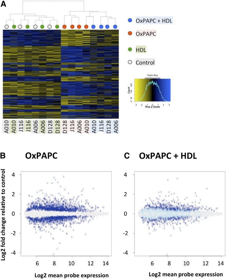
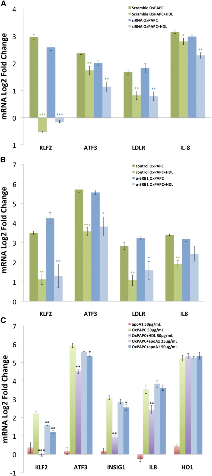
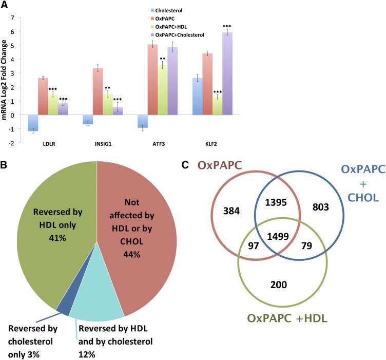
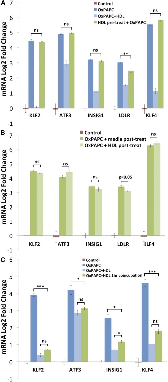
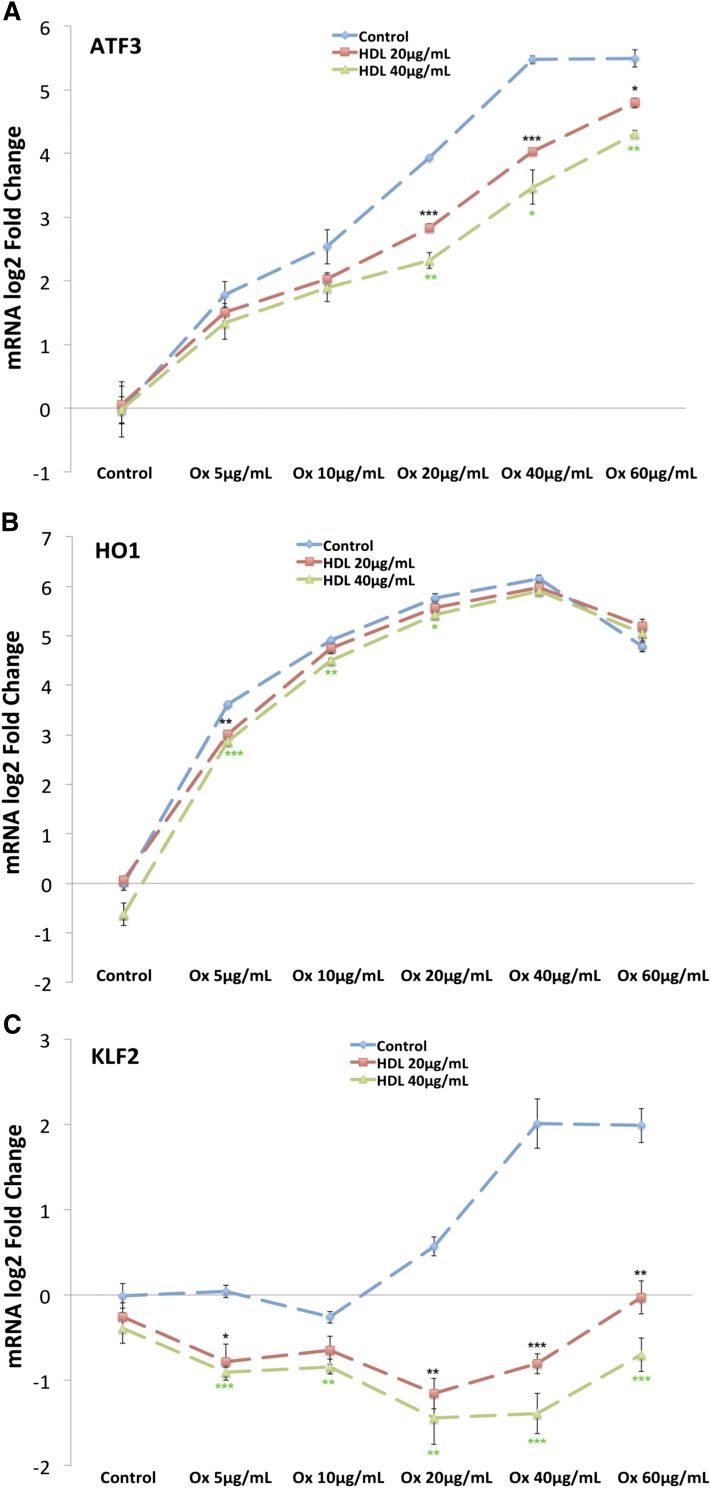
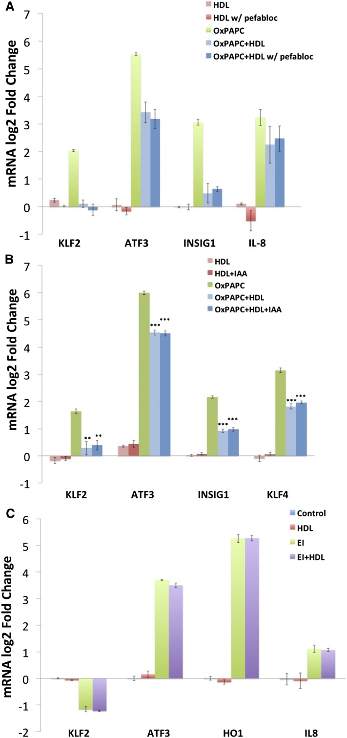
Oxidized 1-palmitoyl-2-arachidonyl-sn-glycero-3-phospholcholine (OxPAPC) and its component phospholipids accumulate in atherosclerotic lesions and regulate the expression of >1,000 genes, many proatherogenic, in human aortic endothelial cells (HAECs). In contrast, there is evidence in the literature that HDL protects the vasculature from inflammatory insult. We have previously shown that in HAECs, HDL attenuates the expression of several proatherogenic genes regulated by OxPAPC and 1-palmitoyl-2-(5,6-epoxyisoprostane E2)-sn-glycero-3-phosphocholine. We now demonstrate that HDL reverses >50% of the OxPAPC transcriptional response. Genes reversed by HDL are enriched for inflammatory and vascular development pathways, while genes not affected by HDL are enriched for oxidative stress response pathways. The protective effect of HDL is partially mimicked by cholesterol repletion and treatment with apoA1 but does not require signaling through scavenger receptor class B type I. Furthermore, our data demonstrate that HDL protection requires direct interaction with OxPAPC. HDL-associated platelet-activating factor acetylhydrolase (PAF-AH) hydrolyzes short-chain bioactive phospholipids in OxPAPC; however, inhibiting PAF-AH activity does not prevent HDL protection. Our results are consistent with HDL sequestering specific bioactive lipids in OxPAPC, thereby preventing their regulation of select target genes. Overall, this work implicates HDL as a major regulator of OxPAPC action in endothelial cells via multiple mechanisms.

Keywords: high density lipoprotein; inflammatory signaling; minimally modified low density lipoprotein; platelet-activating factor acetylhydrolase; scavenger receptor class B type I.


Figures

Fig. 1.
Fig. 1.
HDL affects 52% of genes regulated by OxPAPC. HAECs from four unique donors were treated with HDL (40 μg/ml), OxPAPC (40 μg/ml), or both (OxPAPC+HDL), and mRNA was isolated and subjected to microarray analysis. Differentially expressed genes as compared with control samples (false discovery rate adjusted P < 0.05) were identified in OxPAPC-treated samples and in OxPAPC+HDL-treated samples independently. OxPAPC regulated 1,679 genes out of the 14,899 genes analyzed on the array. A: Heat map showing the pattern of regulation of genes affected by OxPAPC in untreated cells, cells treated with HDL, cells treated with OxPAPC, and cells treated with OxPAPC+HDL. Dendogram on top shows clustering of the samples, where color of the nodes represents treatment group: gray for control, green for HDL, red for OxPAPC, and blue for OxPAPC+HDL. Individual donors are indicated below the heat map. Each sample was hybridized to two arrays, and expression was averaged between the technical replicates. While HDL itself has no apparent effect on gene expression (and on samples clustering), OxPAPC+HDL-treated samples clearly cluster separately from OxPAPC-treated samples, indicating a global effect of HDL on OxPAPC-related changes in gene expression. B: Log2-fold change of probe expression in OxPAPC-treated samples as a function of mean probe expression (x-axis). Genes that were not affected by OxPAPC are in gray. Genes that were significantly induced or repressed by OxPAPC are shown in dark blue. C: Fold change of expression of OxPAPC-affected genes (dark blue in B) in OxPAPC+HDL-treated samples. As in B, genes unaffected by OxPAPC are shown in gray. Genes affected by OxPAPC (dark blue in B) are shown in either dark blue or light blue. Dark blue indicates genes significantly affected by OxPAPC and not reversed by HDL, while light blue indicates genes reversed by HDL.
Fig. 2.
Fig. 2.
The effect of HDL on OxPAPC signaling is not mediated by SR-BI. HAECs were transfected with either siRNA targeting SR-BI or scramble control siRNA and treated 48 h later with media, OxPAPC (50 μg/ml), HDL (50 μg/ml), or OxPAPC and HDL (50 μg/ml) as described in the Materials and Methods. The mRNA expression levels of KLF2, ATF3, INSIG1, IL8, and HO1 were determined by qPCR and normalized to GAPDH expression levels. All data are presented as average log2-fold change of triplicates ± SEM. Asterisks indicate significance level in unpaired Student’s t-test (*P < 0.05, **P < 0.01, ***P < 0.001) A: Despite effective silencing of SR-BI, the expression patterns of KLF2, ATF3, LDLR, and IL-8 after OxPAPC or OxPAPC+HDL treatment were unchanged. Results were confirmed using two separate siRNAs. B: Similarly, blocking of SR-BI using SR-BI antiserum (1:300 dilution) had no effect on the HDL response of KLF2, ATF3, LDLR, or IL-8. Rabbit IgG was used as control. C: HAECs were either untreated or treated with OxPAPC (50 μg/ml) in the presence or absence of HDL (50 μg/ml) or delipidated apoA1 (25–50 μg/ml). HDL and apoA1 were added for 1 h of pretreatment and 4 h of cotreatment. The mRNA expression levels of KLF2, ATF3, INSIG1, IL-8, and HO1 were determined by qPCR and normalized to GAPDH expression levels.
Fig. 3.
Fig. 3.
Cholesterol loading mimics some aspects of the HDL response. To determine the extent to which HDL’s effect on OxPAPC signaling in HAECs is mediated by cholesterol repletion, we compared the effect of cholesterol loading using cholesterol-cyclodextrin with that of HDL. HAECs were pretreated for 1 h with media containing 1% FBS with or without HDL (50 μg/ml) or cholesterol-cyclodextrin (20 μg/ml). Media or OxPAPC was then added to a final concentration of 50 μg/ml. A: After a 4 h treatment, mRNA was isolated and quantified by qPCR. The expression of SREBP target genes (LDLR and INSIG1) was induced by OxPAPC and similarly affected by HDL and cholesterol-cyclodextrin cotreatment. In contrast, HDL inhibited the OxPAPC-mediated induction of ATF3 and KLF2, whereas cholesterol-cyclodextrin had no effect and enhanced the upregulation of ATF3 and KLF2, respectively. Data are presented as average log2-fold change of triplicates ± SEM. * P < 0.05, ** P < 0.01, *** P < 0.001 for comparison with OxPAPC log2-fold changes. P values defined by unpaired Student’s t-test. B, C: To further characterize the similarity between the effects of HDL and cholesterol-cyclodextrin on OxPAPC signaling in HAECs, we repeated the treatment described previously on three additional HAEC donors and quantified global mRNA expression by microarray. B: Pie chart summarizing the overlap between the effects of HDL and cholesterol-cyclodextrin on genes affected by OxPAPC. Consistent with our previous experiment, HDL reversed a majority (53%) of genes affected by OxPAPC. In contrast, cholesterol-cyclodextrin reversed only 23% of genes reversed by HDL. Only 3% of genes affected by OxPAPC were reversed by cholesterol but not by HDL. C: Venn diagram, comparing cholesterol treatment versus HDL treatment. Cholesterol-cyclodextrin, when combined with OxPAPC, had synergistic effects on gene expression as reflected by the number of genes (i.e., 803) affected only in presence of both. OxPAPC and HDL, however, had a limited synergistic effect on gene expression.
Fig. 4.
Fig. 4.
The effect of HDL on the OxPAPC response is mediated predominantly by direct interactions between HDL and OxPAPC. A: HDL pretreatment alone has minimal effect on the OxPAPC response. HAECs were pretreated for 2 h with HDL (50 μg/ml), washed three times with PBS to remove HDL, and then cells treated for 4 h with OxPAPC (50 μg/ml). In contrast to the standard cotreatment (shown as OxPAPC+HDL), pretreatment alone with HDL did not inhibit the effect of OxPAPC on the expression of KLF2, ATF3, INSIG1, and KLF4 and modestly reduced the induction of LDLR. For the standard treatment, cells were incubated without (dark blue bar) or with HDL (light blue bar) for 1 h, and then OxPAPC was added to the media for 4 h. B: Posttreatment with HDL does not inhibit the effect of OxPAPC on HAEC gene expression. HAECs were treated for 3 h with OxPAPC (50 μg/ml), washed three times with PBS to remove OxPAPC, and posttreated with either control media or media containing HDL (50 μg/ml) for an additional 3 h. Posttreatment with HDL did not reduce the induction of KLF2, ATF3, INSIG1, and KLF4 and only marginally affected the induction of LDLR compared with posttreatment with media alone. C: In contrast, coincubation of OxPAPC with HDL reduces the OxPAPC response to a similar extent as our standard HAEC pretreatment with HDL followed by cotreatment. OxPAPC (50 μg/ml) was incubated for 1 h at 37°C in medium M199 + 1% FBS with or without HDL (50 μg/ml). The solution was then used to treat HAECs for 4 h in parallel with standard control, OxPAPC (50 μg/ml), and OxPAPC+HDL (50 μg/ml) treatments. In both cases, OxPAPC was cotreated with HDL for 4 h. Data are presented as average log2-fold change of triplicate samples ± SEM. * P < 0.05, ** P < 0.01, *** P < 0.001 for indicated comparisons. P values defi ned by unpaired Student’s t-test.
Fig. 5.
Fig. 5.
HDL has a gene-specific effect on OxPAPC signaling in HAECs. HAECs were pretreated in the absence or presence of HDL at 20 μg/ml or 40 μg/ml for 1 h as described in the Materials and Methods. Subsequently, OxPAPC was added to a final concentration of 5 μg/ml, 10 μg/ml, 20 μg/ml, 40 μg/ml, or 60 μg/ml. After a 4 h cotreatment, mRNA was isolated from cells and quantified by qPCR. The expression of ATF3 (A) was induced in the presence of OxPAPC, and HDL cotreatment caused a dose-dependent shift in the dose-response curve. B: In contrast, HDL had a minimal effect on HO1 induction at all concentrations of OxPAPC tested. C: Unlike ATF3 and HO1, the expression of KLF2 was induced by OxPAPC and repressed in the presence of both OxPAPC and HDL, indicating a syngergistic effect of cotreatment. Points along the dose-response curve were calculated as average log2-fold change versus media control. All treatments were run in triplicate, and error bars correspond to ± SEM. Asterisks indicate significance level (*P < 0.05, **P < 0.01, ***P < 0.001).
Fig. 6.
Fig. 6.
The effect of HDL on OxPAPC signaling in HAECs is not dependent on the activity of PAF-AH or direct binding to cysteine residues but is specific to phospholipid components of OxPAPC. HDL (5 mg/ml) was incubated with 1 mM Pefabloc at 37°C for 1 h to inactivate lipoprotein-associated PAF-AH. The HDL was then dialyzed overnight and used in cotreatments with OxPAPC. After the inactivation of PAF-AH, HDL (50 μg/ml) reduced the effects of OxPAPC on markers of inflammatory response (IL-8 and KLF2), ER stress (ATF3), and cholesterol depletion (INSIG1) (A). Shown are the results from one of three representative experiments. Data are presented as average log2-fold change of duplicate samples ± (Max − Min)/2. B: HDL (5 mg/ml) was incubated with 100 mM iodoacetamide at 37°C for 1 h to irreversibly block free thiol residues on HDL. The HDL was then dialyzed overnight and used in HAEC treatments as described in the Materials and Methods. Treatment with iodoacetamide did not affect the ability of HDL to inhibit OxPAPC signaling in HAECs indicating that OxPAPC binding to cysteine residues on HDL is not mediating HDL protection. Data are presented as average log2-fold change of triplicates ± SEM. * P < 0.05, ** P < 0.01, *** P < 0.001 for comparison with OxPAPC log2-fold changes. P values defined by unpaired Student’s t-test. C: HAECs were treated with 1 μg/ml EI in the absence or presence of 50 μg/ml HDL for 4 h as described in the Materials and Methods. Unlike for OxPAPC, HDL cotreatment did not mitigate the effects of EI on the expression of IL-8, KLF2, ATF3, or HO1 as determined by qPCR. Data are presented as average log2-fold change versus control treated samples. All treatments were run in triplicate, and error bars correspond to SEM.

References

    1. Kannel W. B., Dawber T. R., Frideman G. D., Glennon W. E., McNamara P. M. 1964. Risk factors in coronary heart disease. An evaluation of several serum lipids as predictors of coronary heart disease: the Framingham Study. Ann. Intern. Med. 61: 888–899. - PubMed
    1. Kannel W. B., Dawber T. R., Kagan A., Revotskie N., Stokes J., III 1961. Factors of risk in the development of coronary heart disease—six-year follow-up experience: the Framingham Study. Ann. Intern. Med. 55: 33–50. - PubMed
    1. Lusis A. J. 2000. Atherosclerosis. Nature. 407: 233–241. - PMC - PubMed
    1. Vaya J. 2013. The association between biomarkers in the blood and carotid plaque composition-focusing on oxidized lipids, oxysterols and plaque status. Biochem Pharmacol. 86: 15–18. - PubMed
    1. Berliner J. A., Watson A. D. 2005. A role for oxidized phospholipids in atherosclerosis. N. Engl. J. Med. 353: 9–11. - PubMed

MeSH terms

LinkOut - more resources