

Increased mitochondrial apoptotic priming of human regulatory T cells after allogeneic hematopoietic stem cell transplantation
- PMID: 24859877
- PMCID: PMC4562540
- DOI: 10.3324/haematol.2014.104166
Increased mitochondrial apoptotic priming of human regulatory T cells after allogeneic hematopoietic stem cell transplantation
Abstract
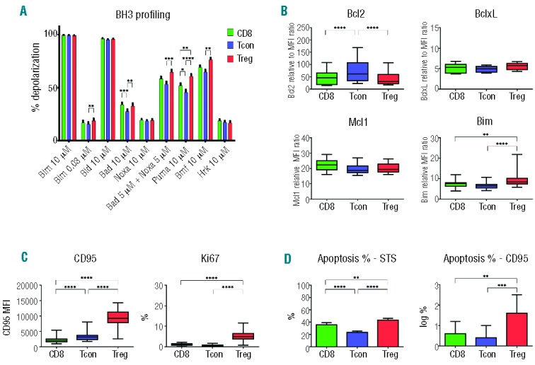
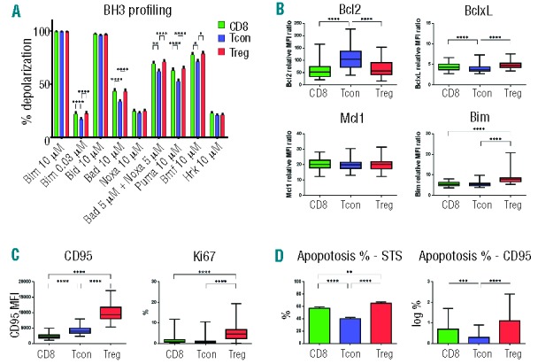
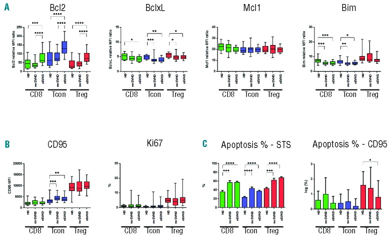
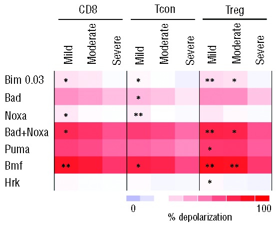
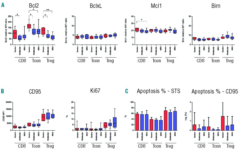
CD4 regulatory T cells play a critical role in establishment of immune tolerance and prevention of graft-versus-host disease after allogeneic hematopoietic stem cell transplantation. The recovery and maintenance of regulatory T cells is dependent on homeostatic factors including the generation of naïve regulatory T cells from hematopoietic precursor cells, the proliferation and expansion of mature regulatory T cells, and the survival of regulatory T cells in vivo. In this study, quantitation of mitochondrial apoptotic priming was used to compare susceptibility of regulatory T cells, conventional CD4 T cells and CD8 T cells to intrinsic pathway apoptosis in 57 patients after allogeneic hematopoietic stem cell transplantation and 25 healthy donors. In healthy donors, regulatory T cells are more susceptible to mitochondrial priming than conventional T cells. Mitochondrial priming is increased after hematopoietic stem cell transplantation in all T-cell subsets and particularly in patients with chronic graft-versus-host disease. Regulatory T cells express high levels of CD95 and are also more susceptible than conventional T cells to apoptosis through the extrinsic pathway. However, CD95 expression and extrinsic pathway apoptosis is not increased after hematopoietic stem cell transplantation. Decreased expression of BCL2 and increased expression of BIM, a mitochondrial cell death activator protein, in regulatory T cells contributes to increased mitochondrial priming in this T-cell subset but additional factors likely contribute to increased mitochondrial priming following hematopoietic stem cell transplantation.
Copyright© Ferrata Storti Foundation.
Figures

Similar articles
-
Unbalanced recovery of regulatory and effector T cells after allogeneic stem cell transplantation contributes to chronic GVHD.Blood. 2016 Feb 4;127(5):646-57. doi: 10.1182/blood-2015-10-672345. Epub 2015 Dec 15. Blood. 2016. PMID: 26670634 Free PMC article.
-
Naive and Stem Cell Memory T Cell Subset Recovery Reveals Opposing Reconstitution Patterns in CD4 and CD8 T Cells in Chronic Graft vs. Host Disease.Front Immunol. 2019 Mar 6;10:334. doi: 10.3389/fimmu.2019.00334. eCollection 2019. Front Immunol. 2019. PMID: 30894856 Free PMC article.
-
CD4+CD25+FOXP3+ T regulatory cells reconstitute and accumulate in the bone marrow of patients with multiple myeloma following allogeneic stem cell transplantation.Haematologica. 2008 Mar;93(3):423-30. doi: 10.3324/haematol.11897. Epub 2008 Feb 20. Haematologica. 2008. PMID: 18287134
-
The role of regulatory T cells in graft-versus-host disease management.Expert Rev Hematol. 2020 Feb;13(2):141-154. doi: 10.1080/17474086.2020.1709436. Epub 2020 Jan 11. Expert Rev Hematol. 2020. PMID: 31874061 Review.
-
Hematopoietic stem cell graft manipulation as a mechanism of immunotherapy.Int Immunopharmacol. 2003 Aug;3(8):1121-43. doi: 10.1016/S1567-5769(03)00014-6. Int Immunopharmacol. 2003. PMID: 12860168 Review.
Cited by
-
Loss of BIM in T cells results in BCL-2 family BH3-member compensation but incomplete cell death sensitivity normalization.Apoptosis. 2020 Apr;25(3-4):247-260. doi: 10.1007/s10495-020-01593-6. Apoptosis. 2020. PMID: 31993851 Free PMC article.
-
PD-1 modulates regulatory T-cell homeostasis during low-dose interleukin-2 therapy.Blood. 2017 Apr 13;129(15):2186-2197. doi: 10.1182/blood-2016-09-741629. Epub 2017 Feb 1. Blood. 2017. PMID: 28151427 Free PMC article.
-
Adapted to Survive: Targeting Cancer Cells with BH3 Mimetics.Cancer Discov. 2022 May 2;12(5):1217-1232. doi: 10.1158/2159-8290.CD-21-1334. Cancer Discov. 2022. PMID: 35491624 Free PMC article. Review.
-
Hematopoietic Stem Cell Niche During Homeostasis, Malignancy, and Bone Marrow Transplantation.Front Cell Dev Biol. 2021 Jan 22;9:621214. doi: 10.3389/fcell.2021.621214. eCollection 2021. Front Cell Dev Biol. 2021. PMID: 33553181 Free PMC article. Review.
-
BAK is a predictive and prognostic biomarker for the therapeutic effect of docetaxel treatment in patients with advanced gastric cancer.Gastric Cancer. 2016 Jul;19(3):827-38. doi: 10.1007/s10120-015-0557-1. Epub 2015 Oct 20. Gastric Cancer. 2016. PMID: 26486506 Clinical Trial.
References
-
- Bhushan V, Collins RH., Jr Chronic graft-vs-host disease. JAMA. 2003;290(19):2599–603. - PubMed
-
- Edinger M, Hoffmann P, Ermann J, Drago K, Fathman CG, Strober S, et al. CD4+CD25+ regulatory T cells preserve graft-versus-tumor activity while inhibiting graft-versus-host disease after bone marrow transplantation. Nat Med. 2003;9(9):1144–50. - PubMed
Publication types
MeSH terms
Substances
Grants and funding
- P01 AI056299/AI/NIAID NIH HHS/United States
- P01 CA065493/CA/NCI NIH HHS/United States
- R01 HL118979/HL/NHLBI NIH HHS/United States
- P01 CA078378/CA/NCI NIH HHS/United States
- T32 CA009172/CA/NCI NIH HHS/United States
- CA142106/CA/NCI NIH HHS/United States
- CA183559/CA/NCI NIH HHS/United States
- R01 AI034495/AI/NIAID NIH HHS/United States
- R01 CA183560/CA/NCI NIH HHS/United States
- CA183560/CA/NCI NIH HHS/United States
- R01 CA183559/CA/NCI NIH HHS/United States
- R01 CA072669/CA/NCI NIH HHS/United States
- P01 CA142106/CA/NCI NIH HHS/United States
- R01 HL056067/HL/NHLBI NIH HHS/United States
- AI056299/AI/NIAID NIH HHS/United States
LinkOut - more resources
Full Text Sources
Other Literature Sources
Research Materials
