

Targeted gene therapy and cell reprogramming in Fanconi anemia
- PMID: 24859981
- PMCID: PMC4203359
- DOI: 10.15252/emmm.201303374
Targeted gene therapy and cell reprogramming in Fanconi anemia
Abstract
Gene targeting is progressively becoming a realistic therapeutic alternative in clinics. It is unknown, however, whether this technology will be suitable for the treatment of DNA repair deficiency syndromes such as Fanconi anemia (FA), with defects in homology-directed DNA repair. In this study, we used zinc finger nucleases and integrase-defective lentiviral vectors to demonstrate for the first time that FANCA can be efficiently and specifically targeted into the AAVS1 safe harbor locus in fibroblasts from FA-A patients. Strikingly, up to 40% of FA fibroblasts showed gene targeting 42 days after gene editing. Given the low number of hematopoietic precursors in the bone marrow of FA patients, gene-edited FA fibroblasts were then reprogrammed and re-differentiated toward the hematopoietic lineage. Analyses of gene-edited FA-iPSCs confirmed the specific integration of FANCA in the AAVS1 locus in all tested clones. Moreover, the hematopoietic differentiation of these iPSCs efficiently generated disease-free hematopoietic progenitors. Taken together, our results demonstrate for the first time the feasibility of correcting the phenotype of a DNA repair deficiency syndrome using gene-targeting and cell reprogramming strategies.
Keywords: Fanconi anemia; cell reprogramming; gene‐targeting; iPSCs; zinc finger nucleases.
© 2014 The Authors. Published under the terms of the CC BY 4.0 license.
Figures

Top: schematic representation of the donor integrase-defective lentiviral vector (IDLV) used to promote insertion of the EGFP/FANCA cassette into the AAVS1 locus. Middle: AAVS1 locus with the zinc finger nucleases (ZFNs) target site. Bottom: AAVS1 locus upon ZFN-mediated targeted insertion of the EGFP/PGK-FANCA cassette. Black arrow shows transcription of the EGFP from the endogenous PPP1R12C promoter. HA, homology arm; SD, splice donor; SA, splice acceptor; BGHpA, bovine growth hormone polyadenylation signal; SV40pA, simian virus 40 polyadenylation signal. Constituents of the LTR (U5-R-ΔU3) are also indicated.
Proliferation advantage of targeted Fanconi anemia (FA) fibroblasts (EGFP+ cells) during in vitro incubation.
Comparative analysis of gene targeting in FA-A fibroblasts, untransduced or transduced with a lentiviral vector expressing hTERT. Analyses were performed 14 days after gene targeting.
In vitro proliferation advantage of targeted FA fibroblasts (EGFP+) previously transduced with hTERT (FA-52T fibroblasts).
Targeted integration analysis of the EGFP/PGK-FANCA cassette into the AAVS1 site by PCR using primers specific for the 5′ or 3′ integration junctions (red arrows in the top schematic) defined as 5′ TI or 3′ TI, respectively.

Top: histogram showing the percentage of FA-A fibroblasts, unstransduced or co-transduced with the donor integrase-defective lentiviral vector (IDLV) and the AdV5/35-ZFNs (geFA-52T Fib), showing FANCD2 foci in the absence or the presence of mitomycin C (MMC). Bottom: representative images of FANCD2 foci (red) in cells shown in the top histogram, after MMC treatment.
Chromosomal instability induced by diepoxybutane (DEB) in untreated (FA-52T) and gene-edited FA fibroblasts (geFA-52T Fib). Left: representative FISH analysis was performed by staining telomeres (in green), centromeres (in pink) and chromosomes (in blue). Right: histogram showing the number of chromosomal aberrations per cell.

Expression of TRA1-60, SSEA-4, OCT4, and NANOG pluripotency markers by immunofluorescence staining of gene-edited FA-iPSCs (geFA-iPSCs; clone 16).
Immunofluorescence analysis of ectoderm (β-II-tubulin), endoderm (Fox2A), and mesoderm (α-SMA and Brachyury) in teratomas generated from geFA-iPSCs (clone 16).
Southern blot analysis of genomic DNA extracted from the indicated gene-corrected FA iPSC clones (geFA-IPSCs) and from parental fibroblasts, either unmanipulated (FA) or after gene editing (ge-FA iPSCs, clones 16, 26 and 31). Genomic DNA was digested with BglI and hybridized with a probe for PPP1R12C. The band of 9.6 kb corresponds to the targeted integration in PPP1R12C, while the 3.3 kb correspond to the untargeted allele.
Southern blot analysis of samples shown in (C) digested with BstXI and hybridized with a probe (P) for EGFP. One single band of 5.1 kb is expected for specific integrations in PPP1R12C.
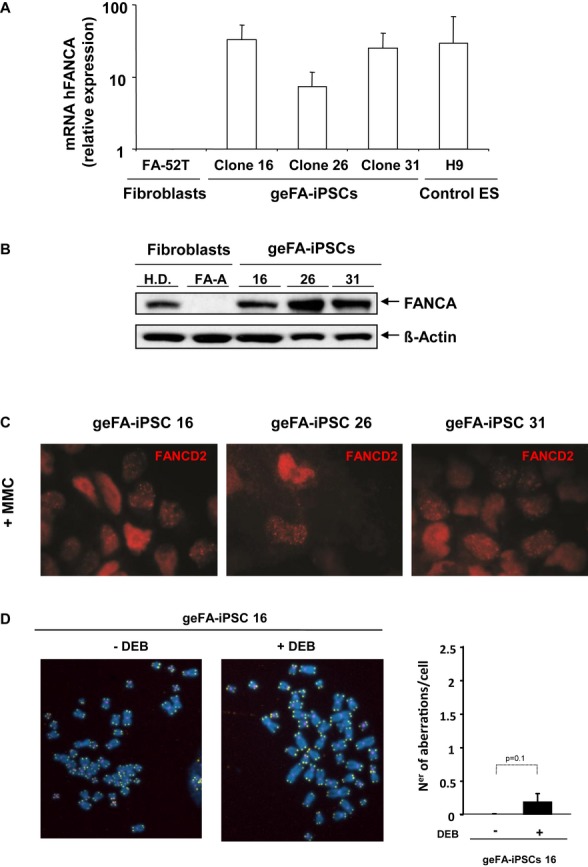
Histogram showing the levels of hFANCA expression in gene-edited FA-iPSC clones and human ES (H9) relative to untreated FA-52T fibroblasts. Data are shown as mean ± s.e. of three different analyses.
Western blot analysis showing FANCA expression in geFA-iPSC clones in comparison with fibroblasts from HD and a FA-A patient.
Representative immunofluorescence analysis of FANCD2 foci in geFA-iPSCs after DNA damage with mitomycin C (MMC).
Chromosomal instability induced by diepoxybutane (DEB) was also tested in geFA-iPSC 16. FISH analysis was performed using probes to detect telomeres (green), centromeres (pink) and chromosomes (blue). Right: histogram showing the number of chromosomal aberrations per cell.

Analysis of the percentage of CD43+CD34+, CD45+CD34+, and CD45+ cells generated by unexcised and excised geFA-iPSCs (clones 16 and Ex 16.1).
Left: Representative pictures of hematopoietic colonies generated by geFA-iPSCs. Right: Analysis of the clonogenic potential of unexcised and excised ge-FAiPSCs (clones 16 and Ex 16.1) in comparison with H.D. cord blood cells.
Survival to mitomycin C (MMC) of CFCs obtained from geFA-IPSCs (clone 16) in comparison with BM CFCs from two different FA patients (FA-664 BM and FA-82 BM) and with CFCs from a healthy cord blood (H.D. CB).
References
MeSH terms
Substances
Grants and funding
LinkOut - more resources
Full Text Sources
Other Literature Sources
Miscellaneous
