Review
. 2014 Jul 18;289(29):19862-8.
doi: 10.1074/jbc.R114.568477. Epub 2014 May 23.

Prions: generation and spread versus neurotoxicity

Affiliations
Review

Prions: generation and spread versus neurotoxicity

Mark Halliday et al. J Biol Chem. .

Abstract

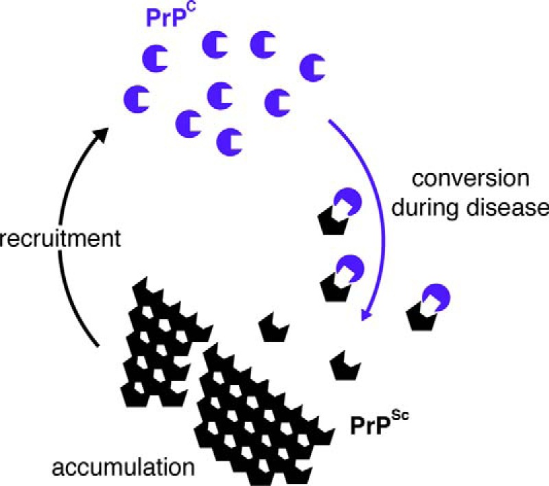
Neurodegenerative diseases are characterized by the aggregation of misfolded proteins in the brain. Among these disorders are the prion diseases, which are transmissible, and in which the misfolded proteins ("prions") are also the infectious agent. Increasingly, it appears that misfolded proteins in Alzheimer and Parkinson diseases and the tauopathies also propagate in a "prion-like" manner. However, the association between prion formation, spread, and neurotoxicity is not clear. Recently, we showed that in prion disease, protein misfolding leads to neurodegeneration through dysregulation of generic proteostatic mechanisms, specifically, the unfolded protein response. Genetic and pharmacological manipulation of the unfolded protein response was neuroprotective despite continuing prion replication, hence dissociating this from neurotoxicity. The data have clear implications for treatment across the spectrum of these disorders, targeting pathogenic processes downstream of protein misfolding.

Keywords: Alzheimer Disease; Alzheimer's; Gene Therapy; Neurodegeneration; Neuroprotection; Prion; Unfolded Protein Response (UPR).


Figures

FIGURE 1.
FIGURE 1.
Schematic of prion conversion. Native prion protein (PrPC; blue circular shapes) is converted into PrPSc (black hexagonal shapes) in an autocatalytic process during prion replication. The two proteins have identical primary but different secondary structure. PrPSc is rich in β-sheet, is protease-resistant, and accumulates, recruiting more PrPC for further cycles of conversion.
FIGURE 2.
FIGURE 2.
Schematic representation of PERK branch of the UPR leading to translational repression and points of intervention. Rising levels of misfolded proteins are detected by binding immunoglobulin protein (BiP) in the ER, activating PERK, which autophosphorylates, and in turn phosphorylates eIF2α, resulting in reduced translation. The decline in protein synthesis leads to the loss of key proteins, and hence synaptic failure and neurodegeneration. The points of action of GSK2606414, (a specific inhibitor of PERK), of lentivirus mediating RNAi of PrP (LV-shPrP), and of lentivirus overexpressing the eIF2α-P phosphatase, GADD34/PP1 (LV-GADD34) are shown. By inhibiting/preventing PERK phosphorylation (GSK2606414 and LV-shPrP) or dephosphorylating eIF2α-P (LV-GADD34), protein synthesis is restored. (Salubrinal prevents dephosphorylation of eIF2α-P, exacerbating the reduction of translation.)
FIGURE 3.
FIGURE 3.
Manipulation of the UPR rescues translation and is neuroprotective in prion-diseased mice. a, lentivirally mediated RNAi against PrP (blue bars) or overexpression of GADD34 (green) reduces levels of eIF2α-P. LV-shPrP, lentivirus mediating RNAi of PrP; LV-GADD34, lentivirus overexpressing the eIF2α-P phosphatase, GADD34/PP1. b, restoring synapse number, global protein synthesis rates, burrowing behavior, and neuronal cell numbers when compared with untreated prion-diseased mice (black) or empty vector controls (gray). Salubrinal (orange) had a detrimental effect in the same experiments. All data in bar charts show mean ± S.E. *, p < 0.01; **, p < 0.001; ***, p < 0.005. c, neuroprotective effects of RNAi of PrP or GADD34 overexpression in CA1 pyramidal cell ribbon of hippocampus from prion-diseased mice. Adapted from Ref. .
FIGURE 4.
FIGURE 4.
PERK inhibition by GSK2606414 prevents clinical disease in prion-infected mice. a, mice were treated with GSK2606414 (blue) or vehicle (red) from 7 wpi. b–c, GSK2606414 restored global protein synthesis rates (b), prevented loss of novel object memory (c), and afforded marked neuroprotection in hippocampus (d). e, levels of total PrP and PrPSc (as shown by proteinase K digestion (PK)) were unaffected by treatment. f and g, clinical cure in treated mice with normal posture and movement of hind legs. All data in bar charts show mean ± S.E. Controls represent mice inoculated with normal brain homogenate (white bar) (n = 12 for each) (*, p < 0.01). Adapted from Ref. .

Similar articles

Cited by

References

    1. Prusiner S. B. (1982) Novel proteinaceous infectious particles cause scrapie. Science 216, 136–144 - PubMed
    1. Griffith J. S. (1967) Self-replication and scrapie. Nature 215, 1043–1044 - PubMed
    1. McKinley M. P., Bolton D. C., Prusiner S. B. (1983) A protease-resistant protein is a structural component of the scrapie prion. Cell 35, 57–62 - PubMed
    1. Oesch B., Westaway D., Wälchli M., McKinley M. P., Kent S. B., Aebersold R., Barry R. A., Tempst P., Teplow D. B., Hood L. E., et al. (1985) A cellular gene encodes scrapie PrP 27–30 protein. Cell 40, 735–746 - PubMed
    1. Chesebro B., Race R., Wehrly K., Nishio J., Bloom M., Lechner D., Bergstrom S., Robbins K., Mayer L., Keith J. M., et al. (1985) Identification of scrapie prion protein-specific mRNA in scrapie-infected and uninfected brain. Nature 315, 331–333 - PubMed

MeSH terms